What is the investigation of choice for chronic pancreatitis?
Which imaging modality is best for detecting an abnormally located placenta?
All of the following are radio-opaque stones except?
What is the most accurate ultrasound method for assessing gestational age?
Reflex nephropathy is diagnosed mainly by:
Spider leg appearance is seen in which of the following conditions?
Imaging studies of a patient with a genetic disorder shows the 'central dot sign'. Which condition is characteristic of this finding?
Free gas in the abdomen, specifically under the diaphragm, can be best diagnosed by which radiographic view?
Which of the following imaging findings is diagnostic of Adenomyosis?
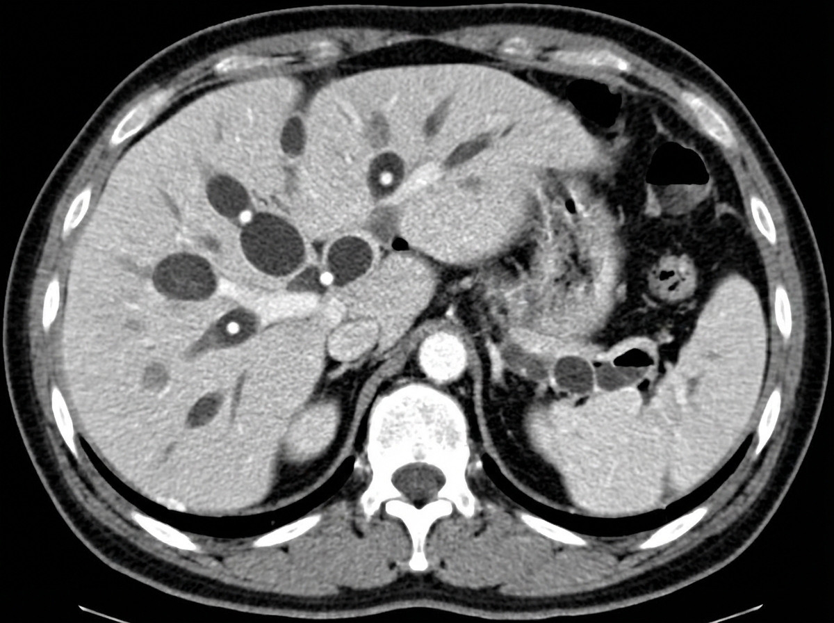
The "cut off" sign seen on imaging is characteristic of which of the following conditions?
Explanation: **Explanation:** **Why ERCP is the Correct Answer:** Endoscopic Retrograde Cholangiopancreatography (ERCP) is traditionally considered the **gold standard** for diagnosing chronic pancreatitis because of its high sensitivity in detecting early ductal changes. It allows for the visualization of the "Chain of Lakes" appearance (alternating segments of dilation and stenosis in the main pancreatic duct). While MRCP is now the preferred non-invasive diagnostic tool, ERCP remains the "investigation of choice" in many classic MCQ formats due to its ability to provide detailed ductal anatomy and its therapeutic potential (stenting/stone removal). **Analysis of Incorrect Options:** * **A. Ultrasound (USG):** Often the initial screening tool. It can show calcifications or pseudocysts but lacks the sensitivity to detect early-stage chronic pancreatitis or subtle ductal changes. * **B. Abdominal X-ray:** Useful only for identifying **pancreatic calcifications** (seen in ~30% of cases), which is a pathognomonic sign. However, a negative X-ray does not rule out the disease. * **C. Barium Study:** This is obsolete for pancreatic pathology. It may indirectly show an "inverted 3 sign" (Frostberg’s sign) on the duodenum due to an enlarged pancreatic head, but it cannot diagnose the parenchyma or ducts. **High-Yield Clinical Pearls for NEET-PG:** * **Most sensitive non-invasive test:** Magnetic Resonance Cholangiopancreatography (MRCP) with Secretin enhancement. * **Most sensitive imaging for early changes:** Endoscopic Ultrasound (EUS). * **Pathognomonic finding:** Pancreatic calcifications on CT or X-ray. * **Classic Sign:** "Chain of Lakes" appearance (dilated duct with strictures). * **Triad of Chronic Pancreatitis:** Steatorrhea, Diabetes Mellitus, and Pancreatic Calcifications.
Explanation: **Explanation:** The gold standard for diagnosing an abnormally located placenta (Placenta Previa) is **Transvaginal Sonography (TVS)**. **1. Why TVS is the Correct Answer:** TVS provides superior resolution and allows for a more accurate measurement of the distance between the internal cervical os and the placental edge. Unlike other methods, it is not affected by maternal obesity or the position of the fetal head. It is proven to be safe and does not increase the risk of bleeding, provided the probe is not inserted into the cervical canal. **2. Why Other Options are Incorrect:** * **Transabdominal Sonography (TAS):** While often the initial screening tool, TAS has a high false-positive rate (up to 25%). It is limited by maternal habitus, acoustic shadowing from the fetal head, and "apparent" previa caused by an overdistended bladder compressing the lower uterine segment. * **Doppler Ultrasonography:** This is primarily used to detect **Placenta Accreta Spectrum** (morbidly adherent placenta) by visualizing hypervascularity at the bladder-uterine interface, rather than just the location of the placenta. * **MRI:** While highly accurate for posterior placentas and assessing the depth of invasion in accreta, it is expensive, not readily available, and reserved as a second-line modality when ultrasound results are inconclusive. **Clinical Pearls for NEET-PG:** * **Distance Rule:** A distance of **<2 cm** from the internal os defines a low-lying placenta; if it covers the os, it is placenta previa. * **Placental Migration:** A diagnosis of previa made in the first or second trimester often resolves by the third trimester due to the differential growth of the lower uterine segment. * **Safety:** TVS is the "Gold Standard" and is safe even in the presence of active vaginal bleeding.
Explanation: In radiology, the visibility of a urinary stone on a plain X-ray (KUB) depends on its effective atomic number and density. Stones with high calcium content or heavy minerals attenuate X-rays and appear **radio-opaque** (white), while those composed of organic compounds without heavy atoms are **radiolucent** (invisible on X-ray). **Explanation of the Correct Answer:** * **C. Uric acid:** These are the classic **radiolucent** stones. They are composed of organic material with a low atomic number, making them invisible on plain radiographs. However, they can be visualized as "filling defects" on IVP or clearly seen on Non-Contrast CT (NCCT), where they typically show low Hounsfield Units (400–600 HU). **Explanation of Incorrect Options:** * **A. Calcium (Oxalate/Phosphate):** These are the **most radio-opaque** stones due to the high atomic number of calcium. They are easily seen on plain X-rays. * **B. Struvite (Triple Phosphate):** Composed of Magnesium Ammonium Phosphate, these are radio-opaque (though less dense than pure calcium). They are often associated with Proteus infections and can form large **Staghorn calculi**. * **C. Cystine:** These are often described as **"faintly radio-opaque"** or having a "ground-glass" appearance. While less dense than calcium, they contain sulfur atoms, which provide enough attenuation to be visible on a plain film. **NEET-PG High-Yield Pearls:** 1. **Mnemonic for Opacity (Decreasing order):** **C**alcium > **S**truvite > **C**ystine > **U**ric acid/Xanthine (**C**an **S**ome **C**ats **U**nderstand?). 2. **Pure Radiolucent Stones:** Uric acid, Xanthine, and Indinavir (protease inhibitor) stones. 3. **Gold Standard Investigation:** **NCCT (Non-Contrast CT) KUB** is the investigation of choice for urolithiasis as it detects all stones (except Indinavir). 4. **Management:** Uric acid stones are unique because they can often be dissolved via **medical alkalization** of urine (pH > 6.5).
Explanation: **Explanation:** The **Crown-Rump Length (CRL)** is the most accurate ultrasound parameter for dating a pregnancy. It is measured from the top of the fetal head (crown) to the bottom of the buttocks (rump) between **7 and 13 weeks + 6 days** of gestation. During this period, fetal growth is rapid and biological variation is minimal, allowing for a dating accuracy of within **±3 to 5 days**. **Analysis of Options:** * **Crown-Rump Length (CRL):** Correct. It is the gold standard for gestational age (GA) assessment in the first trimester. * **Gestational Sac Size (Mean Sac Diameter):** This is the earliest sign of pregnancy on USG (visible at ~5 weeks), but it is less accurate than CRL because sac shape can be distorted by bladder filling or uterine contractions. * **Femur Length (FL):** Along with Biparietal Diameter (BPD) and Abdominal Circumference (AC), this is used in the second and third trimesters. Accuracy decreases as pregnancy progresses (±2–3 weeks in the third trimester) due to genetic growth variations. * **Menstrual History (LMP):** While used to calculate the "Expected Date of Delivery" (Naegele’s rule), it is often unreliable due to irregular cycles, late ovulation, or poor patient recall. **High-Yield Clinical Pearls for NEET-PG:** * **First Trimester:** CRL is the most accurate. * **Second Trimester:** Biparietal Diameter (BPD) is the most common parameter used, but the combination of BPD, HC, AC, and FL is preferred. * **Rule of Thumb:** The earlier the ultrasound is performed, the more accurate the gestational age estimation. * **Transvaginal Sonography (TVS):** Can detect a gestational sac at a β-hCG level of **1,500–2,000 mIU/mL** (Discriminatory Zone).
Explanation: **Explanation:** **Reflux Nephropathy (RN)** is the result of chronic renal scarring caused by the retrograde flow of infected urine from the bladder into the kidneys, secondary to **Vesicoureteral Reflux (VUR)**. **Why Micturating Cystourethrogram (MCUG) is the Correct Answer:** MCUG (also known as Voiding Cystourethrogram/VCUG) is the **gold standard investigation** for diagnosing and grading VUR. It allows for the real-time visualization of the retrograde flow of contrast from the bladder into the ureters and renal pelvis during micturition. Identifying the presence and severity of reflux is the definitive step in diagnosing the underlying cause of reflux nephropathy. **Analysis of Incorrect Options:** * **X-ray KUB:** This is a plain radiograph primarily used to detect radiopaque calculi. It cannot visualize the ureters, bladder dynamics, or the presence of reflux. * **CT Scan:** While CT can show renal scarring or thinning of the cortex, it is not the primary modality for diagnosing VUR and involves high radiation exposure, which is undesirable in the pediatric population where RN is most commonly diagnosed. * **MRI Scan:** MR Urography can show structural changes, but it is expensive, often requires sedation in children, and is not the first-line diagnostic tool for reflux. **High-Yield Clinical Pearls for NEET-PG:** * **DMSA Scan:** While MCUG diagnoses the *reflux*, a **99mTc-DMSA scan** is the most sensitive investigation to detect the resulting **renal scarring** (the "nephropathy" component). * **Classic Presentation:** A child presenting with recurrent Urinary Tract Infections (UTIs) and hypertension. * **Radiological Sign:** Look for "clubbing" of the calyces and overlying cortical thinning on intravenous pyelography (IVP). * **Grading:** VUR is graded I to V based on the extent of contrast ascent and ureteral dilatation seen on MCUG.
Explanation: **Explanation:** The **"Spider leg appearance"** is a classic radiological sign seen on an **Intravenous Pyelogram (IVP)** in patients with **Autosomal Dominant Polycystic Kidney Disease (ADPKD)**. This appearance occurs because multiple large, fluid-filled cysts within the renal parenchyma compress, stretch, and elongate the renal calyces and pelvis. On imaging, these thinned and elongated calyces resemble the long, spindly legs of a spider. **Analysis of Options:** * **A. Adult Polycystic Kidney Disease (Correct):** As described, the bilateral enlargement of the kidneys by numerous cysts distorts the collecting system into the characteristic spider-leg configuration. * **B. Wilm’s Tumor:** Typically presents with a **"Claw sign"** or a "Cup-and-spill" appearance. It causes displacement and distortion of the calyces, but usually in a localized, distorted mass effect rather than the symmetrical elongation seen in ADPKD. * **C. Horseshoe Kidney:** Characterized by the **"Flower vase appearance"** (or Handshaking sign) on IVP due to the abnormal axis of the kidneys and the low-lying, medially placed lower poles. * **D. Vesicoureteral Reflux (VUR):** Best diagnosed via **Voiding Cystourethrogram (VCUG)**. It shows retrograde flow of urine into the ureters and may lead to hydronephrosis or "clubbing" of calyces in chronic cases, but not a spider-leg appearance. **High-Yield Facts for NEET-PG:** * **ADPKD:** Associated with Berry aneurysms (Circle of Willis), hepatic cysts, and mitral valve prolapse. * **Swiss Cheese Appearance:** Seen on Nephrogram phase of IVP in **Autosomal Recessive** Polycystic Kidney Disease (ARPKD). * **Malignant Spider Leg:** Occasionally, large renal cell carcinomas can mimic this sign, but it remains the classic hallmark for ADPKD in exams.
Explanation: ### Explanation **Correct Answer: C. Caroli's disease** The **'Central Dot Sign'** is a pathognomonic radiological finding seen on Contrast-Enhanced Computed Tomography (CECT) or MRI in patients with **Caroli’s disease**. * **Mechanism:** Caroli’s disease is a rare genetic disorder (Type V Choledochal cyst) characterized by multifocal, segmental saccular dilatation of the intrahepatic bile ducts. The "central dot" represents a small **portal vein branch** (and sometimes a tiny hepatic artery) that is completely surrounded by the dilated, fluid-filled bile duct. On imaging, this appears as a small, enhancing dot within the center of a non-enhancing cystic area. --- ### Analysis of Incorrect Options: * **A. Primary Sclerosing Cholangitis (PSC):** Characterized by a **"beaded appearance"** (string of pearls) due to alternating segments of strictures and dilatations of the bile ducts. It does not feature the central dot sign. * **B. Liver Hemangioma:** The classic finding is **peripheral globular enhancement** with centripetal fill-in on delayed scans. * **D. Polycystic Liver Disease (PLD):** While it involves multiple cysts, these are simple liver cysts that **do not communicate** with the biliary tree. In Caroli’s disease, the "cysts" are actually dilated bile ducts that communicate with the biliary system. --- ### High-Yield Clinical Pearls for NEET-PG: 1. **Caroli’s Syndrome:** When Caroli’s disease is associated with **Congenital Hepatic Fibrosis**, it is termed Caroli’s Syndrome (often presenting with portal hypertension). 2. **Genetics:** It is typically inherited in an **Autosomal Recessive** pattern and is frequently associated with Autosomal Recessive Polycystic Kidney Disease (ARPKD). 3. **Complications:** Patients are at high risk for recurrent cholangitis, pigment gallstones, and **Cholangiocarcinoma** (7-15% risk). 4. **Imaging Choice:** **MRCP** is the gold standard for diagnosis as it non-invasively demonstrates the communication between the cysts and the biliary tree.
Explanation: **Explanation:** The detection of pneumoperitoneum (free intraperitoneal gas) is a critical diagnostic step in cases of suspected hollow viscus perforation. **Why the Standing View is Correct:** The **Erect (Standing) Chest X-ray** is the most sensitive radiographic view for diagnosing free gas. Due to the physical principle of buoyancy, air is lighter than abdominal viscera and fluid; therefore, it rises to the highest point in the peritoneal cavity. In a standing position, this gas collects under the domes of the diaphragm, appearing as a thin, radiolucent (black) crescent between the diaphragm and the liver or stomach. **Analysis of Incorrect Options:** * **Left lateral recumbent view (Left Lateral Decubitus):** This is the **second-best** alternative if the patient cannot stand. Gas rises to the highest point, which in this position is the right flank (between the liver and the lateral chest wall). It is preferred over the right lateral view because gas is easier to distinguish against the solid liver than against the gastric bubble. * **Right lateral recumbent view:** This is rarely used because gas would collect over the stomach and spleen, making it difficult to differentiate free air from normal intraluminal gastric gas. * **Sitting position view:** While gas will still rise, the diaphragm is not as clearly visualized as in a true erect chest X-ray, and the sensitivity is lower compared to the standing view. **NEET-PG High-Yield Pearls:** * **Sensitivity:** As little as **1–2 ml** of free gas can be detected on a well-taken erect chest X-ray. * **Technique:** The patient should remain upright for at least **5–10 minutes** before the film is taken to allow gas to migrate upwards. * **Signs of Pneumoperitoneum:** Look for **Rigler’s Sign** (gas on both sides of the bowel wall) and the **Football Sign** (large volume pneumoperitoneum) on a supine film. * **Gold Standard:** While X-ray is the initial screening tool, **Non-contrast CT (NCCT)** is the most sensitive imaging modality overall for detecting pneumoperitoneum.
Explanation: **Explanation:** **Adenomyosis** is a common gynecological condition characterized by the ectopic presence of endometrial glands and stroma within the myometrium, leading to reactive hypertrophy of the surrounding smooth muscle. **Why Option D is the Correct Answer:** In the context of this specific question, **calcifications** are a recognized, though less common, feature of chronic adenomyosis. While MRI is the gold standard for diagnosis, CT can show a globular, enlarged uterus with punctate or coarse calcifications within the myometrium, representing areas of chronic hemorrhage or associated pathology. *Note: In clinical practice, MRI is superior, but for the purpose of this question, calcification is the designated diagnostic marker.* **Analysis of Incorrect Options:** * **Option A:** While a junctional zone (JZ) thickness **>12 mm** on T2W MRI is highly suggestive of adenomyosis, it is generally considered a "highly suggestive" or "major" criterion rather than the sole "diagnostic" feature in isolation, as physiological variations can occur. * **Option B:** The "Rain shower" appearance (acoustic shadowing) and coarsened echotexture are classic **Ultrasound (USG)** findings, but they are often considered non-specific as they can mimic or coexist with uterine fibroids (leiomyomas). * **Option C:** Linear striations (subendometrial lines/buds) and echogenic nodules are indeed USG features of adenomyosis, but they are often subtle and less definitive than the structural changes seen on advanced imaging. **Clinical Pearls for NEET-PG:** * **Gold Standard Imaging:** MRI (T2-weighted sequences). * **Pathognomonic MRI finding:** Junctional zone thickness >12 mm (JZ <8 mm rules it out). * **Classic USG signs:** "Venetian blind" or "Rain shower" shadowing, asymmetrical myometrial thickening, and subendometrial cysts. * **Clinical Triad:** Menorrhagia, Dysmenorrhea, and a symmetrically enlarged, "globular," tender uterus. * **Key Distinction:** Unlike fibroids, adenomyosis has ill-defined margins and does not distort the external contour of the uterus as significantly.
Explanation: ### Explanation The **"Colon Cut-off Sign"** is a classic radiographic finding associated with **Acute Pancreatitis**. **Why it occurs:** In acute pancreatitis, the inflammatory process and exudate spread from the pancreas into the **phrenicocolic ligament** via the transverse mesocolon. This inflammation causes localized spasm and functional obstruction (ileus) of the **splenic flexure** of the colon. On a plain abdominal X-ray, this appears as a gas-filled transverse colon that ends abruptly at the splenic flexure, with an absence of air in the descending and sigmoid colon. **Analysis of Incorrect Options:** * **B. Diverticulitis:** Typically presents with localized bowel wall thickening, pericolic fat stranding, or abscess formation on CT. It does not typically produce a proximal gas-distended colon with an abrupt distal cutoff. * **C. Appendicitis:** Common radiographic signs include a calcified appendicolith or a "sentinel loop" (localized ileus of the terminal ileum/cecum), but not the colon cut-off sign. * **D. Carcinoma of the colon:** While it causes mechanical obstruction with a transition point, the "colon cut-off sign" specifically refers to the **functional** ileus seen in inflammatory pancreatitis, not a physical neoplastic mass. **High-Yield Clinical Pearls for NEET-PG:** * **Sentinel Loop:** Another X-ray sign of acute pancreatitis, representing a localized ileus of a jejunal loop near the inflamed pancreas. * **Renal Halo Sign:** Peripancreatic fluid/edema outlining the kidneys, also seen in pancreatitis. * **Gold Standard Imaging:** Contrast-Enhanced CT (CECT) is the investigation of choice to assess the severity and complications (like necrosis) of pancreatitis, usually performed 48–72 hours after symptom onset.
Imaging of Liver
Practice Questions
Biliary Tract Imaging
Practice Questions
Pancreatic Imaging
Practice Questions
Spleen and Lymphatic System
Practice Questions
Gastrointestinal Tract Imaging
Practice Questions
Renal and Urinary Tract Imaging
Practice Questions
Adrenal Imaging
Practice Questions
Female Pelvic Imaging
Practice Questions
Male Pelvic Imaging
Practice Questions
Abdominal Trauma Imaging
Practice Questions
Acute Abdomen Imaging
Practice Questions
Imaging of Peritoneal Cavity and Retroperitoneum
Practice Questions
Get full access to all questions, explanations, and performance tracking.
Start For Free