
Abdominal and Pelvic Radiology — MCQs

On this page
All are features of ileocecal tuberculosis except?
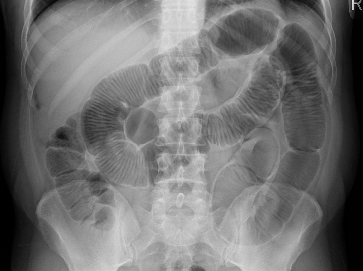
A 40-year-old male presents with colicky abdominal pain, multiple episodes of bilious vomiting, and absence of bowel movements and flatus. An X-ray of the abdomen was performed. Based on the findings, what is the most likely diagnosis?
What is the initial investigation for an amoebic liver abscess?
A patient complains of epigastric pain, radiating to the back off and on. What is the investigation of choice?
Which of the following is a radiological finding of a benign gastric ulcer?
Yoyo reflux is most commonly associated with which of the following conditions?
What is the best method to visualize the proximal bile duct?
What condition is associated with the "comb sign" on CT abdomen?
Intravenous urogram shows a 'flower vase' appearance. What is the most likely diagnosis?
Which is the best investigation to detect pneumoperitoneum?
Practice by Chapter
Imaging of Liver
Practice Questions
Biliary Tract Imaging
Practice Questions
Pancreatic Imaging
Practice Questions
Spleen and Lymphatic System
Practice Questions
Gastrointestinal Tract Imaging
Practice Questions
Renal and Urinary Tract Imaging
Practice Questions
Adrenal Imaging
Practice Questions
Female Pelvic Imaging
Practice Questions
Male Pelvic Imaging
Practice Questions
Abdominal Trauma Imaging
Practice Questions
Acute Abdomen Imaging
Practice Questions
Imaging of Peritoneal Cavity and Retroperitoneum
Practice Questions
Want unlimited practice?
Get full access to all questions, explanations, and performance tracking.
Scan to download app