
Abdominal and Pelvic Radiology — MCQs

On this page
Which of the following are ultrasound signs of fetal death?
A 'cut off' sign on a plain X-ray of the abdomen is indicative of which of the following conditions?
Fleischner sign on barium study is seen in which of the following conditions?
Non-visualization of the kidney is seen in all EXCEPT?
What is the investigation of choice for extra-adrenal pheochromocytoma?
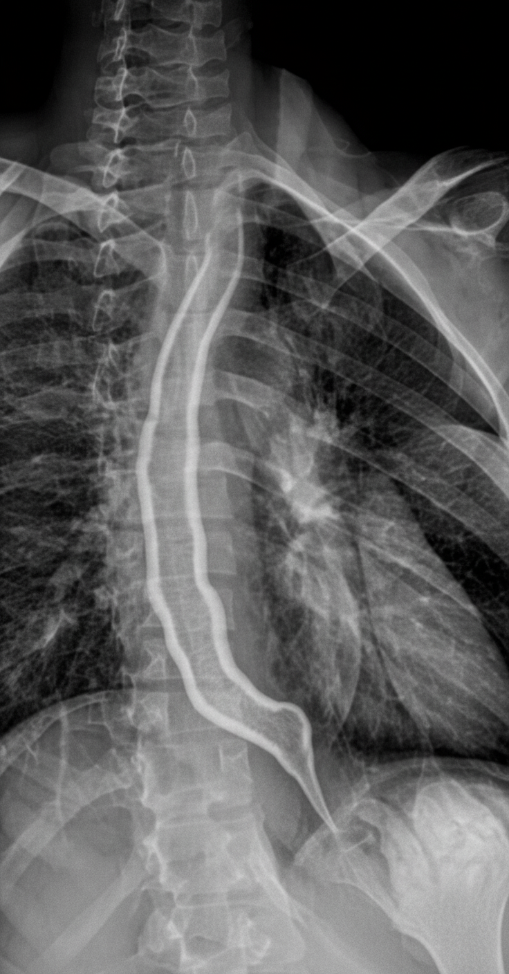
What is the diagnosis demonstrated in the barium esophagogram?
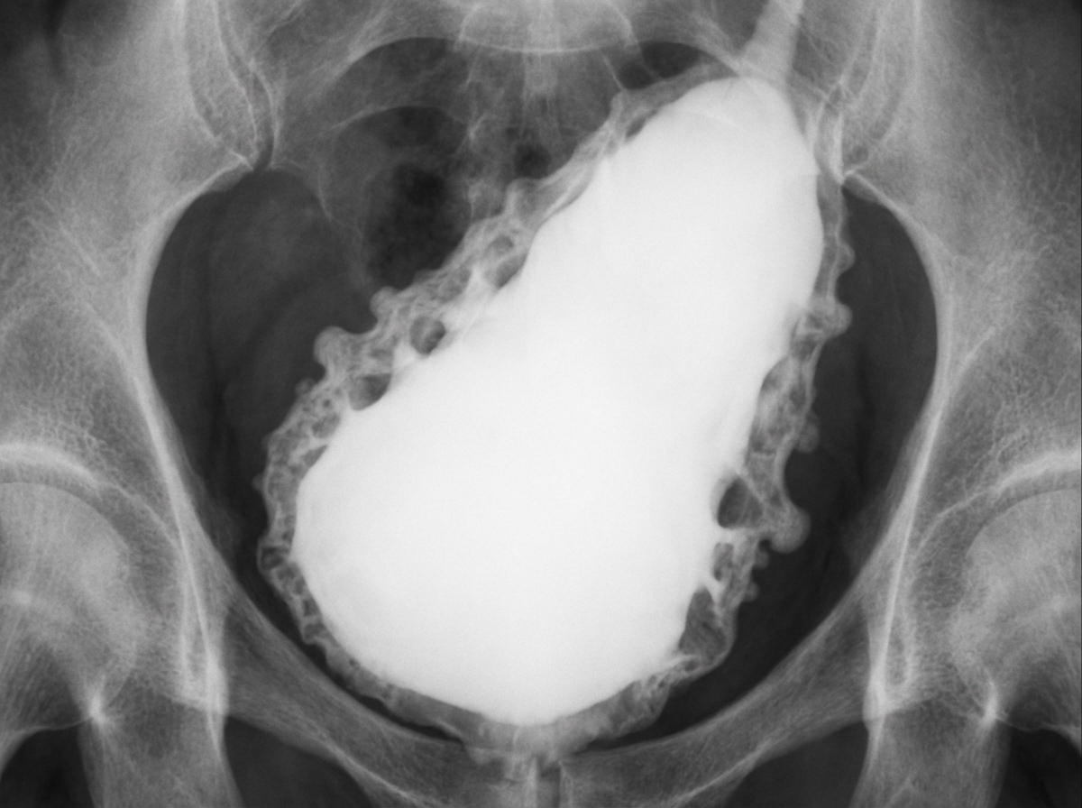
A given cystogram appearance is a characteristic feature of which of the following conditions?
What is the most common cause of widening of the C-loop of the duodenum?
What is the minimum size of a liver metastasis that can be detected by contrast-enhanced computed tomography (CECT)?
What is the investigation of choice for small bowel tumors?
Practice by Chapter
Imaging of Liver
Practice Questions
Biliary Tract Imaging
Practice Questions
Pancreatic Imaging
Practice Questions
Spleen and Lymphatic System
Practice Questions
Gastrointestinal Tract Imaging
Practice Questions
Renal and Urinary Tract Imaging
Practice Questions
Adrenal Imaging
Practice Questions
Female Pelvic Imaging
Practice Questions
Male Pelvic Imaging
Practice Questions
Abdominal Trauma Imaging
Practice Questions
Acute Abdomen Imaging
Practice Questions
Imaging of Peritoneal Cavity and Retroperitoneum
Practice Questions
Want unlimited practice?
Get full access to all questions, explanations, and performance tracking.
Scan to download app