What is the ideal investigation for fistula-in-ano?
Air on either side of the bowel wall on a radiograph is called:
Spider leg appearance on intravenous urography is typically seen in which of the following conditions?
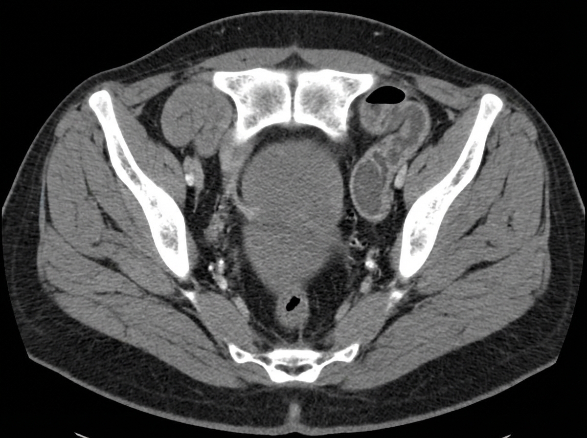
An elderly woman presented with abdominal pain and vomiting for three days. A computed tomogram of the abdomen was obtained. What is the diagnosis?

What is the investigation of choice for gallstones?
In which of the following conditions are fluid levels typically NOT visible on imaging?
Which of the following is the earliest sign of ulcerative colitis on a double contrast barium enema (DCBE) study?
A patient presents with marked loin pain, tenderness, and pyrexia. Initial investigations reveal non-specific findings on IVP. Ultrasound shows a heterogeneous mass with posterior acoustic enhancement and central necrosis and internal debris. CT scan demonstrates marginal enhancement with air densities within. What is the likely diagnosis?
Ultrasound and radionuclide studies are primary imaging modalities used for the assessment of which of the following conditions?
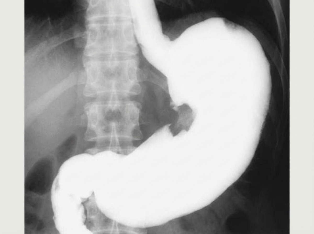
What is indicated by the arrow in the contrast X-ray abdomen?

Explanation: **Explanation:** The gold standard and ideal investigation for fistula-in-ano is **Magnetic Resonance Imaging (MRI)**. **Why MRI is the Correct Choice:** MRI provides superior soft-tissue contrast, allowing for precise visualization of the anal sphincter complex (internal and external sphincters) and the levator ani muscle. It is highly accurate in identifying the primary tract, secondary extensions (branching), and associated abscesses. Most importantly, MRI is essential for classifying the fistula according to the **Parks Classification**, which guides surgical management and helps prevent recurrence or fecal incontinence. **Analysis of Incorrect Options:** * **Endoanal Ultrasound (EAUS):** While useful and portable, it has a limited field of view. It often struggles to differentiate between active inflammation and old scarring and may miss high supralevator extensions. * **Fistulography:** This involves injecting contrast into the external opening. It is largely obsolete because it cannot visualize the anal sphincters and often fails to show the internal opening or secondary tracks if they are plugged with debris. * **CT Scan:** CT has poor soft-tissue resolution for the pelvic floor. It is generally reserved for identifying large perirectal abscesses rather than mapping complex fistulous tracts. **High-Yield Clinical Pearls for NEET-PG:** * **St. James’s University Hospital Classification:** This is the specific MRI-based grading system used to assess the severity of fistulae. * **Goodsall’s Rule:** A clinical rule stating that tracts with an external opening posterior to the transverse anal line follow a curved path to the midline, while anterior openings usually follow a straight radial path. * **Gold Standard for Acute Abscess:** While MRI is best for fistulae, **Clinical Examination (EUA - Examination Under Anesthesia)** remains the primary way to manage acute perianal abscesses.
Explanation: **Explanation:** The presence of air on both the inner (luminal) and outer (serosal) surfaces of the bowel wall on an abdominal radiograph is known as the **Rigler sign** (also called the double-wall sign). Normally, only the inner wall of the bowel is visible because it is outlined by intraluminal gas. The outer wall remains invisible as it blends with the water density of adjacent soft tissues or fluid. When there is **pneumoperitoneum** (free intraperitoneal air), this air outlines the serosal surface, making the bowel wall appear as a distinct, thin line. This is a classic sign of hollow viscus perforation. **Analysis of Incorrect Options:** * **Air under diaphragm sign:** This refers to a crescent-shaped lucency between the liver/spleen and the diaphragm on an upright chest or abdominal X-ray. It is the most common sign of pneumoperitoneum but does not specifically describe the "double-wall" appearance. * **Cupola sign:** This refers to air trapped under the central tendon of the diaphragm, appearing as an inverted cup-shaped lucency overlying the lower thoracic vertebrae on a supine film. * **Doge’s cap sign (Morison’s pouch air):** This is a triangular collection of air in the hepatorenal recess (Morison’s pouch), often seen in supine radiographs. **High-Yield Pearls for NEET-PG:** * **Rigler Sign** is best appreciated on **supine** radiographs when an upright film cannot be obtained. * **Football sign:** A large oval lucency in the abdomen representing massive pneumoperitoneum (the abdominal cavity acts as the "ball" and the falciform ligament as the "laces"). * **Tell-tale Triangle sign:** A small triangular collection of air between three loops of bowel. * The most sensitive view for detecting free air is the **upright chest X-ray**, which can detect as little as 1–2 ml of air.
Explanation: The **"Spider Leg Appearance"** on an Intravenous Urography (IVU) is a classic radiological sign of **Autosomal Dominant Polycystic Kidney Disease (ADPKD)**. ### Why the correct answer is right: In ADPKD, the renal parenchyma is replaced by multiple large, expanding cysts. As these cysts grow, they exert pressure on the internal collecting system. This leads to the **elongation, narrowing, and stretching** of the renal calyces. On an IVU, the attenuated and stretched calyces resemble the long, thin legs of a spider, while the enlarged renal pelvis represents the spider's body. ### Why the other options are incorrect: * **Hydronephrosis:** Characterized by the **dilation** of the renal pelvis and calyces (clubbing), rather than the thinning and stretching seen in ADPKD. * **Renal stone:** Typically presents as a radiopaque shadow on a plain KUB film or a **filling defect** on IVU. It may cause secondary hydronephrosis but not the spider leg deformity. * **Renal cell carcinoma:** Usually presents as a localized mass causing focal displacement or distortion of a single calyx (the **"Dromedary hump"** or space-occupying lesion effect), rather than the diffuse, bilateral stretching seen in polycystic disease. ### High-Yield Clinical Pearls for NEET-PG: * **ADPKD:** Most common inherited kidney disease; associated with **PKD1 (Chr 16)** and **PKD2 (Chr 4)** mutations. * **Extra-renal manifestations:** Berry aneurysms (Circle of Willis), hepatic cysts (most common extra-renal site), and mitral valve prolapse. * **Swiss Cheese Appearance:** Seen on **Nephrotomography** in Polycystic Kidney Disease due to multiple radiolucent cysts. * **Flower Vase Appearance:** Seen in **Horseshoe Kidney** due to the anterior displacement of the lower poles.
Explanation: ***Obturator hernia*** - Classic **CT appearance** shows a **bowel loop** herniating through the **obturator foramen** between the **pectineus** and **obturator externus muscles**. - Typically occurs in **elderly, thin women** and can cause **small bowel obstruction** with the characteristic **Howship-Romberg sign** (medial thigh pain). *Cecal volvulus* - CT shows a **dilated cecum** in an abnormal position, often in the **left upper quadrant** with a **whirl sign** at the point of torsion. - Creates a **closed-loop obstruction** with characteristic **coffee bean** or **kidney-shaped** appearance on imaging. *Intussusception* - CT demonstrates the pathognomonic **target sign** or **bowel-within-bowel** appearance with alternating layers of bowel wall. - More commonly seen in **pediatric patients** and would show a **sausage-shaped mass** rather than herniation through anatomical foramina. *Mesenteric ischemia* - CT findings include **bowel wall thickening**, **pneumatosis**, **portal venous gas**, and **lack of bowel wall enhancement** with contrast. - Would show **mesenteric vessel occlusion** or **bowel wall necrosis** rather than mechanical obstruction from herniation.
Explanation: **Explanation:** **Ultrasonography (USG)** is the investigation of choice (IOC) and the initial screening modality for gallstones (cholelithiasis). It has a high sensitivity and specificity (>95%) for detecting stones. On USG, gallstones typically appear as **hyperechoic (bright) structures** within the gallbladder lumen that demonstrate **posterior acoustic shadowing** and move with changes in patient positioning. **Analysis of Options:** * **X-Ray (Option A):** Only about 10–15% of gallstones are radiopaque (contain enough calcium to be seen). Most stones are cholesterol-based and radiolucent, making X-ray an unreliable screening tool. * **Cholecystography (Option C):** Oral cholecystography was historically used but is now obsolete. It is invasive, time-consuming, and depends on the liver's ability to excrete contrast and the gallbladder's ability to concentrate it. * **CAT Scan (Option D):** While CT is excellent for identifying complications like gallbladder perforation or pancreatitis, it is less sensitive than USG for detecting gallstones. Pure cholesterol stones may be isodense to bile, making them "invisible" on a CT scan. **High-Yield Clinical Pearls for NEET-PG:** * **Gold Standard for Acute Cholecystitis:** HIDA scan (Radionuclide scan). * **IOC for Choledocholithiasis (CBD stones):** MRCP (Non-invasive) or ERCP (Invasive/Therapeutic). * **WES Triad:** (Wall-Echo-Shadow) A sonographic sign seen when the gallbladder is completely filled with stones. * **Sludge:** Low-level echoes without shadowing, often a precursor to stone formation.
Explanation: **Explanation:** The presence of air-fluid levels on an abdominal radiograph requires two components: gas and liquid of low viscosity. **1. Why Meconium Ileus is the Correct Answer:** In **Meconium Ileus** (commonly associated with Cystic Fibrosis), the meconium is pathologically thick, tenacious, and "tar-like" due to a lack of pancreatic enzymes. Because this meconium is extremely viscid, it does not separate into distinct layers of gas and liquid. Consequently, air becomes trapped within the thick meconium, creating a **"soap-bubble" or "ground-glass" appearance (Neuhauser’s sign)** rather than discrete air-fluid levels. **2. Analysis of Incorrect Options:** * **Intussusception:** This is a form of mechanical bowel obstruction. As the proximal bowel dilates and fills with swallowed air and digestive secretions, classic air-fluid levels develop. * **Colon Pouch (Pouch Colon):** Associated with anorectal malformations, this condition involves a massive, localized dilation of the colon. The stagnant fecal matter and gas within the large pouch typically produce a prominent, large air-fluid level. * **Duodenal Obstruction:** Whether due to atresia, webs, or stenosis, this results in the **"Double Bubble" sign**. The dilated stomach and proximal duodenum contain both air and fluid, showing two distinct air-fluid levels on upright films. **Clinical Pearls for NEET-PG:** * **Neuhauser’s Sign:** The characteristic "soap bubble" appearance in the right iliac fossa for Meconium Ileus. * **Microcolon:** On contrast enema, Meconium Ileus typically shows a "disuse microcolon." * **Rule of Thumb:** If a neonate has features of distal intestinal obstruction but **no** fluid levels on X-ray, think Meconium Ileus.
Explanation: **Explanation:** **1. Why "Fine Mucosal Granularity" is correct:** Ulcerative Colitis (UC) is a mucosal disease that starts in the rectum and spreads proximally in a continuous fashion. On a Double Contrast Barium Enema (DCBE), the earliest detectable change is the loss of the normal smooth mucosal outline. This is replaced by a **fine, sand-like granularity** (stippling). This appearance is caused by mucosal edema and hyperemia, which creates a hazy or "pitted" surface before frank ulceration occurs. **2. Why the other options are incorrect:** * **Loss of haustrations:** This occurs as the disease progresses to a chronic stage. The bowel wall becomes thickened and fibrotic, leading to the disappearance of normal folds. * **Collar-button ulcers:** These represent deeper ulcerations where the inflammatory process penetrates through the mucosa into the submucosa, creating a "T-shaped" or "mushroom" appearance. This indicates more advanced, active disease rather than the earliest stage. * **Lead pipe colon:** This is a classic **late-stage/chronic** finding. It refers to a featureless, shortened, and rigid colon caused by chronic inflammation, fibrosis, and loss of haustrations. **3. Clinical Pearls for NEET-PG:** * **Distribution:** UC always involves the rectum (proctitis) and extends proximally without "skip lesions" (unlike Crohn’s). * **Backwash Ileitis:** Seen in ~10% of patients with pancolitis; the terminal ileum appears dilated and patulous. * **Pseudopolyps:** These are islands of regenerating normal mucosa surrounded by areas of denuded, ulcerated mucosa. * **Toxic Megacolon:** A life-threatening complication where the transverse colon diameter exceeds **6 cm** on a plain X-ray.
Explanation: **Explanation:** The clinical presentation of loin pain, pyrexia, and tenderness suggests an acute renal infection. The diagnosis of **Renal Abscess** is confirmed by the specific imaging triad described: 1. **Ultrasound:** A heterogeneous mass with **posterior acoustic enhancement** (indicating a fluid-filled/liquefied core) and internal debris (pus). 2. **CT Scan (Gold Standard):** Shows a well-defined, low-attenuation mass with **marginal (rim) enhancement** (the "ring-sign"). The presence of **air densities (gas)** within the collection is a classic, though not always present, sign of an abscess caused by gas-forming organisms. **Why other options are incorrect:** * **Emphysematous Pyelonephritis:** This is a life-threatening necrotizing infection characterized by *diffuse* gas within the renal parenchyma/collecting system, rather than a localized, rim-enhancing mass. * **Pyonephrosis:** This refers to infected, obstructed hydronephrosis. Imaging would show a dilated pelvicalyceal system with fluid-debris levels, rather than a parenchymal mass. * **Acute Pyelonephritis:** Typically shows an enlarged, edematous kidney with "striated nephrogram" on CT. It does not present as a localized, necrotic mass with rim enhancement unless it progresses to an abscess. **NEET-PG High-Yield Pearls:** * **CT scan** is the investigation of choice for renal abscess. * **Posterior acoustic enhancement** on USG is a key differentiator for fluid-filled structures (abscess/cyst) versus solid tumors. * Commonest causative organism: *Staphylococcus aureus* (via hematogenous spread) or *E. coli* (via ascending infection). * Management: Small abscesses (<3cm) respond to antibiotics; larger ones (>3cm) usually require percutaneous drainage.
Explanation: **Explanation:** **Renal Transplant (Correct Answer):** Ultrasound (USG) and Radionuclide studies are the mainstays for monitoring renal transplants because they are non-invasive, bedside-compatible, and avoid nephrotoxic contrast. * **USG with Color Doppler** is the initial investigation of choice to assess surgical complications (perinephric collections like hematomas or urinomas) and vascular complications (renal artery/vein thrombosis). * **Radionuclide scans (DTPA/MAG3)** are gold standards for assessing the functional status of the graft, helping differentiate between Acute Tubular Necrosis (ATN) and hyperacute/acute rejection based on perfusion and excretion patterns. **Why other options are incorrect:** * **Renal Trauma:** **Contrast-Enhanced CT (CECT)** is the gold standard. It accurately grades the severity of injury, detects active extravasation, and assesses the pedicle. USG (FAST) is used only for initial screening of hemoperitoneum. * **Renovascular Hypertension:** While Doppler can screen for Renal Artery Stenosis, **CT Angiography (CTA)** or **MR Angiography (MRA)** are the preferred diagnostic modalities. Digital Subtraction Angiography (DSA) remains the "gold standard." * **Renal Tuberculosis:** **Intravenous Urography (IVU)** or **CT Urography** are preferred to visualize the characteristic "moth-eaten" calyces, infundibular strictures, and phantom calyx. **High-Yield Clinical Pearls for NEET-PG:** * **Resistive Index (RI):** In a transplanted kidney, an RI > 0.7–0.8 on Doppler is a sensitive (though non-specific) indicator of graft dysfunction (e.g., rejection or obstruction). * **MAG3 Scan:** This is preferred over DTPA in transplants with poor function because it has higher extraction efficiency. * **Putty Kidney:** A classic radiological sign of end-stage Renal TB (autonephrectomy) seen on X-ray/CT.
Explanation: ***Gastric ulcer*** - Shows the classic **niche sign** on barium meal X-ray, appearing as a contrast-filled crater projecting beyond the normal gastric contour. - **Hampton's line** (thin radiolucent line at the base) and **mucosal folds radiating** toward the ulcer crater are characteristic features. *GIST* - Appears as a **submucosal filling defect** with smooth, well-defined margins on contrast studies. - Lacks the **niche sign** and mucosal fold convergence typical of ulcers, presenting as a mass lesion instead. *Carcinoma stomach* - Shows an **irregular filling defect** with **mucosal destruction** and **shouldering** (abrupt transition from normal to abnormal mucosa). - Demonstrates **loss of normal gastric distensibility** and rugal pattern destruction, unlike the focal crater of an ulcer. *Gastric polyp* - Presents as a **smooth, rounded filling defect** that projects into the gastric lumen without crater formation. - Shows **regular margins** and **mobile appearance** on fluoroscopy, contrasting with the fixed ulcer crater.
Imaging of Liver
Practice Questions
Biliary Tract Imaging
Practice Questions
Pancreatic Imaging
Practice Questions
Spleen and Lymphatic System
Practice Questions
Gastrointestinal Tract Imaging
Practice Questions
Renal and Urinary Tract Imaging
Practice Questions
Adrenal Imaging
Practice Questions
Female Pelvic Imaging
Practice Questions
Male Pelvic Imaging
Practice Questions
Abdominal Trauma Imaging
Practice Questions
Acute Abdomen Imaging
Practice Questions
Imaging of Peritoneal Cavity and Retroperitoneum
Practice Questions
Get full access to all questions, explanations, and performance tracking.
Start For Free