
Abdominal and Pelvic Radiology — MCQs

On this page
A 44-year-old man with a history of quadriplegia presented to the emergency department with symptoms of a urinary tract infection. What is the diagnosis?
Which structure is outlined with contrast on a CT scan using intraperitoneal contrast material?
Pseudo-flow Doppler signal (continuous flow) in the hepatic vein in the setting of Budd-Chiari syndrome indicates what?
What is the best imaging modality to differentiate between medical and surgical jaundice?
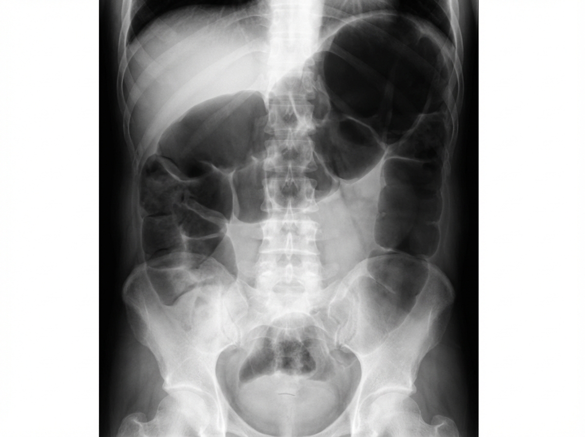
A plain abdominal X-ray in a patient with a sigmoid volvulus will show a distended bowel loop with its apex in which location?
What is the preferred imaging modality for confirming choledocholithiasis?
Which imaging study has the highest pickup for a gastrojejunocolic fistula?
What pattern is described as "paint brush-like" in intravenous urography?
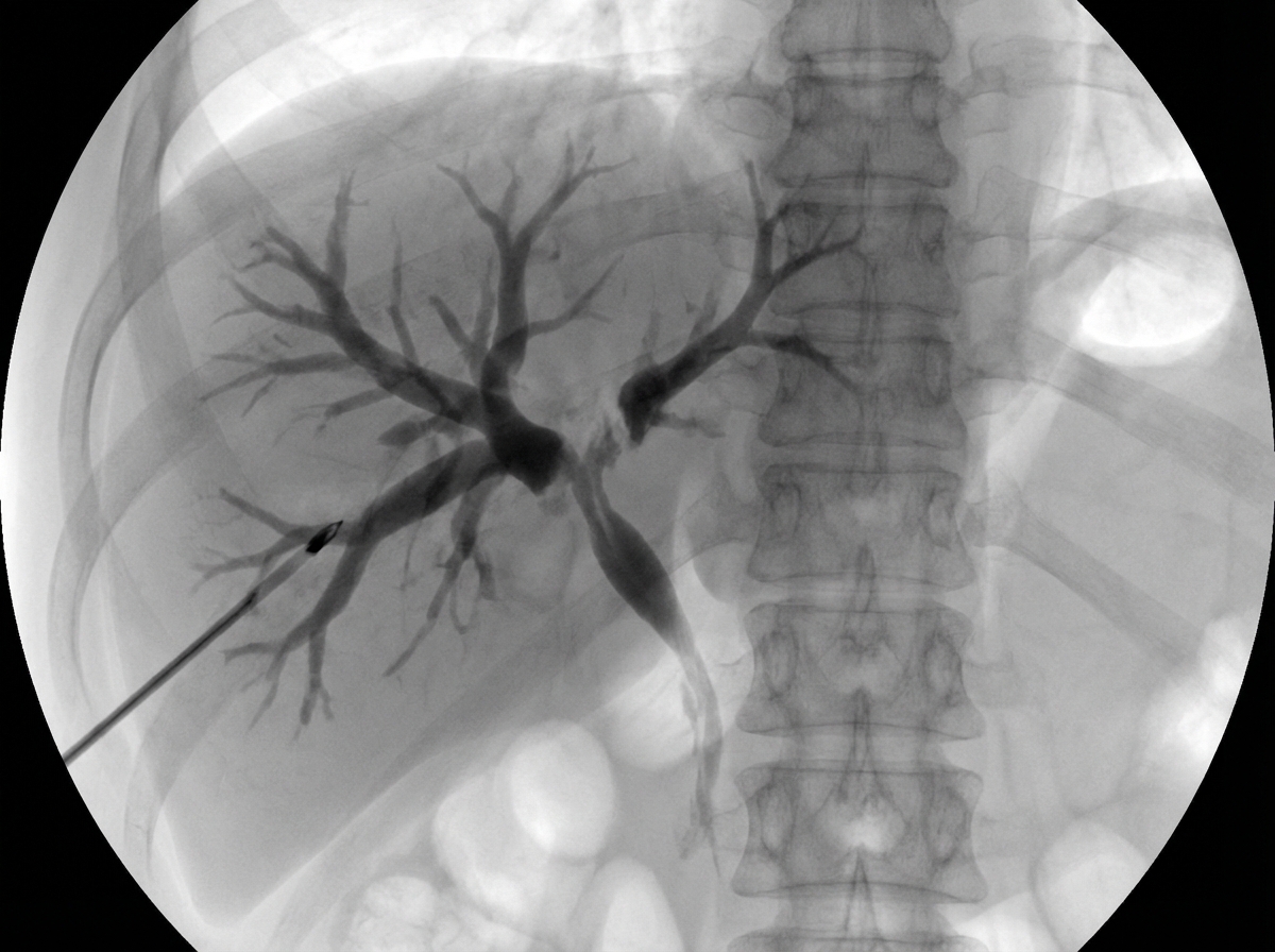
You are shown an AP view from a percutaneous trans-hepatic cholangiogram of a jaundiced patient. What is the MOST likely diagnosis?
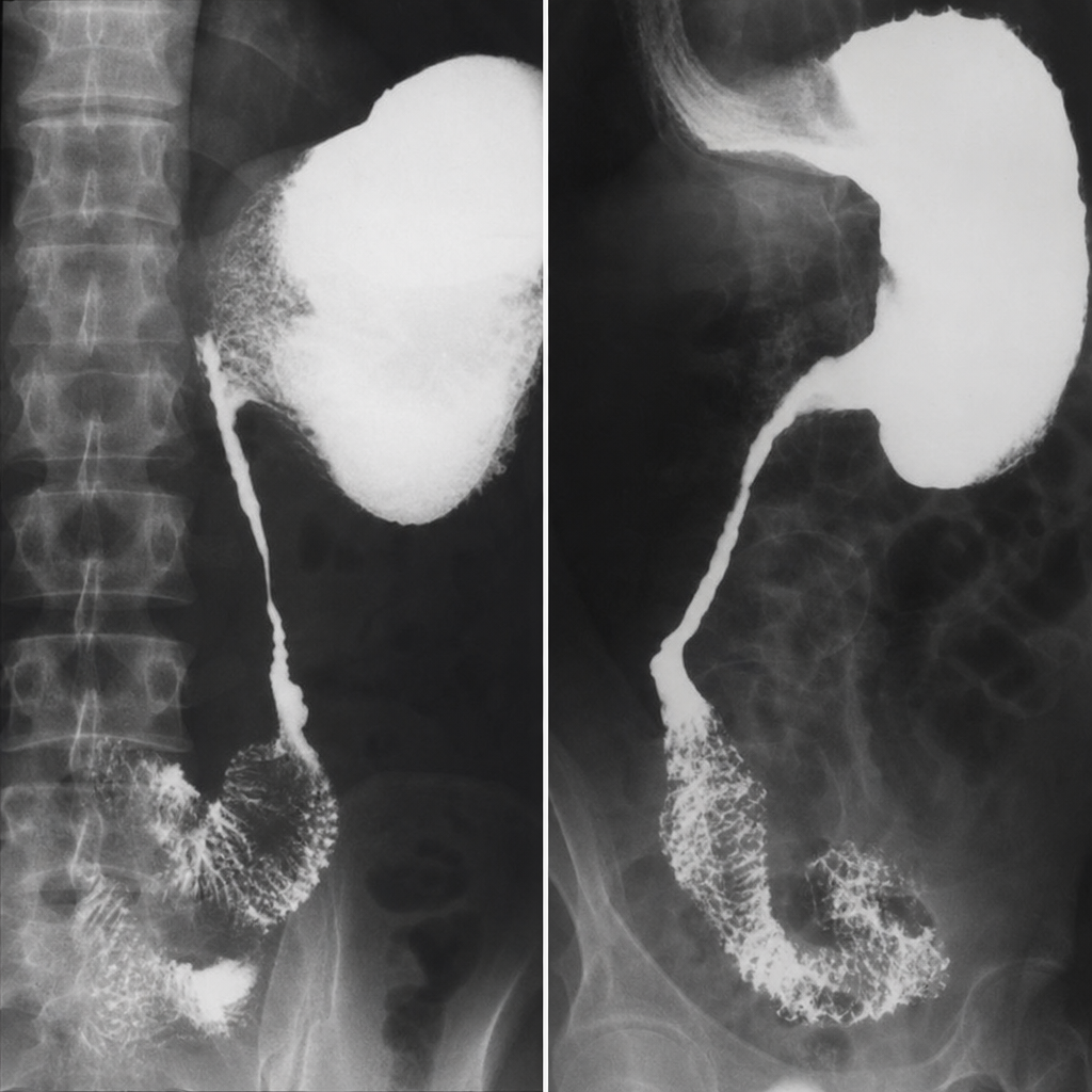
A 35-year-old male presents with a history of recurrent attacks of colicky abdominal pain. A barium meal follow-through was performed. What is the name of this radiological sign?
Practice by Chapter
Imaging of Liver
Practice Questions
Biliary Tract Imaging
Practice Questions
Pancreatic Imaging
Practice Questions
Spleen and Lymphatic System
Practice Questions
Gastrointestinal Tract Imaging
Practice Questions
Renal and Urinary Tract Imaging
Practice Questions
Adrenal Imaging
Practice Questions
Female Pelvic Imaging
Practice Questions
Male Pelvic Imaging
Practice Questions
Abdominal Trauma Imaging
Practice Questions
Acute Abdomen Imaging
Practice Questions
Imaging of Peritoneal Cavity and Retroperitoneum
Practice Questions
Want unlimited practice?
Get full access to all questions, explanations, and performance tracking.
Scan to download app