
Abdominal and Pelvic Radiology — MCQs

On this page
Which of the following imaging modalities is NOT included in the PI-RADS reporting scheme for prostate glands?
What is the spot diagnosis for the given image/description?
What is the investigation of choice to detect a 4 mm nodule in the pancreas?
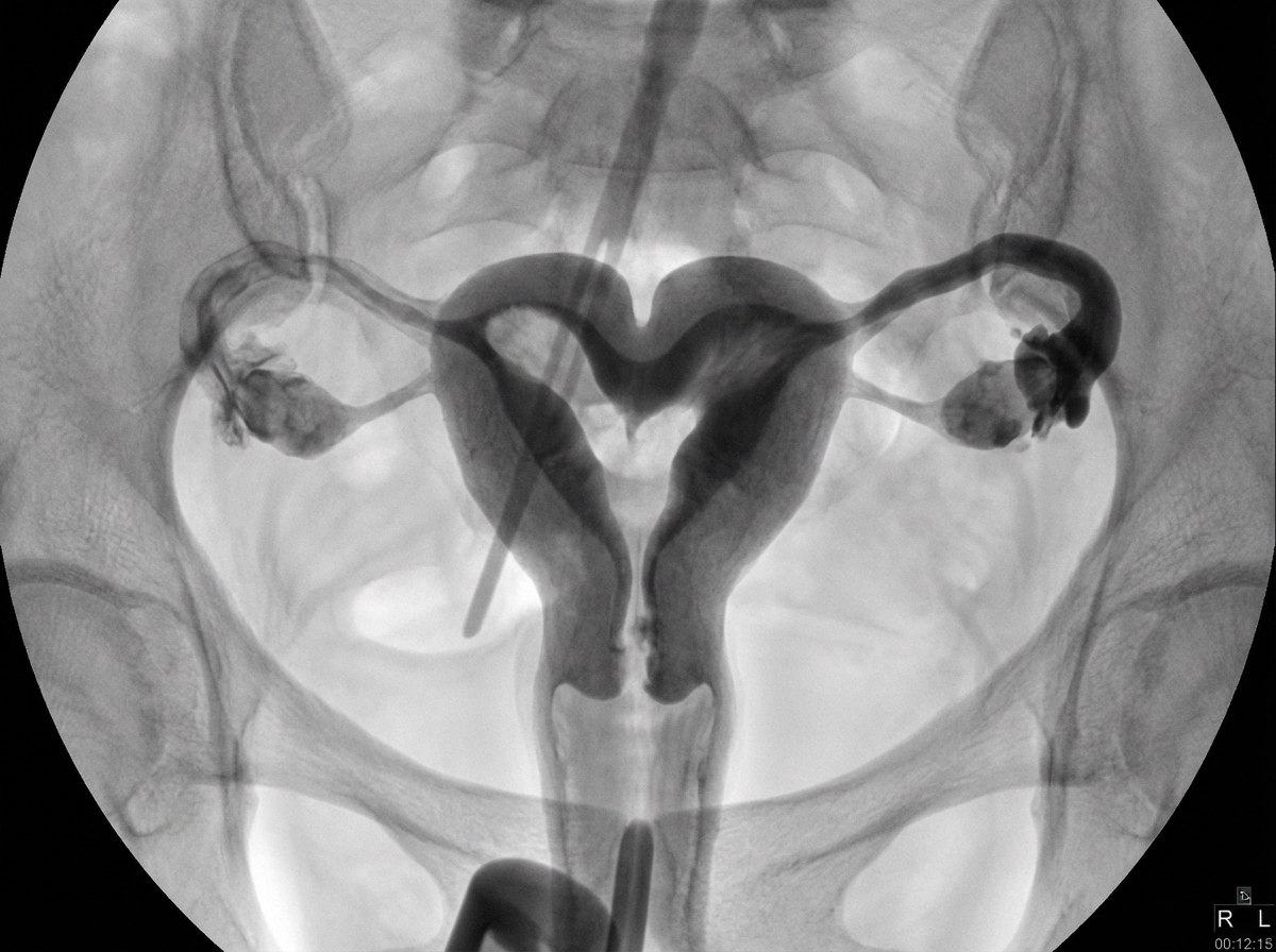
Which of the following conditions is characterized by a trifoliate appearance?
A 72-year-old man presents with increasing dyspepsia and weight loss. His physical examination is unremarkable. A barium meal is performed. The oesophagus is normal, but a 'bull's eye' lesion is noted in the gastric mucosa. Which one of the following is not a recognised cause of this appearance?
A 5-year-old child presents with gas under the diaphragm. What is the most likely diagnosis?
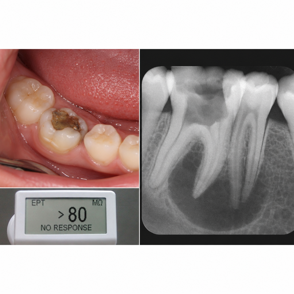
A 13-year-old boy presents with pain in the lower right tooth region with no remarkable medical history. On intraoral examination, tooth 85 was found to be carious and did not respond to EPT. Radiographs show changes in the apical region. What is the most probable diagnosis?
Omental caking on CT scan suggests a high possibility of which of the following conditions?
The CT severity index in acute pancreatitis is described by which scoring system?
What is the investigation of choice (IOC) for polycystic kidney disease?
Practice by Chapter
Imaging of Liver
Practice Questions
Biliary Tract Imaging
Practice Questions
Pancreatic Imaging
Practice Questions
Spleen and Lymphatic System
Practice Questions
Gastrointestinal Tract Imaging
Practice Questions
Renal and Urinary Tract Imaging
Practice Questions
Adrenal Imaging
Practice Questions
Female Pelvic Imaging
Practice Questions
Male Pelvic Imaging
Practice Questions
Abdominal Trauma Imaging
Practice Questions
Acute Abdomen Imaging
Practice Questions
Imaging of Peritoneal Cavity and Retroperitoneum
Practice Questions
Want unlimited practice?
Get full access to all questions, explanations, and performance tracking.
Scan to download app