
Abdominal and Pelvic Radiology — MCQs

On this page
What is the initial imaging modality of choice for suspected pyonephrosis?
Which of the following is NOT a cause of pneumoperitoneum?
What is the primary imaging technique used in a case of suspected acute pancreatitis?
Which of the following is NOT radioopaque?
The triad of vomiting, abdominal distension, and a "string of beads" sign on abdominal X-ray is typically suggestive of what condition?
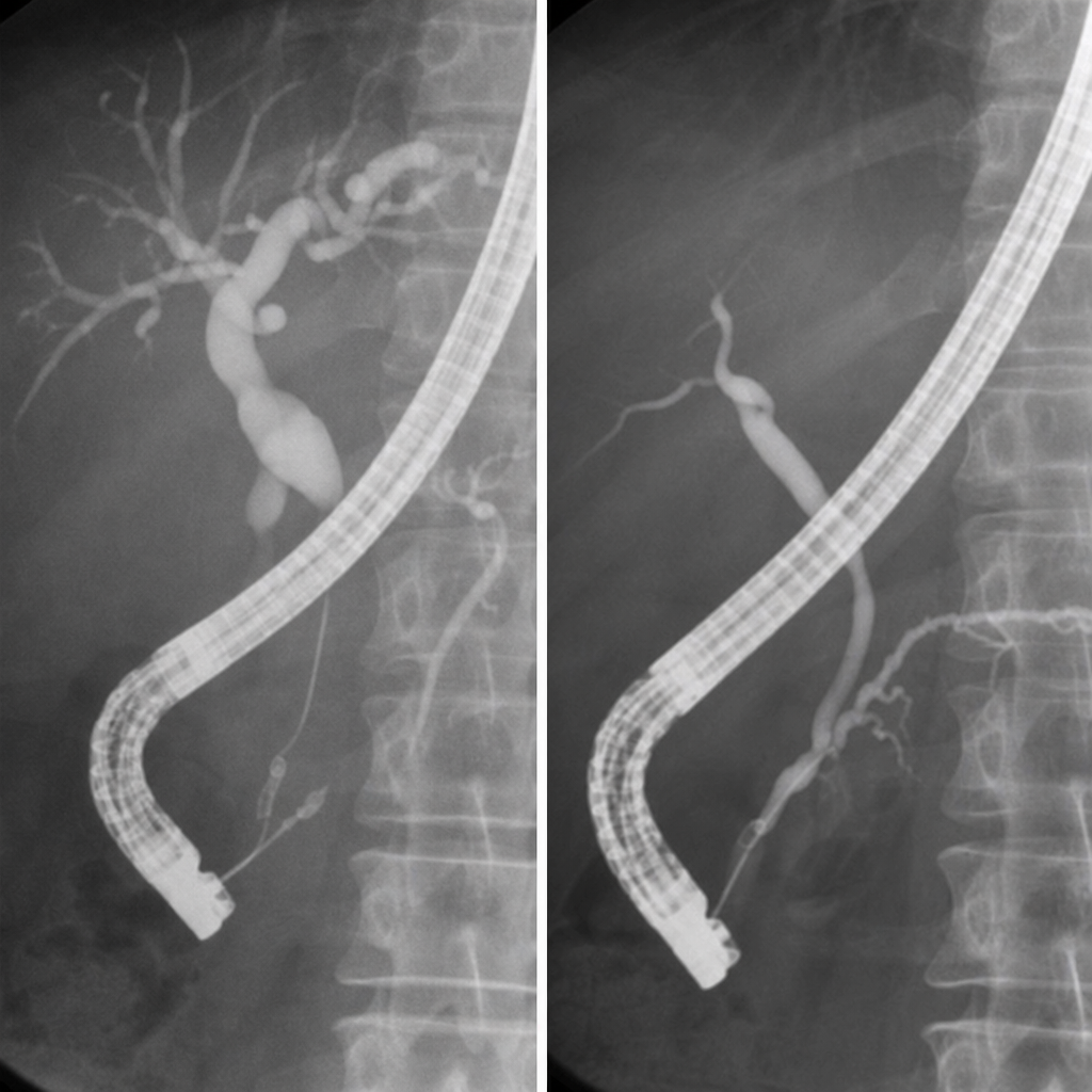
A 40-year-old woman presents with abdominal pain. Two films from an ERCP are shown. What is the MOST likely diagnosis?
What is the investigation of choice for evaluating a renal mass?
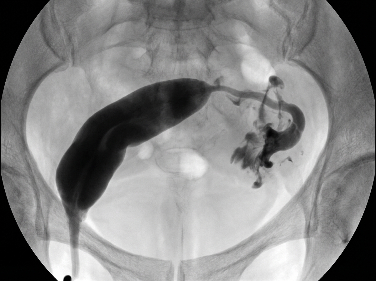
A hysterosalpingogram showing a congenital mullerian anomaly is depicted. What is the most likely diagnosis?
Which of the following is NOT a feature of papillary necrosis?
Which of the following signs is seen in Crohn's disease?
Practice by Chapter
Imaging of Liver
Practice Questions
Biliary Tract Imaging
Practice Questions
Pancreatic Imaging
Practice Questions
Spleen and Lymphatic System
Practice Questions
Gastrointestinal Tract Imaging
Practice Questions
Renal and Urinary Tract Imaging
Practice Questions
Adrenal Imaging
Practice Questions
Female Pelvic Imaging
Practice Questions
Male Pelvic Imaging
Practice Questions
Abdominal Trauma Imaging
Practice Questions
Acute Abdomen Imaging
Practice Questions
Imaging of Peritoneal Cavity and Retroperitoneum
Practice Questions
Want unlimited practice?
Get full access to all questions, explanations, and performance tracking.
Scan to download app