Anatomy
1 questionsWhich nerve is affected in the hand deformity shown in the image at rest? 
Obstetrics and Gynecology
2 questionsIdentify the CTG pattern?
Identify the medical device shown in the image.
Orthopaedics
3 questionsA 20-year-old male patient presented with localized pain, which is gradual in onset and worsened over time. X-ray showed the following finding. What is the diagnosis?
Which nerve will be involved in the following finding at rest?
Identify the instrument shown in the image:
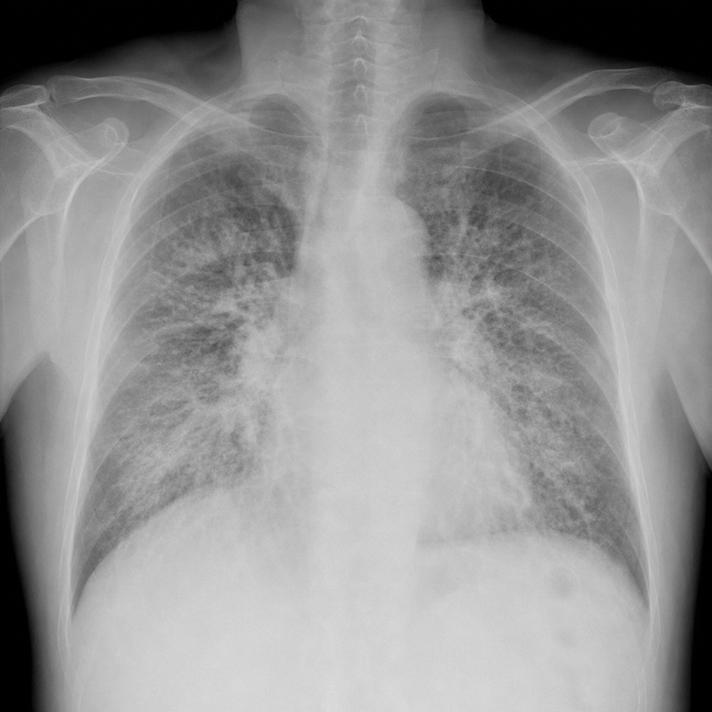
Radiology
3 questionsBased on the provided chest X-ray images, what is the most likely diagnosis?
A 65-year-old patient presents with abdominal distension and constipation. What is the most likely diagnosis based on the abdominal X-ray shown?
Based on the provided X-ray image, identify the most likely diagnosis.
Surgery
1 questionsA 70-year-old patient presents with absolute constipation and abdominal distension. The X-ray abdomen is given below. What is the most likely diagnosis?