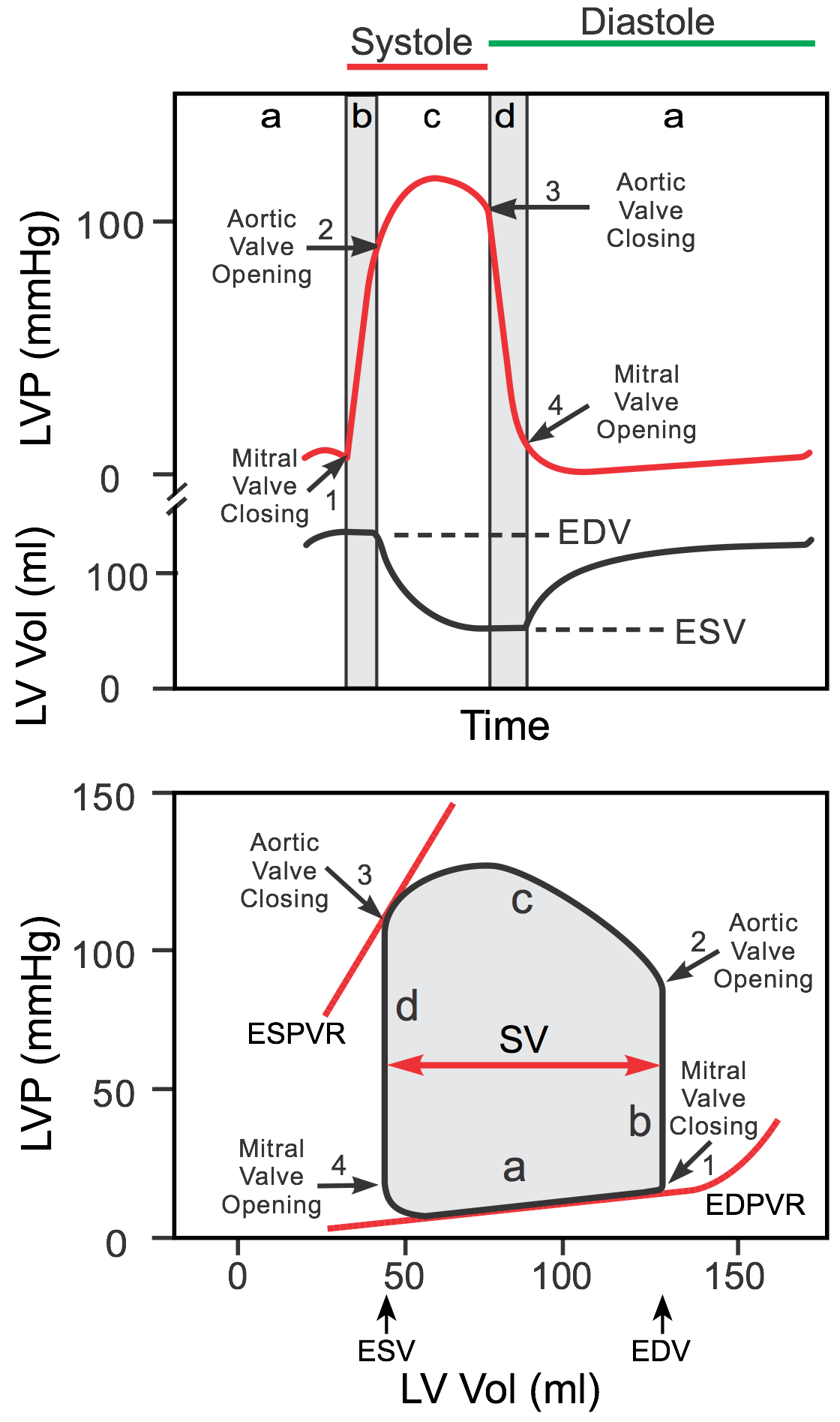
Pressure-Volume loop of cardiac cycle is shown below. What does point C represent?

The primary effect of vagal stimulation on the heart is
A shift of posture from supine to upright posture is associated with cardiovascular adjustments. Which of the following is NOT true in this context?
What is the mean arterial pressure (MAP) for a person with an arterial blood pressure of 125/75 mm Hg?
Which of the following equations correctly represents Poiseuille's Hagen law for fluid flow?
Coronary blood flow is maximum during which phase of the cardiac cycle?
Which heart sound is associated with decreased ventricular compliance?
Which is referred to as peripheral heart?
Which of the following is required for the Direct Fick method of measuring cardiac output?
The 'a' wave in jugular venous pressure is due to?
Explanation: ***Aortic valve opens*** - Point C represents the critical moment when the **left ventricular pressure** surpasses the **aortic pressure**, causing the aortic valve to open. - This event marks the beginning of the **ejection phase**, where blood is pumped from the left ventricle into the aorta. *Mitral valve opens* - The mitral valve opens at point A, signaling the start of **ventricular filling** as blood flows from the left atrium into the left ventricle. - At this point, the left ventricular pressure is at its lowest, and the volume begins to increase. *Mitral valve closes* - The mitral valve closes at point B, indicating the end of **ventricular filling** and the start of **isovolumetric contraction**. - This closure prevents backflow of blood into the left atrium as the ventricle begins to contract. *Aortic valve closes* - The aortic valve closes at point F, which signifies the end of **ejection** and the beginning of **isovolumetric relaxation**. - At this point, the left ventricular pressure falls below aortic pressure, and the ventricle begins to relax without changing volume.
Explanation: ***Decreased heart rate*** - **Vagal stimulation** releases **acetylcholine**, which activates muscarinic receptors on the sinoatrial (SA) node, leading to a decrease in its firing rate and thus a slower heart rate. - This parasympathetic effect primarily targets the **SA node** and **AV node**, influencing chronotropy (heart rate) more significantly than inotropy (contractility). *Increased P-R interval in ECG* - While vagal stimulation does slow **AV node conduction**, increasing the P-R interval, this is a more specific electrophysiological effect rather than the primary overall physiological outcome of vagal stimulation on the heart. - The most direct and immediate consequence of vagal nerve activity is the slowing of the heart's rhythm, which manifests as a **decreased heart rate**. *Decreased force of heart contraction* - The **vagus nerve** has a relatively weak effect on ventricular contractility, as parasympathetic innervation is far less dense in the ventricles compared to the atria and nodal tissues. - Therefore, a significant **decrease in the force of contraction** is not a primary or direct result of typical vagal stimulation in a healthy heart. *Decreased cardiac output* - While a markedly decreased heart rate *could* lead to a decreased cardiac output (CO = HR x SV), this is not the most direct or immediate physiological effect of vagal stimulation. - The primary action is on the heart rate, and changes in cardiac output would be a **secondary consequence** depending on the extent of bradycardia and compensatory mechanisms.
Explanation: ***Rise in central venous pressure*** - When a person moves from a supine to an upright posture, gravity causes **blood pooling in the lower extremities**, leading to a *decrease* in venous return to the heart, not a rise in central venous pressure. - A decrease in central venous pressure is an expected physiological response to orthostasis due to the aforementioned venous pooling. *Decrease in central venous pressure* - This statement is physiologically *true* because gravity causes blood to pool in the lower limbs, reducing venous return and subsequently lowering the central venous pressure. - The **baroreflex** responds to this fall, attempting to restore blood pressure. *Rise in heart rate* - This is a normal physiological response to orthostatic stress, mediated by the **baroreflex**, to maintain cardiac output and blood pressure against gravity. - The sympathetic nervous system increases **heart rate** and contractility to compensate for reduced venous return. *Decrease in cardiac output* - Upon standing, the initial reduction in venous return leads to a transient decrease in **stroke volume**, which, despite the compensatory rise in heart rate, often results in a net *decrease* in cardiac output. - This is a normal and expected cardiovascular adjustment as the body adapts to the upright position.
Explanation: ***The mean arterial pressure is 92 mm Hg*** - **Mean arterial pressure (MAP)** is calculated using the formula: **MAP = Diastolic Pressure + 1/3 (Systolic Pressure - Diastolic Pressure)**. - Given a blood pressure of **125/75 mm Hg** (systolic/diastolic), MAP = 75 + 1/3 (125 - 75) = 75 + 1/3 (50) = 75 + 16.67 ≈ **91.67 mm Hg**, which rounds to 92 mm Hg. *The pulse pressure is 50 mm Hg* - **Pulse pressure** is the difference between **systolic and diastolic pressure**. - In this case, 125 mm Hg (systolic) - 75 mm Hg (diastolic) = **50 mm Hg**, which is accurate but not the MAP. *Diastolic pressure is 75 mm Hg* - The **diastolic pressure** is the lower number in a blood pressure reading, representing the pressure during cardiac relaxation. - For a blood pressure of 125/75 mm Hg, the **diastolic pressure is indeed 75 mm Hg**, but this is only one component of MAP. *Systolic pressure is 125 mm Hg* - The **systolic pressure** is the upper number in a blood pressure reading, representing the pressure during cardiac contraction. - For a blood pressure of 125/75 mm Hg, the **systolic pressure is indeed 125 mm Hg**, but this alone does not represent MAP.
Explanation: ***F = (PA-PB) X 3.14 X r^4/8nl*** - This equation correctly represents Poiseuille's Hagen law, which describes the **volumetric flow rate** (F) of an incompressible fluid through a rigid cylindrical tube. - It shows that flow is directly proportional to the **pressure difference** ($P_A - P_B$) and the **fourth power of the radius** (r⁴), and inversely proportional to the fluid's **viscosity** (n) and the **length of the tube** (l). *F = (PA-PB) X 3.14 X r^3/8nl* - This option incorrectly uses **r³** instead of **r⁴** in the numerator. - Poiseuille's law explicitly states that the flow rate is proportional to the **fourth power of the radius**, highlighting the significant impact of vessel diameter on fluid flow. *F = (PA-PB) X 3.14 X r^4/8n* - This equation omits the **length of the tube (l)** from the denominator. - Flow rate is inversely proportional to the **length of the tube** because a longer tube implies greater resistance to flow. *F = (PA-PB) X 3.14 X r^2/8nl* - This option incorrectly uses **r²** instead of **r⁴** in the numerator. - The **fourth power dependence** on radius is a critical aspect of Poiseuille's law, demonstrating that even small changes in vessel radius have a large effect on flow.
Explanation: ***Maximum during diastole*** [1] - During **diastole**, the ventricular myocardium **relaxes**, reducing extravascular compression on the intramural coronary arteries [1] - This allows **maximum coronary blood flow** to perfuse the myocardium (approximately 70-80% of total coronary flow occurs during diastole) [1] - During **systole**, strong ventricular contraction compresses coronary vessels, significantly **impeding blood flow** (especially in the subendocardium) [1] - The left coronary artery flow is almost completely interrupted during systole due to high intraventricular pressure [1] *70 mL/min* - This represents a numerical value for coronary blood flow but does not specify the **phase of the cardiac cycle** - Average resting coronary blood flow is approximately 225-250 mL/min (about 5% of cardiac output) *Adenosine increases it* - While adenosine is a potent **coronary vasodilator**, this describes a regulatory mechanism, not the **phase** when flow is naturally maximal *Less than skin* - This is a comparative statement about regional blood flow distribution, not the **timing during the cardiac cycle**
Explanation: **S4** - An **S4 heart sound**, also known as an **atrial gallop**, is heard just before S1. It is caused by the atria contracting forcefully to push blood into a stiff, non-compliant ventricle. - This sound is commonly associated with conditions causing decreased ventricular compliance, such as **ventricular hypertrophy** or **ischemia**. *S1* - The **S1 heart sound** marks the beginning of systole and is produced by the closure of the **mitral and tricuspid valves**. - It reflects the timing of AV valve closure rather than ventricular compliance itself. *S2* - The **S2 heart sound** marks the beginning of diastole and is produced by the closure of the **aortic and pulmonic valves**. - Its components (A2 and P2) can be affected by changes in pressure and flow across the semilunar valves, but not directly by ventricular compliance. *S3* - An **S3 heart sound**, or **ventricular gallop**, is heard just after S2 during early diastole. It is caused by rapid passive filling of a dilated, often volume-overloaded, ventricle. - Unlike S4, S3 is associated with increased ventricular compliance due to volume overload and is often normal in children or young adults.
Explanation: ***Soleus*** - The **soleus muscle** is often referred to as the "peripheral heart" or "second heart" due to its crucial role in **venous return** from the lower limbs. - Its contractions, particularly during walking and running, help pump deoxygenated blood against gravity back towards the heart, preventing **venous pooling** and **edema**. *Popliteus* - The **popliteus muscle** is a small muscle located behind the knee, primarily responsible for **unlocking the knee joint** from full extension. - While important for knee stability and movement, it does not have a significant role in **venous return**. *Plantaris* - The **plantaris muscle** is a small, slender muscle in the calf, often absent or rudimentary in humans. - It assists weakly in **plantarflexion** of the foot and flexion of the knee, but has no significant role in **venous pump function**. *None of the options* - This option is incorrect because the **soleus muscle** is indeed known as the "peripheral heart" due to its vital role in **venous blood circulation**.
Explanation: ***All of the options*** - The **Direct Fick method** calculates **cardiac output (CO)** using the formula: **CO = VO₂ / (CaO₂ - CvO₂)**, where VO₂ is oxygen consumption, CaO₂ is arterial oxygen content, and CvO₂ is mixed venous oxygen content. - Therefore, all three measurements—**O₂ content of arterial blood**, **O₂ consumption per unit time**, and **O₂ content of venous blood**—are essential components required for this calculation. - Each component plays a critical role in determining cardiac output: **O₂ content of arterial blood (CaO₂)** - Represents the oxygen delivered by the **arterial circulation** to the tissues - Essential for calculating the **arteriovenous oxygen difference (A-V O₂ difference)**, which reflects oxygen extraction by tissues - Typically measured from a systemic arterial sample **O₂ consumption per unit time (VO₂)** - Measures the body's **total oxygen utilization** per minute - Typically obtained through **spirometry** or metabolic cart measurements - Forms the **numerator** of the Fick equation, representing total oxygen uptake by tissues **O₂ content of venous blood (CvO₂)** - Indicates the **oxygen remaining in the blood** after tissue extraction - Must be measured from **mixed venous blood** (typically from pulmonary artery via right heart catheterization) - Combined with arterial O₂ content to determine the **A-V O₂ difference** (denominator of the equation) *Why other individual options are incomplete* - Selecting only one or two components would provide insufficient data to calculate cardiac output using the Direct Fick principle - The method fundamentally requires measuring both oxygen delivery (arterial content) and return (venous content), plus total consumption, to determine flow rate
Explanation: ***Atrial contraction*** - The **a wave** in jugular venous pressure (JVP) corresponds to the increase in right atrial pressure due to **atrial systole** (contraction). - This pushes blood against the closed tricuspid valve and back into the great veins, causing a visible pulsation. *Atrial filling* - **Atrial filling** occurs during diastole and contributes to the overall increase in atrial pressure but is not the primary cause of the a wave. - This phase is more related to the **v wave** (due to ventricular contraction pushing blood into the atria against a closed tricuspid valve) and the **y descent** (due to tricuspid valve opening and rapid ventricular filling). *Atrial relaxation* - **Atrial relaxation** follows **atrial contraction** and is associated with the **x descent** on the JVP waveform, representing the decrease in right atrial pressure as the atrium relaxes and the tricuspid valve moves away from the atrium during ventricular systole. - It results in a fall in pressure not a rise, so it cannot be the a wave. *Ventricular relaxation* - **Ventricular relaxation** (early diastole) is primarily responsible for the **y descent**, which occurs as the tricuspid valve opens and blood rapidly empties from the atrium into the ventricle. - This phase of the cardiac cycle is related to falling pressures within the right atrium and ventricle, not the initial pressure rise seen in the **a wave**.
Cardiac Electrophysiology
Practice Questions
Cardiac Cycle
Practice Questions
Cardiac Output and Its Regulation
Practice Questions
Hemodynamics and Blood Flow
Practice Questions
Arterial System Physiology
Practice Questions
Microcirculation and Lymphatics
Practice Questions
Venous Return and Central Venous Pressure
Practice Questions
Cardiovascular Reflexes
Practice Questions
Regional Circulations
Practice Questions
Cardiovascular Responses to Exercise and Stress
Practice Questions
Get full access to all questions, explanations, and performance tracking.
Start For Free