
Cardiovascular System — MCQs

On this page
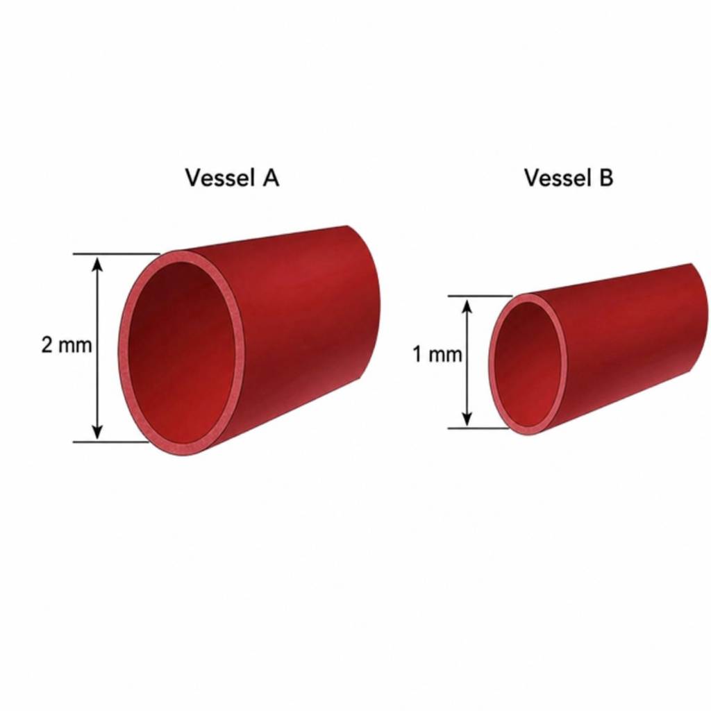
Below are two vessels shown. Assuming the pressure along both the vessels is the same and both of them follow linear flow pattern, what will be the amount of blood flow in vessel A compared to vessel B?
Willem Einthoven got Nobel Prize for:
Which heart sound is associated with decreased ventricular compliance?
Coronary blood flow is maximum during which phase of the cardiac cycle?
Which of the following causes coronary vasodilation?
Which of the following is required for the Direct Fick method of measuring cardiac output?
The 'a' wave in jugular venous pressure is due to?
What is the definition of autoregulation in the context of blood flow?
What does the first heart sound correspond to?
At which point in the cardiac cycle does the closure of the mitral valve begin? (Refer to the diagram showing points A, B, C, and D)

Practice by Chapter
Cardiac Electrophysiology
Practice Questions
Cardiac Cycle
Practice Questions
Cardiac Output and Its Regulation
Practice Questions
Hemodynamics and Blood Flow
Practice Questions
Arterial System Physiology
Practice Questions
Microcirculation and Lymphatics
Practice Questions
Venous Return and Central Venous Pressure
Practice Questions
Cardiovascular Reflexes
Practice Questions
Regional Circulations
Practice Questions
Cardiovascular Responses to Exercise and Stress
Practice Questions
Want unlimited practice?
Get full access to all questions, explanations, and performance tracking.
Scan to download app