Cardiac muscle is able to function as a syncytium because of the structural presence of which of the following?
What is the equilibrium pressure in the absence of flow called?
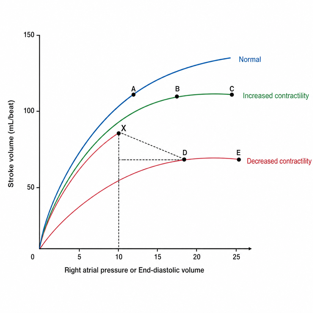
What is the typical cardiac output of a newborn?
In a standard Electrocardiogram, an augmented limb lead measures the electrical potential difference between which points?
Acetylcholine decreases heart rate by:
Which of the following is the last part of the ventricle to be depolarized?
The ventricular repolarization in ECG is best represented by which wave?
Increase in heart rate just before starting exercise is due to?
Massage of the carotid sinus results in all of the following, EXCEPT:
What is true about baroreceptors?
Explanation: **Explanation:** **1. Why Gap Junctions are correct:** Cardiac muscle functions as a **functional syncytium**, meaning that when one cell is excited, the action potential spreads rapidly to all cells, causing the heart to contract as a single unit. This is made possible by **Gap Junctions** (communicating junctions). These are protein channels (formed by connexins) that provide low-resistance pathways for the flow of ions and electrical impulses directly from the cytoplasm of one cardiomyocyte to the next. **2. Analysis of Incorrect Options:** * **A. Branching fibres:** While cardiac muscle is histologically characterized by branching, this structural arrangement provides mechanical strength and a network-like appearance but does not facilitate the electrical coupling required for a syncytium. * **B. Intercalated discs:** This is a common distractor. Intercalated discs are the entire complex at the cell-to-cell junction. They contain *both* mechanical links (Desmosomes/Fascia adherens) and electrical links (Gap junctions). While gap junctions are *located within* the intercalated discs, the specific structure responsible for the syncytial function is the gap junction. * **C. Protoplasmic bridges:** This is a legacy concept. Cardiac cells are discrete individual units separated by cell membranes; they do not share continuous cytoplasm (unlike a true anatomical syncytium like skeletal muscle). **3. NEET-PG High-Yield Pearls:** * **Functional vs. Anatomical Syncytium:** Skeletal muscle is an *anatomical* syncytium (multinucleated cells), while cardiac muscle is a *functional* syncytium. * **Location:** Gap junctions are found in the **longitudinal** portions of the intercalated discs, while desmosomes are in the **transverse** portions. * **Clinical Correlation:** Mutations in gap junction proteins (e.g., Connexin 43) are linked to arrhythmogenic disorders. * **All-or-None Law:** Due to the syncytial nature, cardiac muscle follows the "All-or-None Law" at the level of the entire organ, unlike skeletal muscle which follows it at the single-fiber level.
Explanation: **Explanation:** **Mean Circulatory Filling Pressure (MCFP)** is the correct answer. It represents the pressure that would exist in the entire cardiovascular system if the heart were stopped and the blood volume was redistributed evenly throughout all vessels. In the absence of flow, the pressure gradient between arteries and veins disappears, reaching an equilibrium. This pressure (normally ~7 mmHg) is a measure of the "fullness" of the circulatory system and is the primary upstream force driving venous return to the right atrium. **Analysis of Incorrect Options:** * **Critical Closing Pressure:** This is the minimum arterial pressure required to keep a small blood vessel open. Below this pressure, the vessel collapses due to the surrounding tissue pressure and surface tension, stopping flow. * **Perfusion Pressure:** This is the pressure gradient that drives blood flow through an organ or tissue. It is calculated as the difference between the arterial inflow pressure and the venous outflow pressure (e.g., Mean Arterial Pressure minus Central Venous Pressure). * **Pulse Pressure:** This is the difference between the systolic and diastolic blood pressures (SBP - DBP). It reflects the stroke volume and arterial compliance, rather than a static equilibrium. **High-Yield Pearls for NEET-PG:** * **Determinants:** MCFP is primarily determined by **blood volume** and **venous tone** (compliance). * **Venous Return:** The rate of venous return is determined by the gradient: **MCFP – Right Atrial Pressure (RAP)**. * **Clinical Correlation:** In cases of hemorrhage, MCFP decreases; in cases of sympathetic stimulation or fluid overload, MCFP increases. * **Mean Systemic Filling Pressure (MSFP):** While often used interchangeably with MCFP, MSFP specifically refers to the equilibrium pressure in the systemic circulation alone (excluding pulmonary circulation).
Explanation: **Explanation:** The correct answer is **350 ml/kg/min**. In newborns, the metabolic demand per unit of body mass is significantly higher than in adults to support rapid growth, thermogenesis, and high oxygen consumption. To meet these demands, the neonatal heart must maintain a much higher weight-adjusted cardiac output. **Why 350 ml/kg/min is correct:** In a healthy term neonate, the resting cardiac output is approximately **350 ml/kg/min**. This is nearly 4 to 5 times higher than the adult weight-adjusted cardiac output (which is roughly 70–80 ml/kg/min). Because the neonatal myocardium is less compliant and has fewer contractile elements, the stroke volume is relatively fixed. Consequently, the newborn is highly dependent on a **high heart rate** (120–160 bpm) to maintain this elevated cardiac output. **Analysis of incorrect options:** * **A & B (200–250 ml/kg/min):** These values are too low for a newborn. While these figures might represent the cardiac output in older infants or children as they grow and their metabolic rate per kg decreases, they do not meet the peak demands of the immediate neonatal period. * **C (300 ml/kg/min):** While closer, this value still underestimates the physiological norm of 350 ml/kg/min typically cited in standard pediatric physiology textbooks (like Guyton or Nelson). **High-Yield Clinical Pearls for NEET-PG:** * **Stroke Volume:** In neonates, the stroke volume is small and relatively "fixed" due to high non-contractile protein content in the heart. * **Heart Rate Dependency:** Bradycardia in a neonate is a clinical emergency because it leads to a direct and precipitous drop in cardiac output. * **Total Output:** While the *weight-adjusted* output is high (350 ml/kg/min), the *absolute* cardiac output of a 3kg newborn is only about 1 liter/min.
Explanation: ### Explanation In a standard 12-lead ECG, the **augmented limb leads (aVR, aVL, and aVF)** are unipolar leads. Unlike bipolar leads that measure the potential difference between two specific electrodes, augmented leads measure the electrical potential at one "exploring" electrode relative to a **composite reference point**. **Why Option B is correct:** To create an augmented lead, the ECG machine uses one limb as the positive electrode (e.g., the Right Arm for aVR). The reference point (negative pole) is created by connecting the **other two limbs** together. For example, in lead **aVR**, the potential is measured at the Right Arm relative to the average potential of the Left Arm and Left Leg. The term "augmented" refers to the fact that the device disconnects the central terminal's connection to the exploring limb to increase (augment) the voltage signal by 50%, making it readable. **Why other options are incorrect:** * **Option A:** This describes **Bipolar Limb Leads (I, II, and III)**, which measure the difference between exactly two limbs (e.g., Lead I: Left Arm minus Right Arm). * **Option C:** This describes the **Wilson Central Terminal (WCT)** used for precordial (chest) leads (V1–V6), where one limb is measured against a near-zero reference formed by all three limb electrodes combined. * **Option D:** This does not correspond to any standard lead configuration in clinical electrocardiography. ### NEET-PG High-Yield Pearls * **Einthoven’s Law:** Lead II = Lead I + Lead III. * **Goldberger’s Terminals:** These are the specific central terminals used for augmented leads (aVR, aVL, aVF). * **Heart Vector:** aVR is the only lead that normally shows a completely inverted P-wave and QRS complex in a healthy heart because the depolarization wave moves directly away from the right shoulder. * **Hexaxial Reference System:** The limb leads (3 bipolar + 3 augmented) view the heart in the **frontal plane** at 30° intervals.
Explanation: **Explanation:** The heart rate is primarily regulated by the autonomic nervous system’s effect on the **Sinoatrial (SA) node**. Acetylcholine (ACh) is the primary neurotransmitter of the parasympathetic system (vagus nerve). **1. Why Option A is Correct:** ACh binds to **Muscarinic (M2) receptors** in the SA node. This leads to the activation of a specific type of G-protein-coupled inward-rectifier potassium channel known as **$K_{ACh}$ channels**. Activation of these channels causes an **increased efflux (outward movement) of $K^+$ ions**. This loss of positive charge results in **hyperpolarization** of the nodal cells, making the resting membrane potential more negative. Consequently, it takes longer for the prepotential to reach the threshold, thereby decreasing the heart rate (negative chronotropic effect). **2. Why Other Options are Incorrect:** * **Option B:** $Ca^{2+}$ ions are in higher concentration extracellularly; therefore, they do not "efflux" to decrease heart rate. Furthermore, ACh actually *decreases* $Ca^{2+}$ conductance. * **Option C:** $K^+$ ions move out of the cell (efflux) down their concentration gradient, not into the cell (influx). * **Option D:** Increasing $Ca^{2+}$ influx would cause depolarization and increase the heart rate (the mechanism of Sympathetic/Catecholamine action). ACh inhibits $I_{Ca}$ (calcium current) and $I_f$ (funny current). **Clinical Pearls for NEET-PG:** * **Vagal Tone:** The resting heart rate is lower than the intrinsic SA node rate (100 bpm) due to continuous vagal "tone" mediated by ACh. * **Atropine:** A competitive antagonist of M2 receptors; it blocks ACh action, leading to tachycardia. It is the drug of choice for symptomatic bradycardia. * **Mechanism Summary:** ACh $\rightarrow$ M2 Receptor $\rightarrow$ $\uparrow$ $K^+$ conductance + $\downarrow$ cAMP $\rightarrow$ Hyperpolarization $\rightarrow$ $\downarrow$ Heart Rate.
Explanation: ### Explanation The sequence of ventricular depolarization is a high-yield concept in cardiac physiology, determined by the specialized conduction system (Purkinje fibers). **1. Why Option A is Correct:** Ventricular depolarization follows a specific anatomical sequence: * **Direction 1 (Septum to Apex):** It begins at the left side of the interventricular septum and moves toward the apex. * **Direction 2 (Endocardium to Epicardium):** Because Purkinje fibers are located in the subendocardial layer, the electrical impulse travels from the **inner (endocardial) surface to the outer (epicardial) surface.** * **Direction 3 (Apex to Base):** The wave of depolarization sweeps from the apex toward the base of the heart. Consequently, the **posterobasal portion of the left ventricle** (specifically the epicardial surface) is the furthest point from the initial septal activation and the last to receive the impulse. **2. Why Other Options are Incorrect:** * **B & D (Endocardium):** The endocardium is always depolarized *before* the epicardium because the Purkinje system initiates the impulse from the subendocardial layer. * **C & D (Apical regions):** The apex is one of the earliest regions to depolarize as the Bundle of His branches reach the papillary muscles and apical walls first. **3. NEET-PG High-Yield Pearls:** * **First part to depolarize:** Left side of the interventricular septum (moving left to right). * **First part to repolarize:** Epicardium (Repolarization occurs in the *opposite* direction of depolarization: Epicardium $\rightarrow$ Endocardium). * **Septal Q-wave:** Initial septal depolarization (left to right) is responsible for the small physiological Q-waves seen in lateral leads (V5, V6). * **Total Ventricular Conduction Time:** Approximately 0.06 seconds.
Explanation: ### Explanation **Correct Option: D. T wave** The **T wave** represents **ventricular repolarization**, which is the recovery phase of the ventricular myocardium. During this period, the ventricles return to their resting electrical state. It is typically an asymmetrical, upright deflection. The T wave is longer in duration than the QRS complex because repolarization is a slower process than depolarization. **Analysis of Incorrect Options:** * **A. P wave:** Represents **atrial depolarization**, which triggers atrial contraction. (Note: Atrial repolarization occurs during the QRS complex but is buried and not visible). * **B. Q wave:** The first downward deflection after the P wave; it represents the **depolarization of the interventricular septum**. * **C. R wave:** The first upward deflection of the QRS complex; it represents the **depolarization of the main mass of the ventricles**. **High-Yield Clinical Pearls for NEET-PG:** * **T-wave Inversion:** A classic sign of myocardial ischemia or old infarction. * **Tall Tented T-waves:** Pathognomonic for **Hyperkalemia**. * **Flat T-waves/U-waves:** Often seen in **Hypokalemia**. * **QT Interval:** Represents the total time for ventricular depolarization and repolarization. Prolonged QT is a risk factor for *Torsades de Pointes*. * **Directionality:** Although repolarization is the electrical opposite of depolarization, the T wave is normally in the same direction as the QRS complex because repolarization occurs from the epicardium to the endocardium (reverse order of depolarization).
Explanation: **Explanation:** The increase in heart rate just before the commencement of exercise is known as the **Anticipatory Rise**. This phenomenon is primarily mediated by the **limbic system** and the **cerebral cortex**, which trigger the sympathetic nervous system and the adrenal medulla. **Why "Release of Adrenaline" is correct:** Before physical exertion begins, psychological anticipation triggers a sympathetic "fight-or-flight" response. This leads to the release of **epinephrine (adrenaline)** from the adrenal medulla and norepinephrine from sympathetic nerve endings. Adrenaline acts on the **$\beta_1$ receptors** of the SA node, increasing the rate of depolarization and resulting in tachycardia even before any muscle movement occurs. **Why other options are incorrect:** * **Proprioceptors (Option B):** These are mechanoreceptors located in joints and muscles. They stimulate the cardiorespiratory centers only **at the onset** of movement. While they contribute to the rapid rise in heart rate during the initial seconds of exercise, they do not account for the rise *before* exercise starts. * **Stretch Receptors (Option A):** Atrial stretch receptors (involved in the **Bainbridge reflex**) respond to increased venous return. During exercise, the skeletal muscle pump increases venous return, which then increases heart rate. However, this occurs **during** exercise, not before. **High-Yield Facts for NEET-PG:** * **Bainbridge Reflex:** Increased right atrial pressure $\rightarrow$ Increased heart rate (to prevent pooling of blood in veins). * **Marey’s Law:** Heart rate is inversely proportional to blood pressure (mediated by baroreceptors). * **Vagal Tone:** The resting heart rate is lower than the intrinsic SA node rate (100 bpm) due to dominant parasympathetic (vagal) tone. * **Anticipatory Response:** Also involves an increase in rate and depth of ventilation (hyperpnea) before exercise.
Explanation: **Explanation:** The carotid sinus is a dilated area at the base of the internal carotid artery containing **baroreceptors** (stretch receptors). These receptors monitor arterial blood pressure and communicate with the medullary cardiovascular centers via the **Hering’s nerve** (a branch of the Glossopharyngeal nerve, CN IX). **Why Option B is correct:** Carotid sinus massage involves external manual pressure on the neck. This mechanical stimulation mimics an increase in arterial blood pressure. Baroreceptors are stretch-sensitive; therefore, the massage increases the stretch on these receptors, leading to an **increased firing rate** of the carotid sinus nerve fibers. Option B states there is a "decreased firing rate," which is physiologically incorrect, making it the "EXCEPT" answer. **Analysis of Incorrect Options:** * **Option A:** Massage physically compresses the sinus, which the baroreceptors interpret as **increased transmural pressure**. * **Option C:** The brain interprets the increased firing from CN IX as "high blood pressure." To compensate, it inhibits the vasomotor center, leading to a **decreased firing rate of cardiac sympathetic fibers** (reducing heart rate and contractility). * **Option D:** To further lower the perceived high pressure, the medullary center stimulates the cardioinhibitory center (Vagus nucleus), leading to an **increased firing rate of the vagus nerve** (parasympathetic output), causing bradycardia. **High-Yield Clinical Pearls for NEET-PG:** * **Afferent Pathway:** Carotid Sinus → Glossopharyngeal Nerve (CN IX) → Nucleus Tractus Solitarius (NTS). * **Efferent Pathway:** Vagus Nerve (CN X) to the heart and decreased sympathetic flow to blood vessels. * **Clinical Use:** Carotid sinus massage is used to terminate **Paroxysmal Supraventricular Tachycardia (PSVT)** by increasing vagal tone. * **Precaution:** Always auscultate for carotid bruits before massage to avoid dislodging an atheromatous plaque (risk of stroke).
Explanation: **Explanation:** **Why Option C is Correct:** Baroreceptors are specialized **mechanoreceptors** (stretch receptors) located in the walls of the carotid sinus and the aortic arch. They respond to the mechanical stretching of the vessel wall caused by changes in blood pressure. When blood pressure rises, the vessel wall stretches, increasing the firing rate of these receptors to initiate the baroreceptor reflex. **Analysis of Incorrect Options:** * **Option A:** Baroreceptors **stimulate** (not inhibit) the **Nucleus Tractus Solitarius (NTS)** in the medulla. The NTS then excites the caudal ventrolateral medulla (CVLM), which inhibits the rostral ventrolateral medulla (RVLM), leading to decreased sympathetic outflow and increased parasympathetic (vagal) tone. * **Option B:** The baroreceptor reflex is most sensitive at pressures near the normal mean arterial pressure (approx. 100 mmHg). They generally do not function or respond effectively when the mean arterial pressure drops **below 60 mmHg**. At very low pressures (below 50-60 mmHg), the CNS Ischemic Response takes over as the "last ditch stand." * **Option D:** Baroreceptors are highly sensitive to **pulse pressure** (the rate of change in pressure). They fire more vigorously to a pulsating pressure than to a steady mean pressure of the same value. **High-Yield Clinical Pearls for NEET-PG:** * **Afferents:** Carotid sinus baroreceptors send impulses via the **Hering’s nerve** (branch of Glossopharyngeal/CN IX). Aortic arch receptors travel via the **Vagus nerve** (CN X). * **Resetting:** Baroreceptors "reset" to a higher threshold in chronic hypertension within 1-2 days, making them effective for short-term regulation but ineffective for long-term blood pressure control. * **Location:** The carotid sinus is a dilation at the base of the **Internal Carotid Artery**, just above the bifurcation of the common carotid.
Cardiac Electrophysiology
Practice Questions
Cardiac Cycle
Practice Questions
Cardiac Output and Its Regulation
Practice Questions
Hemodynamics and Blood Flow
Practice Questions
Arterial System Physiology
Practice Questions
Microcirculation and Lymphatics
Practice Questions
Venous Return and Central Venous Pressure
Practice Questions
Cardiovascular Reflexes
Practice Questions
Regional Circulations
Practice Questions
Cardiovascular Responses to Exercise and Stress
Practice Questions
Get full access to all questions, explanations, and performance tracking.
Start For Free