Which of the following factors tends to increase the lymph flow?
Which of the following substances crosses capillary walls primarily through water-filled clefts between the endothelial cells?
Regulation of blood flow is maintained by which of the following structures?
What is the cardiac cycle duration in seconds for a person with a heart rate of 75 beats per minute?
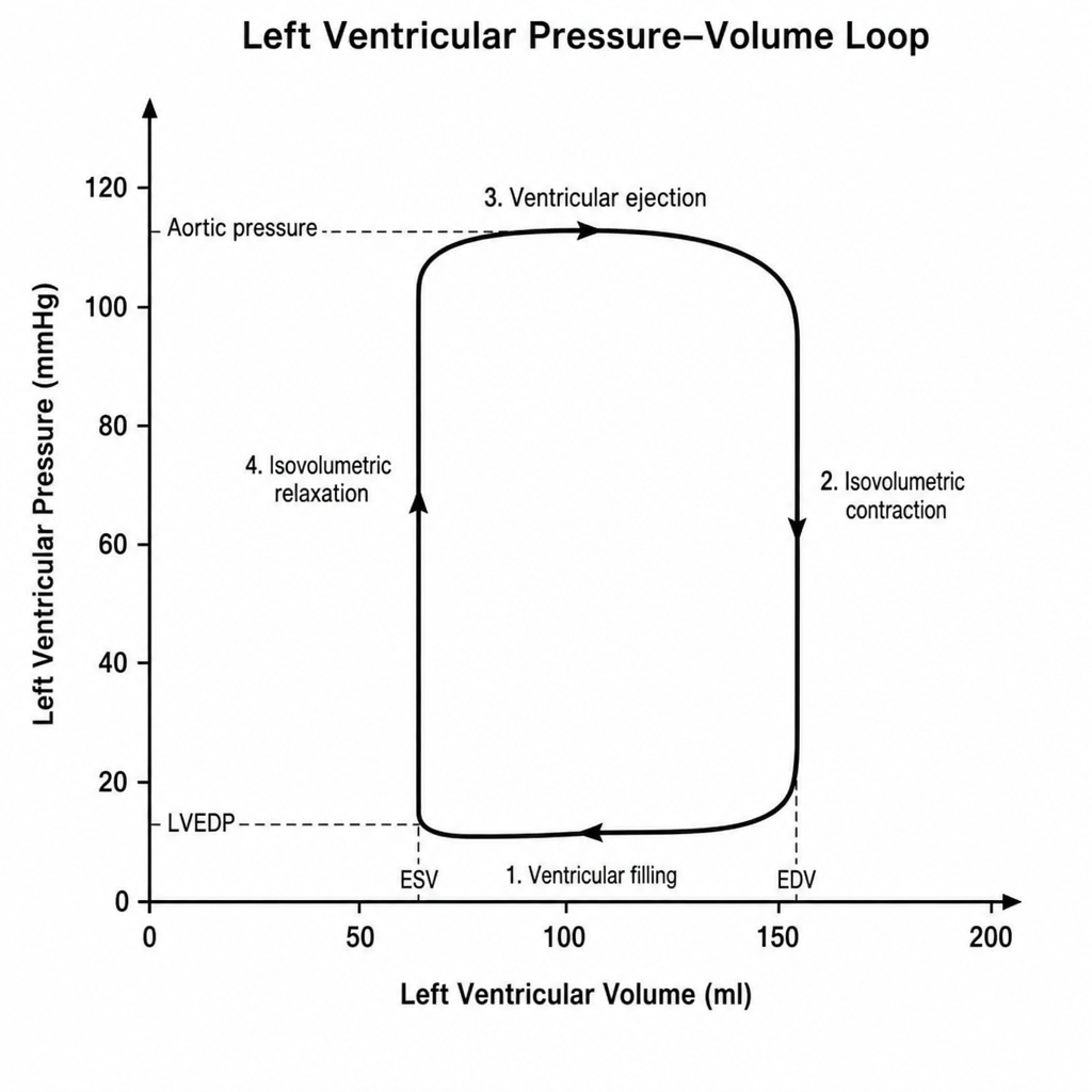
What is the action of Atrial Natriuretic Peptide (ANP)?
How does chronic hypertension affect the range of arterial pressure over which the cerebral circulation can maintain relatively constant blood flow?
Which statement is true regarding heart sounds?
Albumin exerts high oncotic pressure because?
The following ECG findings are seen in hypokalemia:
Which of the following is a feature of the Hering-Breuer reflex?
Explanation: **Explanation:** The rate of lymph flow is primarily determined by the **net filtration pressure** at the arterial end of the capillary. According to **Starling’s Law**, fluid movement is governed by the balance between hydrostatic and oncotic pressures. **1. Why Option A is Correct:** **Increased capillary hydrostatic pressure** (e.g., due to venous obstruction or heart failure) forces more fluid out of the capillaries into the interstitial space. This increase in interstitial fluid volume raises the **interstitial fluid hydrostatic pressure**, which pushes fluid into the lymphatic capillaries, thereby increasing lymph flow. **2. Why the Other Options are Incorrect:** * **B. Increased plasma oncotic pressure:** Plasma proteins (mainly albumin) exert an inward "pulling" force. Increasing this pressure keeps fluid inside the vessel, decreasing filtration and subsequently reducing lymph flow. * **C. Decreased capillary permeability:** Lymph is essentially filtered plasma and proteins. Decreased permeability (e.g., due to certain drugs) prevents fluid and proteins from leaking into the interstitium, thus reducing lymph formation. * **D. Precapillary constriction:** Constricting the arterioles reduces the blood flow and hydrostatic pressure within the downstream capillaries, leading to decreased fluid filtration and lower lymph flow. **High-Yield Clinical Pearls for NEET-PG:** * **Maximum Lymph Flow:** Lymph flow increases as interstitial pressure rises, but it plateaus when the pressure reaches **0 to +2 mmHg**. This is because high tissue pressure eventually compresses the lymphatic vessels themselves. * **The Lymphatic Pump:** The primary "intrinsic" factor for lymph flow is the rhythmic contraction of smooth muscles in the lymphatic vessel walls (**lymphangions**). * **Edema Safety Factor:** Lymph flow can increase up to **10–20 fold** to prevent edema when interstitial fluid pressure rises.
Explanation: ### Explanation The transport of substances across the capillary wall depends primarily on their **lipid solubility**. **1. Why Glucose is the Correct Answer:** Glucose is a **water-soluble (lipid-insoluble)** molecule. Because it cannot dissolve in the lipid bilayer of the endothelial cell membrane, it cannot pass directly through the cells. Instead, it must cross the capillary wall through **intercellular clefts** (water-filled channels between adjacent endothelial cells) or fenestrations. Other substances using this route include water, electrolytes (Na+, Cl-), and amino acids. The rate of diffusion for these substances is limited by their molecular size and the total surface area of the clefts. **2. Why the Other Options are Incorrect:** * **A, B, and C (O2, CO2, and CO):** These are all **lipid-soluble gases**. Lipid-soluble substances can diffuse directly through any portion of the endothelial cell membrane. Because the entire surface area of the capillary (not just the clefts) is available for their transport, they diffuse much more rapidly than water-soluble substances. Their transport is "flow-limited" rather than "diffusion-limited." **Clinical Pearls & High-Yield Facts for NEET-PG:** * **Blood-Brain Barrier (BBB):** In the brain, endothelial cells are joined by **tight junctions**, eliminating these intercellular clefts. This is why glucose requires specific transporters (GLUT-1) to enter the brain, while lipid-soluble gases still pass freely. * **Starling Forces:** While small solutes like glucose move via diffusion through clefts, the movement of water is governed by the balance of hydrostatic and oncotic pressures across these same clefts. * **Permeability:** The permeability of intercellular clefts varies by organ; they are largest in the **liver** (discontinuous capillaries) and smallest in the **brain** (continuous capillaries).
Explanation: **Explanation:** The correct answer is **Arterioles**. **Why Arterioles are the correct answer:** Arterioles are known as the **"Resistance Vessels"** of the cardiovascular system. They possess a thick layer of smooth muscle in their walls which is richly innervated by sympathetic nerve fibers. By undergoing vasoconstriction or vasodilation, arterioles can significantly alter their diameter. According to **Poiseuille’s Law**, resistance is inversely proportional to the fourth power of the radius ($R \propto 1/r^4$). Therefore, even small changes in the arteriolar caliber result in large changes in peripheral resistance, making them the primary site for regulating blood flow to specific organs and maintaining systemic arterial blood pressure. **Why other options are incorrect:** * **Venules:** These are primarily **"Capacitance Vessels."** Their main function is to act as a reservoir for blood (holding about 60-70% of total blood volume) rather than regulating active flow resistance. * **Capillaries:** These are **"Exchange Vessels."** While they are the site of nutrient and gas exchange, they lack smooth muscle fibers in their walls and thus cannot actively contract or dilate to regulate blood flow independently. Flow through capillaries is instead controlled by the upstream arterioles and precapillary sphincters. **High-Yield Clinical Pearls for NEET-PG:** * **Highest Resistance:** The maximum peripheral resistance to blood flow occurs in the **arterioles**. * **Largest Pressure Drop:** The steepest decline in mean arterial pressure occurs as blood passes through the **arterioles**. * **Velocity of Flow:** The velocity of blood flow is **lowest in the capillaries** due to their largest total cross-sectional area, allowing adequate time for exchange. * **Total Peripheral Resistance (TPR):** Arterioles are the main determinants of TPR; their diameter is regulated by local metabolites (autoregulation) and the autonomic nervous system.
Explanation: ### Explanation **1. Why Option B is Correct:** The cardiac cycle duration is inversely proportional to the heart rate. It represents the time taken for one complete heartbeat, including both systole and diastole. The mathematical formula to calculate the duration is: **Cardiac Cycle Duration (seconds) = 60 / Heart Rate (bpm)** For a heart rate of 75 bpm: $60 \div 75 = 0.8 \text{ seconds}$ In a standard 0.8s cycle, atrial systole lasts 0.1s, ventricular systole lasts 0.3s, and total diastole (quiescent period) lasts 0.4s. **2. Why Other Options are Incorrect:** * **Option A (0.4s):** This would correspond to a heart rate of 150 bpm ($60/0.4$). This is a state of significant tachycardia. * **Option C (1.0s):** This corresponds to a heart rate of 60 bpm ($60/1.0$), which is the lower limit of a normal resting heart rate. * **Option D (1.6s):** This corresponds to a heart rate of 37.5 bpm ($60/1.6$), indicating severe bradycardia (e.g., complete heart block). **3. Clinical Pearls & High-Yield Facts for NEET-PG:** * **Inverse Relationship:** When the heart rate increases, the duration of the cardiac cycle decreases. * **Diastole is Vulnerable:** When the heart rate increases, the **diastolic phase shortens significantly more** than the systolic phase. Since coronary perfusion occurs primarily during diastole, extreme tachycardia can lead to myocardial ischemia. * **Formula for Systole:** A common rule of thumb is that ventricular systole occupies approximately 1/3 of the cycle, while diastole occupies 2/3 at resting rates. * **Atrial vs. Ventricular:** Remember that atrial and ventricular cycles overlap; they do not occur in a simple linear sequence of 0.1 + 0.3 + 0.4.
Explanation: **Explanation:** Atrial Natriuretic Peptide (ANP) is a hormone released by the atrial myocytes in response to **atrial stretch** (increased preload/volume overload). Its primary physiological role is to decrease blood pressure and blood volume. **Why Option A is Correct:** ANP acts as a potent vasodilator and promotes natriuresis (sodium excretion) and diuresis (water excretion), which directly leads to **hypotension**. Interestingly, while most vasodilators cause reflex tachycardia, ANP inhibits the baroreceptor reflex and decreases sympathetic outflow while increasing vagal tone. This results in a paradoxical **bradycardia** (the Bezold-Jarisch-like effect), making "bradycardia and hypotension" the correct physiological profile. **Why Other Options are Incorrect:** * **Options B & C (Hypertension):** ANP is an antagonist to the Renin-Angiotensin-Aldosterone System (RAAS). It inhibits renin and aldosterone secretion, leading to a decrease in blood pressure, not an increase. * **Options B & D (Tachycardia):** Although hypotension usually triggers the baroreflex to increase heart rate, ANP specifically blunts this sympathetic response, leading to a lower heart rate (bradycardia) rather than tachycardia. **High-Yield NEET-PG Pearls:** * **Mechanism:** ANP acts via **membrane-bound Guanylyl Cyclase**, increasing intracellular **cGMP**. * **Kidney Effects:** It dilates afferent arterioles and constricts efferent arterioles, thereby **increasing GFR** while promoting sodium loss. * **Clinical Marker:** **BNP (Brain Natriuretic Peptide)**, secreted by ventricles, is a more stable clinical marker used to diagnose and monitor Heart Failure. * **Antagonist:** ANP is the "natural antagonist" to Aldosterone and Angiotensin II.
Explanation: ### Explanation **1. Why Option D is Correct: The Concept of Autoregulation Shift** Cerebral blood flow (CBF) is maintained constant between a Mean Arterial Pressure (MAP) of approximately **60 to 150 mmHg** via cerebral autoregulation. In chronic hypertension, the cerebral arterioles undergo structural remodeling, specifically **medial hypertrophy** and increased wall-to-lumen ratio. This thickening of the vessel walls increases vascular resistance and protects the brain from high-pressure surges. Consequently, the entire autoregulatory curve **shifts to the right**. This means the brain requires a higher MAP to maintain adequate perfusion, but it can also tolerate much higher pressures before developing hypertensive encephalopathy. **2. Why Other Options are Incorrect:** * **Option A:** Incorrect because the cerebral vasculature is highly dynamic; chronic exposure to high pressure triggers structural adaptation (remodeling). * **Option B:** While the vasculature does adapt to higher pressures, this is only half the story. The shift is global, meaning the lower limit also rises. * **Option C:** This is a consequence of the shift, not the primary description of the phenomenon. In hypertensive patients, the "lower limit" of autoregulation might be 100 mmHg instead of 60 mmHg. If their BP is dropped rapidly to "normal" levels (e.g., 120/80), they may actually suffer cerebral ischemia. **3. High-Yield Clinical Pearls for NEET-PG:** * **The "Lower Limit" Danger:** In chronic hypertensives, a rapid therapeutic reduction in blood pressure can lead to **watershed infarcts** because their "new" lower limit of autoregulation is higher than a normotensive person's. * **Mechanism:** Autoregulation is primarily mediated by the **Myogenic mechanism** (Bayliss effect) and metabolic factors (CO2 being the most potent vasodilator). * **Sympathetic Influence:** Chronic sympathetic overactivity in hypertension contributes to the rightward shift, further protecting the brain against high-pressure breakthroughs.
Explanation: ### Explanation The heart sounds are discrete auditory events produced by the vibrations of the cardiac valves and blood flow. Understanding their physical characteristics is essential for clinical diagnosis. **Why Option B is Correct:** The **First Heart Sound (S1)** is produced by the closure of the Atrioventricular (AV) valves (Mitral and Tricuspid) at the onset of ventricular systole. Compared to the second heart sound (S2), S1 is characterized as being **longer in duration** (approx. 0.10–0.17 seconds), lower in pitch (frequency), and softer (the "Lubb" sound). Its longer duration is attributed to the relatively slower closure of the AV valves and the prolonged vibrations of the chordae tendineae and ventricular walls. **Analysis of Incorrect Options:** * **Option A:** S1 has a **lower frequency** (25–45 Hz) compared to S2 (50 Hz). S2 is higher-pitched and "snappier" (the "Dupp" sound). * **Option C:** S1 is due to the closure of **AV valves**, not semilunar valves. * **Option D:** S2 is due to the closure of **Semilunar valves** (Aortic and Pulmonary) at the onset of ventricular diastole. **High-Yield NEET-PG Pearls:** * **S1 Splitting:** Usually narrow and best heard at the tricuspid area; M1 (Mitral) precedes T1 (Tricuspid). * **S2 Splitting:** Physiological splitting increases during **inspiration** (increased venous return to the right heart delays P2). * **S3 (Ventricular Gallop):** Occurs during the rapid filling phase; normal in children/athletes but indicates **Heart Failure** in adults. * **S4 (Atrial Gallop):** Occurs during atrial contraction; always pathological, indicating a **stiff ventricle** (e.g., LV hypertrophy).
Explanation: **Explanation:** The oncotic pressure (colloid osmotic pressure) of plasma is primarily determined by the **number of particles** in a solution rather than the size of the particles. This is based on Van't Hoff’s Law. **1. Why Option B is Correct:** Albumin accounts for approximately **75-80% of the total plasma oncotic pressure** (about 22 out of 28 mmHg). This is due to two factors: * **High Concentration:** Albumin is the most abundant plasma protein (3.5–5.0 g/dL). Since osmotic pressure is a colligative property, the sheer number of albumin molecules exerts the greatest pull on water. * **Low Molecular Weight:** Compared to other plasma proteins like globulins (MW ~90,000–150,000 Da) or fibrinogen (MW ~340,000 Da), albumin has a relatively **low molecular weight (~69,000 Da)**. For a given mass, a lower molecular weight means more individual molecules are present, further increasing the osmotic effect. **2. Why Other Options are Incorrect:** * **Options A & C:** High molecular weight would mean fewer molecules per unit gram, resulting in *lower* oncotic pressure. * **Options A & D:** Low concentration would significantly decrease the osmotic gradient, leading to fluid leakage into the interstitium (edema). **High-Yield Clinical Pearls for NEET-PG:** * **Gibbs-Donnan Effect:** Albumin is negatively charged at physiological pH. It attracts cations (mainly $Na^+$) into the capillaries, which accounts for about **1/3rd of its total oncotic effect**. * **Hypoalbuminemia:** When serum albumin falls below **2.0–2.5 g/dL** (e.g., in Nephrotic syndrome or Liver Cirrhosis), oncotic pressure drops, leading to generalized edema and ascites. * **Starling’s Forces:** Oncotic pressure is the primary force "holding" fluid inside the vascular compartment, opposing the Hydrostatic pressure which pushes fluid out.
Explanation: **Explanation:** Hypokalemia (low serum potassium) significantly affects the electrical activity of the heart by altering the resting membrane potential and prolonging the repolarization phase. **1. Why Option A is Correct:** In hypokalemia, the resting membrane potential becomes more negative (hyperpolarized), which slows down conduction through the AV node, leading to an **increased PR interval**. Furthermore, low potassium levels affect the repolarization phase (Phase 3), causing **ST-segment depression**, flattening or inversion of T waves, and the appearance of prominent **U waves**. **2. Why the Other Options are Incorrect:** * **Option B:** Peaked T waves are the hallmark of **Hyperkalemia**, not hypokalemia. In hyperkalemia, the PR interval may also increase, but the T wave morphology is the distinguishing factor. * **Option C:** While T wave inversion occurs in hypokalemia, the "prolonged QT interval" is often a misinterpretation. In hypokalemia, the T wave flattens and merges with the U wave, creating a **"QU interval"** that appears long. True QT prolongation is more characteristic of hypocalcemia. * **Option D:** Decreased QT interval is typically seen in **Hypercalcemia** or Digitalis effect, not hypokalemia. **High-Yield Clinical Pearls for NEET-PG:** * **ECG Sequence in Hypokalemia:** T wave flattening → ST depression → Prominent U waves (best seen in V2-V4) → Apparent prolongation of QT (actually QU interval). * **U Wave:** It is the most characteristic finding. It represents the delayed repolarization of Purkinje fibers. * **Danger:** Severe hypokalemia can predispose patients to **Torsades de Pointes** and ventricular arrhythmias. * **Mnemonic:** "Hypo-K has a low T (flat), a low ST (depression), and a big U."
Explanation: The **Hering-Breuer Inflation Reflex** is a protective mechanism triggered by the over-inflation of the lungs. When tidal volume exceeds approximately 1.5 liters (in adults), stretch receptors in the smooth muscles of the bronchi and bronchioles are activated. ### **Mechanism and Explanation** 1. **Respiratory Effect (Option A):** Impulses travel via the **Vagus nerve (CN X)** to the solitary tract nucleus (NTS) in the medulla. This inhibits the dorsal respiratory group (DRG) and the apneustic center, prematurely terminating inspiration. This results in a shorter inspiratory phase, leading to **rapid, shallow breathing** (tachypnea) to prevent alveolar over-distension. 2. **Cardiovascular Effects (Options B & C):** The reflex involves "Vagal cross-talk." Stimulation of the vagal afferents from the lungs leads to a concomitant increase in parasympathetic outflow to the heart and a decrease in sympathetic tone. This results in: * **Bradycardia:** Due to increased vagal (parasympathetic) discharge to the SA node. * **Hypotension:** Due to systemic vasodilation and decreased cardiac output. Since the reflex triggers all three physiological responses, **Option D (All of the above)** is the correct answer. ### **High-Yield Clinical Pearls for NEET-PG** * **Receptors:** Slowly Adapting Stretch Receptors (SARs). * **Afferent Pathway:** Vagus Nerve. * **Physiological Role:** In neonates, it is active during normal tidal breathing. In adults, it is mainly a protective mechanism during exercise or high-volume states. * **Hering-Breuer Deflation Reflex:** A separate reflex where lung deflation triggers a shorter expiratory phase to increase respiratory rate (preventing lung collapse).
Cardiac Electrophysiology
Practice Questions
Cardiac Cycle
Practice Questions
Cardiac Output and Its Regulation
Practice Questions
Hemodynamics and Blood Flow
Practice Questions
Arterial System Physiology
Practice Questions
Microcirculation and Lymphatics
Practice Questions
Venous Return and Central Venous Pressure
Practice Questions
Cardiovascular Reflexes
Practice Questions
Regional Circulations
Practice Questions
Cardiovascular Responses to Exercise and Stress
Practice Questions
Get full access to all questions, explanations, and performance tracking.
Start For Free