
Neurology — MCQs

On this page
What is the best prophylaxis for a 4-year-old male child experiencing febrile seizures?
All of the following statements regarding Sturge Weber syndrome are true, EXCEPT:
A 7-year-old male patient presented with skin lesions on the face and the lumbosacral region. The patient also has a history of frequent seizures. Which of the following chromosomes is involved in the disease?
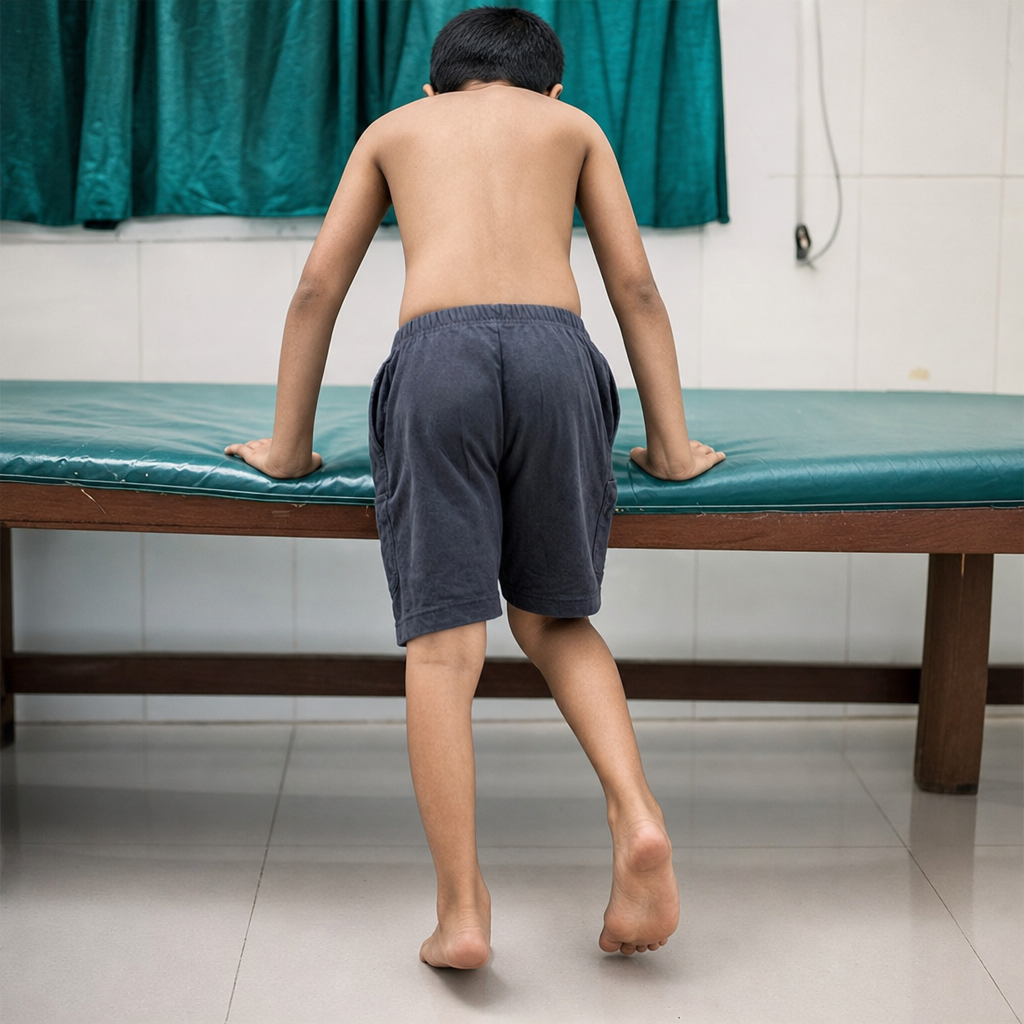
Which of the following disorders is NOT associated with the sign shown in the image?
What is the investigation of choice for the diagnostic evaluation of hydrocephalus in a one-month-old child?
A 9-year-old boy presented with difficulty in climbing stairs and combing. On examination, his calves are swollen and the child uses his feet to stand up on his legs. What is the next diagnostic step?
Continuous prophylactic anticonvulsant therapy is not needed in a child with febrile convulsions with which of the following conditions?
Atypical febrile seizures are associated with which of the following?
A child presented to the casualty with seizures. On examination, oval hypo-pigmented macules were noted on the trunk, along with stib-noo:il. What is the probable diagnosis in this child?
A 7-year-old girl presents with recurrent staring spells lasting a few seconds, after which she returns to her previous activities. Her EEG shows 3 Hz spike-and-wave discharges. Which of the following drugs is NOT indicated for treating this child?
Practice by Chapter
Seizure Disorders and Epilepsy
Practice Questions
Febrile Seizures
Practice Questions
Headache Disorders
Practice Questions
Cerebral Palsy
Practice Questions
Neural Tube Defects
Practice Questions
Neuromuscular Disorders
Practice Questions
Neurodegenerative Disorders
Practice Questions
CNS Infections
Practice Questions
Hydrocephalus
Practice Questions
Movement Disorders
Practice Questions
Traumatic Brain Injury
Practice Questions
Neuroimaging in Pediatrics
Practice Questions
Want unlimited practice?
Get full access to all questions, explanations, and performance tracking.
Scan to download app