Grade of tumor denotes
In which condition is Calretinin primarily used as a diagnostic marker?
Which syndrome is associated with an inherited mutation of the p53 tumor suppressor gene?
A 59-year-old lady presents with a progressive, painless lump in the breast. What is the cause of the skin change associated with breast cancer?

An elderly male is known as a smoker presented with chronic cough, significant weight loss, and fatigue. Serum calcium level is raised. A lung biopsy was done, and it showed large atypical cells with hyperchromasia. What is the probable diagnosis?
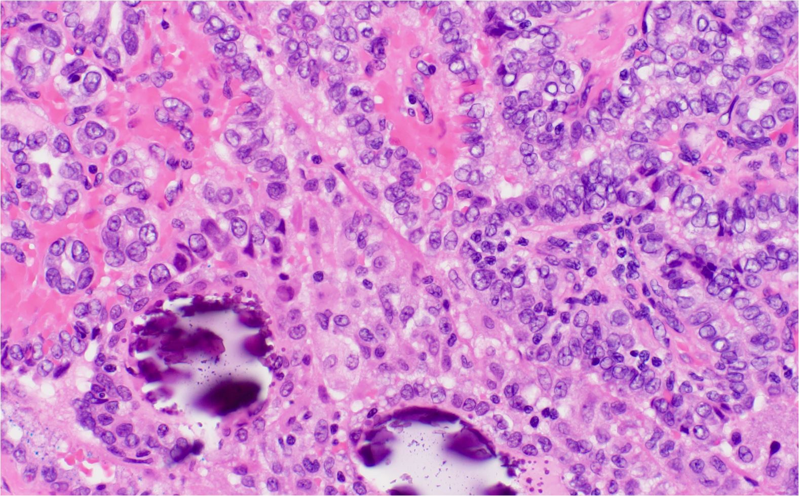
A 25-year-old male presented with a 2cm thyroid nodule. A thyroidectomy was done. The histology picture is given below. What could be the diagnosis based on the histological findings?

What is the T stage classification for a lung carcinoma measuring 2.5 cm and not involving the pleura?
RET proto-oncogene is associated with the development of
Anaplasia is
Which is the most common type of male breast cancer?
Explanation: ***Degree of differentiation*** - The grade of a tumor is primarily defined by the **degree of differentiation** of the cancer cells, indicating how much the tumor cells resemble normal cells [2]. - Grading helps predict the **aggressiveness** of cancer and its behavior in the body. *Vascular invasion* - Vascular invasion refers to the presence of cancer cells within blood vessels and is more associated with tumor **stage** rather than the **grade**. - It indicates the potential for cancer to metastasize, but it does not reflect the differentiation of cells. *Stage of disease* - The stage of disease encompasses the extent of tumor spread and includes aspects such as **size** and **lymph node involvement**, rather than the cellular characteristics of differentiation. - Staging is about **how advanced** the cancer is, while grading focuses on the **appearance** of the tumor cells. *Degree of anaplasia* - Anaplasia refers to the loss of differentiation and organization of cells [1][2], which can relate to tumor **grade**, but it is not synonymous with grading itself. - While higher grades indicate greater anaplasia, not all anaplastic tumors are graded the same due to other factors influencing classification. **References:** [1] Kumar V, Abbas AK, et al.. Robbins and Cotran Pathologic Basis of Disease. 9th ed. Neoplasia, pp. 278-280. [2] Kumar V, Abbas AK, et al.. Robbins and Cotran Pathologic Basis of Disease. 9th ed. Neoplasia, pp. 276-278.
Explanation: ***Mesothelioma*** - **Calretinin** is a sensitive marker used specifically for diagnosing **mesothelioma**, particularly in differentiating it from adenocarcinomas [1]. - It is expressed in **mesothelial cells**, making it a crucial tool in immunohistochemical staining for tumor identification [1]. *Hamartoma* - **Hamartomas** typically do not express **calretinin**, and their diagnosis requires different markers based on the tissue type involved. - They are benign and can occur in various organs but do not show mesothelial characteristics. *Chordoma* - **Chordomas** are malignant tumors originating from notochordal remnants, and they are not associated with **calretinin** positivity. - They are usually **mucins-positive** and identified using other specific markers, such as **brachyury**. *Choristoma* - **Choristomas** are benign lesions of ectopic tissue, and like hamartomas, they do not express **calretinin**. - These tumors are often diagnosed based on the specific type of tissue they contain rather than immunohistochemistry. **References:** [1] Kumar V, Abbas AK, et al.. Robbins and Cotran Pathologic Basis of Disease. 9th ed. The Lung, p. 731.
Explanation: ***Li-Fraumeni syndrome*** - This syndrome is characterized by an inherited mutation in the **TP53 tumor suppressor gene**, which encodes the p53 protein. - Individuals with Li-Fraumeni syndrome have a significantly increased risk of developing various cancers at a young age, including **osteosarcoma**, soft tissue sarcomas, breast cancer, brain tumors, and adrenocortical carcinoma. *Familial adenomatous polyposis syndrome* - This syndrome is caused by an inherited mutation in the **APC tumor suppressor gene**, not p53. - It is characterized by the development of hundreds to thousands of **colorectal adenomatous polyps**, which inevitably progress to colorectal cancer if untreated. *Retinoblastoma syndrome* - This syndrome is caused by an inherited mutation in the **RB1 tumor suppressor gene**, not p53. - It primarily affects the eye, leading to the development of **retinoblastoma**, a malignant tumor of the retina, often in childhood. *Osteosarcoma syndrome* - While osteosarcoma is a common cancer type seen in Li-Fraumeni syndrome, "Osteosarcoma syndrome" itself is not a distinct inherited syndrome specifically defined by a p53 mutation. - **Osteosarcoma** can arise from various genetic predispositions, including Li-Fraumeni syndrome, but it's not the sole defining characteristic of a p53-related syndrome.
Explanation: ***Infiltration of subdermal lymphatics*** - The skin change described often refers to **peau d'orange**, which is caused by the **blockage of superficial lymphatic vessels** by tumor cells [1]. - This lymphatic obstruction leads to **edema** and dimpling of the skin, resembling an orange peel [1]. *Infiltration of the lactiferous duct* - While cancer can arise in or infiltrate lactiferous ducts, this primarily causes symptoms like **nipple discharge**, inversion, or a palpable mass. - It does not directly explain the specific **skin dimpling** or **peau d'orange** appearance. *Involvement of Cooper's ligament* - Involvement of **Cooper's ligaments** (suspensory ligaments of the breast) can cause skin dimpling or retraction [1]. - However, the image depicts widespread **peau d'orange**, which is more commonly associated with lymphatic involvement than direct ligamentous retraction [1]. *Spread of the tumor to the anterior chest wall* - Tumor extension to the **anterior chest wall** signifies advanced disease and can cause **fixation of the breast** to underlying structures or skin ulceration [2]. - This typically results in **immobility of the breast** or an ulcerated lesion, not the widespread edematous and dimpled appearance of **peau d'orange**. **References:** [1] Cross SS. Underwood's Pathology: A Clinical Approach. 6th ed. Common Clinical Problems From Liver And Biliary System Disease, pp. 453-454. [2] Cross SS. Underwood's Pathology: A Clinical Approach. 6th ed. Common Clinical Problems From Liver And Biliary System Disease, pp. 456-457.
Explanation: ***Squamous cell carcinoma*** - The presence of **hyperchromatic atypical cells** in the lung biopsy [2] and **elevated serum calcium levels** are indicative of paraneoplastic hypercalcemia often seen in squamous cell carcinoma. - This type of lung cancer typically occurs in **smokers** [1] and is associated with **chronic cough** and **weight loss**. *Large cell neuroendocrine tumor* - While it may show large atypical cells, this tumor type is less commonly associated with **hypercalcemia** or the smoking history highlighted here. - Characteristically, it tends to have a more aggressive course and distinct histological features not indicated in the vignette. *Small cell carcinoma* - This type typically presents with **central lung lesions** and is associated with secretory syndromes like **SIADH**, not hypercalcemia. - Histologically, it features small round blue cells rather than the large atypical cells described in this case. *Adenocarcinoma* - Often arises peripherally in the lung and typically does not present with hypercalcemia. - Histological features include **gland formation**, which contrasts with the **large atypical cells** noted in the biopsy. **References:** [1] Cross SS. Underwood's Pathology: A Clinical Approach. 6th ed. Common Clinical Problems From Respiratory Tract Disease, pp. 336-337. [2] Kumar V, Abbas AK, et al.. Robbins and Cotran Pathologic Basis of Disease. 9th ed. The Lung, pp. 723-724.
Explanation: ***Papillary carcinoma thyroid*** [1] - Typically presents as a **thyroid nodule** with distinctive histological features such as **papillary structures** and nuclear **grooves or inclusions** [1]. - Often associated with a history of radiation exposure and is the most common type of **thyroid cancer** in young adults. *Graves disease* - Characterized by **hyperthyroidism** and usually presents with diffuse **goiter** rather than a solitary nodule. - Histological findings typically show **diffuse hyperplasia** rather than a well-circumscribed nodule. *Adenomatous goitre* - Usually presents as a **multinodular goiter** with no malignant potential. - Histologically characterized by **colloid accumulation** and enlarged follicles, not indicative of malignancy. *Follicular adenoma* [2] - Characterized by a well-encapsulated nodule with **uniform follicular cells** and abundant **colloid** [1,2]. - Does not exhibit the **invasive features** seen in malignancies like papillary carcinoma [1]. **References:** [1] Kumar V, Abbas AK, et al.. Robbins and Cotran Pathologic Basis of Disease. 9th ed. The Endocrine System, pp. 1099-1101. [2] Cross SS. Underwood's Pathology: A Clinical Approach. 6th ed. Common Clinical Problems From Liver And Biliary System Disease, pp. 428-429.
Explanation: ***T1c (tumor > 2 cm but ≤ 3 cm)*** - A lung carcinoma measuring 2.5 cm falls within the **T1c classification** because it is greater than 2 cm but no larger than 3 cm. - The absence of **pleural involvement** confirms this is not a higher T stage (T2 or above) [1]. - According to the **8th Edition TNM Classification**, T1c specifically denotes tumors in this size range [1]. *T1a (tumor ≤ 1 cm)* - This classification is for tumors that are **1 cm or less in greatest dimension**, which is much smaller than the 2.5 cm tumor in the question. - T1a represents the **smallest category** of primary lung tumors [1]. *T2 (tumor > 3 cm)* - A T2 classification applies to tumors that are **larger than 3 cm** (T2a: > 3-4 cm, T2b: > 4-5 cm), which does not apply to a 2.5 cm tumor [1]. - T2 can also include smaller tumors with specific features like **visceral pleural invasion**, **main bronchus involvement**, or causing **atelectasis/obstructive pneumonitis**, none of which are present here [1]. *T1b (tumor > 1 cm but ≤ 2 cm)* - This classification applies to tumors **greater than 1 cm but not exceeding 2 cm** in greatest dimension. - The 2.5 cm tumor in the question **exceeds this size range**, making T1b incorrect [1]. - T1b represents an intermediate size between T1a and T1c [1]. **References:** [1] Kumar V, Abbas AK, et al.. Robbins and Cotran Pathologic Basis of Disease. 9th ed. The Lung, p. 725.
Explanation: ***Medullary carcinoma thyroid*** - The **RET proto-oncogene** mutation is strongly linked to medullary thyroid carcinoma, particularly in Multiple Endocrine Neoplasia (MEN) type 2 syndrome [1]. - This carcinoma originates from **C-cells** of the thyroid, which produce calcitonin, and its presence is typically assessed via genetic testing. *Hurthle cell tumor thyroid* - Hurthle cell tumors, while associated with the thyroid, are not typically linked to **RET proto-oncogene** mutations but rather are considered a variant of follicular thyroid carcinoma. - They primarily appear as **eosinophilic cells** and are related to **radioactive iodine resistance**, which differs from medullary carcinoma. *Paraganglioma* - Paragangliomas arise from **neuroendocrine cells** and are associated with mutations in genes such as **SDHB** or **SDHD**, not RET. - They commonly present as **extra-adrenal pheochromocytomas** and are not related to thyroid pathology. *Astrocytoma* - Astrocytomas are primary **central nervous system tumors** that originate from astrocytic cells and have no known association with RET proto-oncogene mutations. - The mutations in this type of tumor are different, often involving genes like **TP53** and **IDH1**. **References:** [1] Kumar V, Abbas AK, et al.. Robbins and Cotran Pathologic Basis of Disease. 9th ed. The Endocrine System, pp. 1102-1103.
Explanation: ***Lack of differentiation*** - Anaplasia refers to a **loss of differentiation** in cells, making them more primitive and less specialized [1]. - It is often seen in **malignant tumors**, indicating a poor prognosis and aggressive behavior [1]. *Morphological changes* - While anaplasia involves **morphological changes**, this term is too broad and can relate to various cellular alterations, not exclusively anaplasia [1]. - Anaplasia specifically emphasizes **lack of differentiation**, distinct from general changes in cell appearance [1]. *Changing one type of epithelium to another* - This describes a process known as **metaplasia**, where one adult cell type transforms into another, not anaplasia. - Anaplasia signifies a **de-differentiation** rather than a change to a different epithelial type [1]. *Nuclear chromatin* - While changes in **nuclear chromatin** can occur in anaplastic cells, this does not define anaplasia itself [1]. - Anaplasia primarily refers to **loss of cell differentiation**, making this option insufficient to describe the concept [1]. **References:** [1] Kumar V, Abbas AK, et al.. Robbins and Cotran Pathologic Basis of Disease. 9th ed. Neoplasia, pp. 276-280.
Explanation: ***Invasive Ductal Carcinoma*** - This is by far the most common type of breast cancer in men, accounting for approximately **80-90%** of all male breast cancer cases. - It originates in the **milk ducts** and then invades the surrounding breast tissue. *Invasive Lobular Carcinoma* - This type of cancer originates in the **milk-producing glands (lobules)**. - It is **extremely rare** in men due to the underdeveloped and non-functional lobules in the male breast. *Mucinous Carcinoma* - This is a **rare subtype** of invasive ductal carcinoma characterized by cancer cells that produce **mucin**. - It accounts for only a small percentage of male breast cancers, typically **2-3%** of cases. *Inflammatory Breast Cancer* - This is a **rare and aggressive** form of breast cancer characterized by rapid onset of redness, swelling, and warmth in the breast, often mistaken for an infection. - Although it can occur in men, it is not the most common type and represents a very small fraction of male breast cancer diagnoses.
Nomenclature and Classification of Tumors
Practice Questions
Characteristics of Benign and Malignant Neoplasms
Practice Questions
Molecular Basis of Cancer
Practice Questions
Carcinogenesis and Carcinogens
Practice Questions
Tumor Progression and Metastasis
Practice Questions
Tumor Markers
Practice Questions
Paraneoplastic Syndromes
Practice Questions
Genetic Basis of Cancer
Practice Questions
Tumor Immunity
Practice Questions
Cancer Epidemiology and Prevention
Practice Questions
Get full access to all questions, explanations, and performance tracking.
Start For Free