All of the following are major subtypes of breast cancer based on Gene array analysis EXCEPT:
How do high-risk HPV types contribute to cervical carcinogenesis?
A patient has a cerebellar mass, renal tumor, and a family history of similar conditions. Which of the following mutations is most likely present in the family?
TTF-1 is a tumor marker for which of the following?
A 49 year old female presents with a breast lump. Which of the following findings is in accordance with basal-like breast cancer?
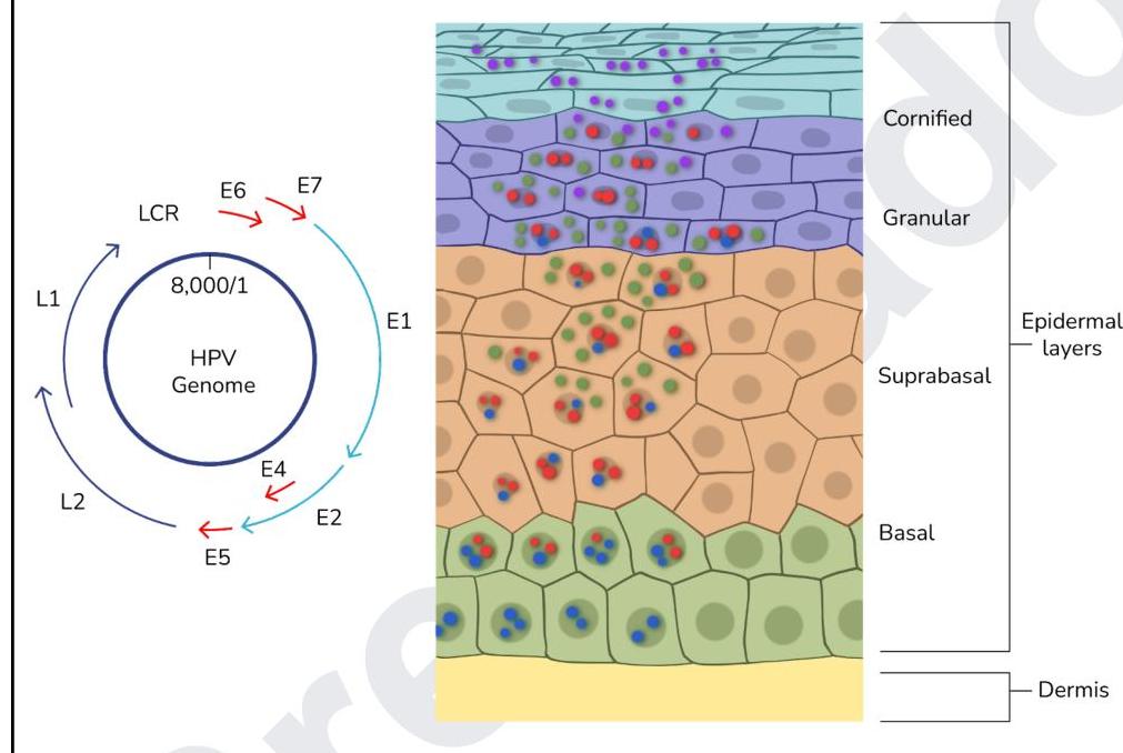
The image below shows the life cycle of a virus. Which of the proteins of the virus act as oncogenes?

Which of the following conditions is associated with perineural invasion?
What is the correct statement about thymoma?
Which of the following is an Anti-apoptotic gene?
A 32-year-old HIV-positive man presents with multiple flesh-colored, pedunculated lesions on his penis. Biopsy shows koilocytes and increased mitotic figures. Which of the following viral proteins is responsible for the cellular changes observed?
Explanation: ***Oestrogen receptor positive*** - While **estrogen receptor (ER) positivity** is a critical prognostic and predictive marker in breast cancer, it is a single marker, not a distinct intrinsic subtype encompassing broader genomic categorization [1]. - The intrinsic subtypes are based on gene expression profiles that cluster tumors into biologically distinct groups, such as Luminal A, Luminal B, HER2-enriched, and Basal-like (including Triple Negative) [3]. *Luminal A and Luminal B* - These are major intrinsic subtypes characterized by the expression of **hormone receptors** (ER and/or PR) and differing in their proliferation rates [2]. - **Luminal A** generally has high ER, low proliferation, and a good prognosis, while **Luminal B** often has higher proliferation (e.g., higher Ki-67) and a slightly worse prognosis [2]. *Triple negative* - This is a major intrinsic subtype (**Basal-like**) defined by the absence of **estrogen receptors (ER)**, **progesterone receptors (PR)**, and **HER2 overexpression** [1], [4]. - It often correlates with a more aggressive clinical course and specific treatment approaches [4]. *Her-2 receptor positive* - This is a major intrinsic subtype characterized by the **overexpression or amplification of the HER2 gene** [3]. - These cancers are often aggressive but respond well to targeted therapies like trastuzumab [3], [4]. **References:** [1] Kumar V, Abbas AK, et al.. Robbins and Cotran Pathologic Basis of Disease. 9th ed. The Breast, pp. 1064-1066. [2] Kumar V, Abbas AK, et al.. Robbins and Cotran Pathologic Basis of Disease. 10th ed. The Breast, pp. 1059-1060. [3] Kumar V, Abbas AK, et al.. Robbins and Cotran Pathologic Basis of Disease. 9th ed. The Breast, p. 1060. [4] Cross SS. Underwood's Pathology: A Clinical Approach. 6th ed. (Basic Pathology) introduces the student to key general principles of pathology, both as a medical science and as a clinical activity with a vital role in patient care. Part 2 (Disease Mechanisms) provides fundamental knowledge about the cellular and molecular processes involved in diseases, providing the rationale for their treatment. Part 3 (Systematic Pathology) deals in detail with specific diseases, with emphasis on the clinically important aspects., pp. 258-259.
Explanation: ***By integration into host genome and expression of E6/E7 oncoproteins*** - High-risk HPV types integrate their **viral DNA** into the host cell's genome, leading to the sustained expression of the **E6 and E7 oncoproteins** [1]. - **E6 targets p53** for degradation, impairing apoptosis, while **E7 targets Rb**, disrupting cell cycle control and promoting uncontrolled cell proliferation [1]. *Through direct mutagenic effects on host DNA* - HPV itself does not directly cause mutations through its own enzymatic activity on host DNA. - Its oncogenic potential stems from the **disruption of host cell regulatory proteins** rather than direct DNA alteration [1]. *Through chronic inflammation and oxidative damage* - While chronic inflammation can contribute to carcinogenesis, it is not the primary or sole mechanism by which high-risk HPV types induce cervical cancer. - HPV-mediated carcinogenesis is more specifically linked to the **oncoprotein activity** that overrides cellular tumor suppressor pathways [2]. *By inducing apoptosis resistance through capsid proteins* - HPV **capsid proteins (L1 and L2)** are primarily involved in viral assembly and entry, not in inducing apoptosis resistance. - The **E6 oncoprotein** is responsible for inactivating **p53** and conferring apoptosis resistance [1]. **References:** [1] Kumar V, Abbas AK, et al.. Robbins and Cotran Pathologic Basis of Disease. 9th ed. Neoplasia, pp. 334-335. [2] Kumar V, Abbas AK, et al.. Robbins and Cotran Pathologic Basis of Disease. 9th ed. The Female Genital Tract, pp. 1007-1008.
Explanation: ***VHL*** - **Von Hippel-Lindau (VHL) disease** is an inherited disorder characterized by the development of tumors and cysts in various parts of the body, including **hemangioblastomas** in the cerebellum and retina, **renal cell carcinomas**, and pheochromocytomas [1]. - The combination of a **cerebellar mass**, renal tumor, and a family history strongly points to VHL disease, which is caused by a germline mutation in the **VHL tumor suppressor gene** [1]. *Neurofibromatosis (NF1)* - **Neurofibromatosis type 1 (NF1)** typically presents with multiple neurofibromas, **café-au-lait spots**, optic pathway gliomas, and Lisch nodules in the iris. - While NF1 can cause tumors, the specific combination of a cerebellar mass and renal tumor is not typical of NF1, and the characteristic skin findings are not mentioned. *Tuberous Sclerosis Complex (TSC)* - **Tuberous Sclerosis Complex (TSC)** is characterized by the growth of benign tumors in the brain (e.g., **subependymal giant cell astrocytomas**), kidneys (e.g., **angiomyolipomas**), heart, lungs, and skin (e.g., facial angiofibromas) [2]. - While TSC can involve brain and kidney tumors, the typical brain tumors are different (astrocytomas vs. hemangioblastomas), and hemangioblastomas are not a common feature of TSC [2]. *Li-Fraumeni syndrome* - **Li-Fraumeni syndrome** is a rare inherited cancer predisposition syndrome characterized by a high risk of developing various cancers, including **sarcomas**, breast cancer, brain tumors (often astrocytomas or medulloblastomas), and adrenocortical carcinoma. - While brain tumors are part of Li-Fraumeni syndrome, renal cell carcinoma is not a primary feature, and the classic cerebellar hemangioblastoma is not typical for this syndrome. **References:** [1] Cross SS. Underwood's Pathology: A Clinical Approach. 6th ed. Common Clinical Manifestations Of Central And Peripheral Nervous System Disease, pp. 724-727. [2] Kumar V, Abbas AK, et al.. Robbins and Cotran Pathologic Basis of Disease. 9th ed. The Central Nervous System, pp. 1318-1319.
Explanation: ***Small cell carcinoma*** - **Thyroid transcription factor 1 (TTF-1)** is a nuclear transcription protein expressed in lung and thyroid neoplasms - It is positive in **85-90% of small cell lung carcinomas**, making it a key immunohistochemical marker [1] - TTF-1 helps differentiate small cell carcinoma from other neuroendocrine tumors and extrapulmonary small cell carcinomas [1] *Adenocarcinoma* - TTF-1 is also **strongly positive in 75-80% of lung adenocarcinomas** - It is a primary marker for lung adenocarcinoma, often used with **Napsin A** and **Cytokeratin 7 (CK7)** *Thymoma* - Thymomas are neoplasms of the **thymus gland** and typically express **cytokeratins** but **not TTF-1** - Characteristic markers include **CD5**, **CD117**, and epithelial markers *Melanoma* - Melanomas are cancers of **melanocytes** and express melanocytic markers like **S-100**, **HMB-45**, **Melan-A**, and **SOX10** - **TTF-1** is not expressed in melanoma and helps exclude lung primary when evaluating metastatic disease **References:** [1] Cross SS. Underwood's Pathology: A Clinical Approach. 6th ed. Common Clinical Problems From Respiratory Tract Disease, pp. 337-338.
Explanation: ***ER-, PR-, HER2-*** - **Basal-like breast cancer** is characterized by its **triple-negative** status, meaning it does not express estrogen receptor (ER), progesterone receptor (PR), or human epidermal growth factor receptor 2 (HER2) [1]. - This specific immunophenotype is crucial for diagnosis and influences treatment strategies, as these cancers do not respond to therapies targeting these receptors [1]. *ER+, PR-, HER2-* - This profile describes a **hormone-sensitive** cancer (ER positive) but without PR or HER2 expression. - While it responds to endocrine therapies, it is distinct from basal-like cancer due to its ER positivity. *ER-, PR-, HER2+* - This profile indicates a cancer that is **HER2-positive**, meaning it overexpresses HER2, and can be targeted with anti-HER2 therapies like trastuzumab. - This is a separate molecular subtype of breast cancer often referred to as HER2-enriched, which is distinct from basal-like [2]. *ER+, PR+, HER2-* - This is the most common subtype, known as **luminal A** or **luminal B** depending on grade and Ki-67 expression, characterized by sensitivity to endocrine therapy [2]. - This hormone receptor-positive and HER2-negative profile is very different from the triple-negative basal-like breast cancer. **References:** [1] Kumar V, Abbas AK, et al.. Robbins and Cotran Pathologic Basis of Disease. 9th ed. The Breast, pp. 1064-1066. [2] Kumar V, Abbas AK, et al.. Robbins and Cotran Pathologic Basis of Disease. 9th ed. The Breast, p. 1060.
Explanation: ***E6, E7*** - The **E6** and **E7** proteins of high-risk human papillomaviruses (HPVs) are considered **oncogenes** because they interfere with critical tumor suppressor pathways [1][2]. - **E6** promotes the degradation of **p53**, a tumor suppressor protein, while **E7** inactivates **retinoblastoma protein (pRb)**, leading to uncontrolled cell proliferation and increased risk of malignant transformation [2]. *L1, L2* - **L1** and **L2** are **late proteins** (structural proteins) that form the **viral capsid** (outer shell) of the HPV virion. - They are essential for assembling new viral particles but do not directly contribute to the oncogenic process by disrupting host cell cycle regulation. *E1, E2, E5* - **E1** is involved in **viral DNA replication**, acting as a helicase and ATPase. - **E2** regulates **viral gene expression** and DNA replication, while **E5** is a small transmembrane protein that can contribute to cell growth but is generally considered less potent in oncogenesis than E6 and E7, and its exact role varies by HPV type. *E1, E2* - **E1** is critical for **viral DNA replication**, and **E2** regulates viral gene transcription and DNA replication. - While important for the viral life cycle, neither E1 nor E2 are the primary drivers of oncogenesis in the way E6 and E7 are, as they do not directly target key tumor suppressor proteins like p53 and pRb. **References:** [1] Kumar V, Abbas AK, et al.. Robbins and Cotran Pathologic Basis of Disease. 9th ed. The Female Genital Tract, pp. 1007-1008. [2] Kumar V, Abbas AK, et al.. Robbins and Cotran Pathologic Basis of Disease. 9th ed. Neoplasia, pp. 334-335.
Explanation: ***Adenoid cystic carcinoma*** - **Adenoid cystic carcinoma** is the **most notoriously characterized** by its strong propensity for **perineural invasion**, which contributes to its high recurrence rate and poor prognosis [1]. - This invasion allows the tumor cells to spread along nerve sheaths, extending beyond the visible tumor margins, often for considerable distances. - It is the **classic example** of perineural invasion among salivary gland tumors [1]. *Mucoepidermoid tumor* - While mucoepidermoid tumors can be locally aggressive, **perineural invasion** is not a characteristic feature that defines this tumor type. - They are more commonly associated with cystic degeneration and mucin production. *Pancreatic cancer* - **Pancreatic adenocarcinoma** does show **significant perineural invasion** (present in 70-90% of cases) and is an important feature contributing to its poor prognosis and pain symptoms. - However, in the context of this question, **adenoid cystic carcinoma** is considered the **most characteristic** or **prototypical** example of perineural invasion, particularly among head and neck neoplasms. - Both are associated with perineural invasion, but adenoid cystic carcinoma is the textbook example. *Pleomorphic adenoma* - A **pleomorphic adenoma** is a benign mixed tumor of the salivary glands and usually does not exhibit **perineural invasion** [2]. - Malignant transformation into a carcinoma ex pleomorphic adenoma can occur, but the benign form primarily grows as an encapsulated mass [2]. **References:** [1] Kumar V, Abbas AK, et al.. Robbins and Cotran Pathologic Basis of Disease. 9th ed. Head and Neck, pp. 753-755. [2] Kumar V, Abbas AK, et al.. Robbins and Cotran Pathologic Basis of Disease. 9th ed. Head and Neck, pp. 751-753.
Explanation: ***Thymoma is the most common neoplasia of the thymus.*** [1] - **Thymoma** is the most common primary tumor of the thymus, accounting for approximately **40-50% of anterior mediastinal masses** in adults. - It is a slow-growing tumor originating from the **epithelial cells** of the thymus [1]. *Chest X-ray is the investigation of choice for the diagnosis of thymoma.* - While a **chest X-ray** may show a widened mediastinum or an anterior mediastinal mass, it is not the investigation of choice for definitive diagnosis or staging [2]. - **CT scan** of the chest with contrast is the preferred imaging modality for evaluating thymomas, providing better anatomical detail and assessing invasiveness [2]. *Thymoma is typically asymptomatic and rarely causes any symptoms.* - Approximately **30-50% of patients with thymoma are asymptomatic** at diagnosis, with the tumor discovered incidentally on imaging [2]. - However, the remaining **50-70% of patients present with symptoms** related to **mass effect** (e.g., chest pain, dyspnea, cough) or **paraneoplastic syndromes** like myasthenia gravis, pure red cell aplasia, or hypogammaglobulinemia [2]. - Therefore, it is incorrect to say thymoma "rarely" causes symptoms. *Thymoma is primarily located in the posterior mediastinum.* - **Thymoma** is characteristically located in the **anterior mediastinum**, which is the most common site for thymic tissue. - Tumors primarily found in the posterior mediastinum are more commonly **neurogenic tumors**. **References:** [1] Cross SS. Underwood's Pathology: A Clinical Approach. 6th ed. Common Clinical Problems From Diseases Of The Urinary And Male Genital Tracts, pp. 571-574. [2] Cross SS. Underwood's Pathology: A Clinical Approach. 6th ed. Common Clinical Problems From Diseases Of The Urinary And Male Genital Tracts, pp. 572-574.
Explanation: ***MCL-1*** - **MCL-1 (myeloid cell leukemia sequence 1)** is a pro-survival protein belonging to the **Bcl-2 family**, which inhibits apoptosis by binding to and sequestering pro-apoptotic proteins [1]. - Its overexpression is frequently observed in various cancers, contributing to **chemoresistance** and tumor survival [2]. *BAK* - **BAK (Bcl-2 antagonist killer 1)** is a **pro-apoptotic protein** that belongs to the Bcl-2 family. - Upon activation, BAK undergoes **oligomerization** on the mitochondrial outer membrane, leading to its permeabilization and the release of pro-apoptotic factors into the cytoplasm. *BIN* - **BIN1 (Bridging Integrator 1)** is a **tumor suppressor gene** that can promote apoptosis in certain contexts, particularly when associated with DNA damage or cellular stress. - It is not primarily known as an anti-apoptotic gene but rather as a protein involved in **membrane dynamics** and cellular signaling, with roles in endocytosis and cytoskeletal regulation. *NOX-Q* - **NOX-Q** is not a commonly recognized or established gene in the context of apoptosis regulation. - The **NOX (NADPH oxidase) family** of enzymes is primarily involved in the production of **reactive oxygen species (ROS)**, which can either induce or inhibit apoptosis depending on the cellular context and concentration. **References:** [1] Kumar V, Abbas AK, et al.. Robbins and Cotran Pathologic Basis of Disease. 9th ed. Neoplasia, p. 310. [2] Kumar V, Abbas AK, et al.. Robbins and Cotran Pathologic Basis of Disease. 9th ed. Neoplasia, pp. 310-311.
Explanation: ***E6 and E7 proteins*** - The presence of **koilocytes** and **increased mitotic figures** in penile lesions, particularly in an HIV-positive individual, strongly suggests **Human Papillomavirus (HPV)** infection leading to condyloma acuminata [2]. - **HPV E6 and E7 oncoproteins** are critical for HPV-induced cell proliferation and immortalization; E6 degrades **p53** (a tumor suppressor), and E7 inactivates **Rb protein** (retinoblastoma protein), leading to uncontrolled cell division and the observed cellular changes [1]. *Tax protein* - The **Tax protein** is associated with **Human T-lymphotropic virus type 1 (HTLV-1)**, which causes Adult T-cell Leukemia/Lymphoma and HTLV-1-associated myelopathy/tropical spastic paraparesis, not HPV-related lesions. - Tax acts as a transcriptional activator, promoting viral gene expression and cellular proliferation but through different mechanisms than HPV oncoproteins. *L1 protein* - The **L1 protein** is a major **capsid protein** of HPV and is used in HPV vaccines to induce protective antibodies. - While essential for viral structure and assembly, L1 itself does not directly cause the cellular proliferative changes or koilocytic atypia seen in infected cells; these are driven by the E6 and E7 oncoproteins. *EBNA-1* - **EBNA-1** (Epstein-Barr Nuclear Antigen 1) is a protein produced by the **Epstein-Barr virus (EBV)**, which is associated with various lymphomas (e.g., Burkitt lymphoma, Hodgkin lymphoma) and nasopharyngeal carcinoma. - It is crucial for the maintenance of the EBV episome in latently infected cells and does not cause penile lesions with koilocytes. **References:** [1] Kumar V, Abbas AK, et al.. Robbins and Cotran Pathologic Basis of Disease. 9th ed. The Female Genital Tract, pp. 1007-1008. [2] Kumar V, Abbas AK, et al.. Robbins and Cotran Pathologic Basis of Disease. 9th ed. The Lower Urinary Tract and Male Genital System, pp. 974-975.
Nomenclature and Classification of Tumors
Practice Questions
Characteristics of Benign and Malignant Neoplasms
Practice Questions
Molecular Basis of Cancer
Practice Questions
Carcinogenesis and Carcinogens
Practice Questions
Tumor Progression and Metastasis
Practice Questions
Tumor Markers
Practice Questions
Paraneoplastic Syndromes
Practice Questions
Genetic Basis of Cancer
Practice Questions
Tumor Immunity
Practice Questions
Cancer Epidemiology and Prevention
Practice Questions
Get full access to all questions, explanations, and performance tracking.
Start For Free