The malignancy associated with Stewart-Treves syndrome is
Which one of the following is NOT associated with BRCA1/BRCA2 genes?
In which of the following is the term low and high grade squamous intraepithelial neoplasia used?
Which one of the following is not an epithelial tumour of the ovary?
Which of the following pairs is not correctly matched?
Presence of signet-ring cells in a cellular or myxomatous stroma is diagnostic of:
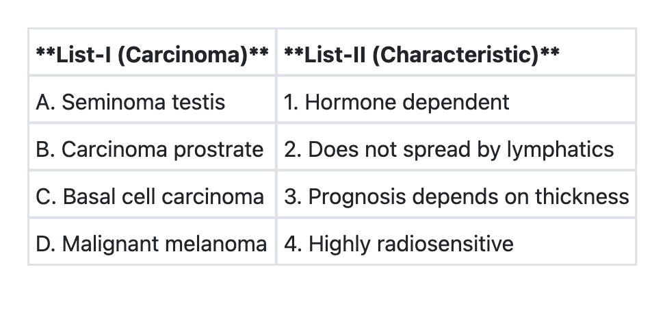
Match List-I with List-II and select the correct answer using the code given below the Lists:

Which of the following statements regarding Paget's disease of nipple are correct? 1. It represents benign pathology of nipple areola complex 2. It is eczema like condition of nipple and areola 3. Erosion of nipple is seen 4. Nipple biopsy is required for definitive diagnosis Select the correct answer using the code given below:
No increased relative risk of invasive breast carcinoma based on histopathological examination of benign breast tissue is for all of the following EXCEPT:
Which of the statements regarding Salivary gland neoplasms are correct? 1. 80–90% of parotid tumors are benign 2. 90% of sublingual gland tumors are malignant 3. 60–70% of submandibular gland tumors are benign 4. Parotid gland is most common site for salivary gland tumors Select the correct answer using the code given below:
Explanation: ***Lymphangiosarcoma*** - **Stewart-Treves syndrome** is characterized by the development of **lymphangiosarcoma** in a setting of chronic lymphedema, most commonly following a radical mastectomy for breast cancer [2]. - The chronic lymphatic obstruction and subsequent lymphedema [2] are thought to create an environment conducive to the development of this rare and aggressive **vascular malignancy** [1]. *Basal cell carcinoma* - This is a common **skin cancer** that typically arises from the basal cells of the epidermis and is primarily associated with **ultraviolet (UV) radiation exposure**, not chronic lymphedema [4]. - While it can occur in individuals with a history of radiation therapy (which might be part of breast cancer treatment), it is not the specific malignancy defining Stewart-Treves syndrome [3]. *Liposarcoma* - **Liposarcoma** is a malignant tumor of **adipose tissue** and does not have a direct association with chronic lymphedema or Stewart-Treves syndrome. - It arises from fat cells and can occur in various locations but is not typically linked to impaired lymphatic drainage. *Malignant melanoma* - This highly aggressive **skin cancer** arises from **melanocytes** and is strongly associated with **UV radiation exposure** and genetic predisposition. - While skin cancers can occur in patients with breast cancer, **melanoma** is not the specific malignancy that defines Stewart-Treves syndrome. **References:** [1] Kumar V, Abbas AK, et al.. Robbins and Cotran Pathologic Basis of Disease. 9th ed. Diseases of Infancy and Childhood, pp. 527-528. [2] Cross SS. Underwood's Pathology: A Clinical Approach. 6th ed. (Basic Pathology) introduces the student to key general principles of pathology, both as a medical science and as a clinical activity with a vital role in patient care. Part 2 (Disease Mechanisms) provides fundamental knowledge about the cellular and molecular processes involved in diseases, providing the rationale for their treatment. Part 3 (Systematic Pathology) deals in detail with specific diseases, with emphasis on the clinically important aspects., pp. 125-126. [3] Kumar V, Abbas AK, et al.. Robbins and Cotran Pathologic Basis of Disease. 9th ed. The Skin, pp. 1157-1158. [4] Cross SS. Underwood's Pathology: A Clinical Approach. 6th ed. Disorders Involving Inflammatory And Haemopoietic Cells, pp. 643-644.
Explanation: ***Liver cancer*** - **BRCA1/BRCA2 mutations** are primarily associated with an increased risk of breast, ovarian, and prostate cancers, but not typically with **liver cancer**. - While liver cancer does have genetic predispositions, they are generally linked to other genes and environmental factors like chronic viral hepatitis or alcohol abuse. *Ovarian cancer* - **BRCA1/BRCA2 gene mutations** significantly increase the risk of developing **hereditary ovarian cancer**, particularly serous ovarian carcinoma [2]. - Individuals with these mutations often undergo prophylactic oophorectomy to reduce their risk [2]. *Prostate cancer* - **BRCA1/BRCA2 mutations**, especially **BRCA2**, are associated with an increased risk of developing **prostate cancer**, often an aggressive form, particularly in younger men. - Screening guidelines for men with BRCA mutations may include earlier and more frequent PSA testing. *Breast cancer* - **BRCA1 and BRCA2 genes** are tumor suppressor genes, and mutations in these genes are the most common cause of **hereditary breast cancer** [1], [2]. - Individuals with BRCA mutations have a significantly higher lifetime risk of developing breast cancer, and often have a higher incidence of bilateral breast cancer [2]. **References:** [1] Kumar V, Abbas AK, et al.. Robbins and Cotran Pathologic Basis of Disease. 9th ed. The Breast, p. 1058. [2] Kumar V, Abbas AK, et al.. Robbins and Cotran Pathologic Basis of Disease. 9th ed. The Breast, pp. 1058-1059.
Explanation: ***Bethesda classification*** - The Bethesda classification system is used for reporting **cervical cytology** results (Pap test) [1]. - It categorizes squamous cell abnormalities into **low-grade squamous intraepithelial lesion (LSIL)** and **high-grade squamous intraepithelial lesion (HSIL)** [1]. *Shaw's classification* - **Shaw's classification** is not a recognized system for reporting cervical cytopathology. - This term does not apply to the categorization of squamous intraepithelial neoplasia. *FIGO staging* - **FIGO (International Federation of Gynecology and Obstetrics) staging** is used for the clinical staging of **gynecologic cancers**, not for initial cytological screening results [1]. - It describes the extent of cancer progression, not intraepithelial lesions. *Papanicolaou method* - The **Papanicolaou (Pap) method** refers to the staining technique and the general cytological test for cervical cancer screening [1]. - While it's the test itself, the **interpretation and reporting** of results, including terms like LSIL and HSIL, fall under the Bethesda classification system [1]. **References:** [1] Kumar V, Abbas AK, et al.. Robbins and Cotran Pathologic Basis of Disease. 9th ed. The Female Genital Tract, pp. 1006-1010.
Explanation: ***Endodermal sinus tumour*** - This is a type of **germ cell tumor** of the ovary, not an epithelial tumor. - It is characterized by the presence of **Schiller-Duval bodies** and elevated **alpha-fetoprotein (AFP)** levels. *Clear cell tumour* - This is a well-recognized sub-type of **epithelial ovarian cancer**, often associated with **endometriosis** [1]. - The histology typically shows cells with clear cytoplasm, sometimes arranged in glandular or tubulocystic patterns [1]. *Serous cystadenoma* - This is a common **benign epithelial tumor** of the ovary, characterized by cysts lined by serous epithelium [2]. - It arises from the **surface epithelium** of the ovary. *Brenner's tumour* - This is a less common but distinct type of **epithelial ovarian tumor**, characterized by nests of **transitional epithelial cells** resembling bladder urothelium [3]. - It is usually **benign** and often discovered incidentally [3]. **References:** [1] Kumar V, Abbas AK, et al.. Robbins and Cotran Pathologic Basis of Disease. 9th ed. The Female Genital Tract, p. 1032. [2] Cross SS. Underwood's Pathology: A Clinical Approach. 6th ed. Common Clinical Problems From Female Genital Tract Disease, pp. 478-480. [3] Kumar V, Abbas AK, et al.. Robbins and Cotran Pathologic Basis of Disease. 9th ed. The Female Genital Tract, pp. 1032-1033.
Explanation: ***Sister Joseph's nodule ……… Solitary secondary deposit in the liver*** - **Sister Joseph's nodule** is a **periumbilical metastatic nodule**, not a solitary secondary deposit in the liver. - It signifies metastases, often from gastrointestinal or pelvic malignancies, via the **lymphatic system** to the umbilicus [1]. *Krukenberg tumour ……… Peritoneal seeding involving ovaries* - **Krukenberg tumors** are characteristic **metastases to the ovary**, typically originating from gastrointestinal carcinomas (e.g., stomach, colon) through **peritoneal dissemination** [1]. - They often present as **bilateral, solid ovarian masses**, characterized histologically by **signet-ring cells**. *Virchow's node ……… Palpable node in left supraclavicular space* - **Virchow's node** refers to a palpable, **enlarged lymph node in the left supraclavicular fossa**, which is a classic sign of metastatic cancer, especially from the **stomach or pancreas** [1]. - This node receives lymphatic drainage from the abdominal cavity through the **thoracic duct** [1]. *Blumer's shelf ……… Secondary deposits in pelvic cul-de-sac* - **Blumer's shelf** is a palpable **rectal shelf** caused by metastatic spread from an abdominal or pelvic malignancy to the **rectovesical or rectouterine pouch** (cul-de-sac) [1]. - These deposits settle in this lowest part of the peritoneal cavity due to **gravity**, forming a hard, nodular mass felt on digital rectal examination [1]. **References:** [1] Cross SS. Underwood's Pathology: A Clinical Approach. 6th ed. (Basic Pathology) introduces the student to key general principles of pathology, both as a medical science and as a clinical activity with a vital role in patient care. Part 2 (Disease Mechanisms) provides fundamental knowledge about the cellular and molecular processes involved in diseases, providing the rationale for their treatment. Part 3 (Systematic Pathology) deals in detail with specific diseases, with emphasis on the clinically important aspects., pp. 233-235.
Explanation: ***Krukenberg tumour*** - **Krukenberg tumours** are characterized by mucin-filled **signet-ring cells** within a fibrous or myxomatous stroma. [1] - They represent metastatic adenocarcinomas, commonly originating from the **gastrointestinal tract**, particularly the stomach. [1] *Gynandroblastoma* - This is a rare **sex cord-stromal tumour** of the ovary that contains both female **(granulosa/theca cells)** and male **(Sertoli/Leydig cells)** components. - It does not typically feature signet-ring cells. *Hilus cell tumour* - **Hilus cell tumours** are **Leydig cell tumours** found in the ovarian hilum, characterized by cells containing **Reinke crystals**. - These tumours are associated with **androgen production** and virilization, and do not contain signet-ring cells. *Struma ovarii* - **Struma ovarii** is a specialized form of **ovarian teratoma** in which **thyroid tissue** is the predominant component (more than 50%). - While it can be functional and cause hyperthyroidism, it is not characterized by the presence of signet-ring cells. **References:** [1] Kumar V, Abbas AK, et al.. Robbins and Cotran Pathologic Basis of Disease. 9th ed. The Gastrointestinal Tract, p. 779.
Explanation: ***A→4 B→1 C→2 D→3*** - This option correctly matches each carcinoma with its characteristic feature. **Seminoma testis** is **highly radiosensitive** (A→4), making radiation therapy an effective treatment modality with excellent cure rates. **Carcinoma of the prostate** is **hormone-dependent** (B→1), relying on androgens for growth, which is why androgen deprivation therapy is a key treatment [1]. **Basal cell carcinoma** is a locally invasive tumor that **does not significantly spread by lymphatics** (C→2), which contributes to its excellent prognosis despite being the most common skin cancer [2, 4]. **Malignant melanoma** has a prognosis that depends on **Breslow thickness** (D→3), which measures the depth of invasion and is the most important prognostic factor. *A→4 B→2 C→1 D→3* - This option incorrectly states that **carcinoma of the prostate does not spread by lymphatics** (B→2) and that **basal cell carcinoma is hormone-dependent** (C→1). Prostate cancer commonly metastasizes to pelvic lymph nodes via lymphatic spread [1], and BCC is not influenced by hormones. *A→3 B→2 C→1 D→4* - This option incorrectly matches **seminoma testis** with prognosis depending on thickness (A→3) and **malignant melanoma** with being highly radiosensitive (D→4). Seminoma is radiosensitive (not melanoma), and melanoma's prognosis depends on Breslow thickness (not seminoma). *A→3 B→1 C→2 D→4* - This option correctly identifies that **prostate cancer is hormone-dependent** (B→1) and **basal cell carcinoma has limited lymphatic spread** (C→2), but incorrectly associates **seminoma testis** with prognosis depending on thickness (A→3) and **malignant melanoma** with being highly radiosensitive (D→4). These last two matches are reversed. **References:** [1] Kumar V, Abbas AK, et al.. Robbins and Cotran Pathologic Basis of Disease. 9th ed. The Lower Urinary Tract and Male Genital System, pp. 993-994. [2] Kumar V, Abbas AK, et al.. Robbins and Cotran Pathologic Basis of Disease. 9th ed. The Skin, pp. 1157-1160. [3] Cross SS. Underwood's Pathology: A Clinical Approach. 6th ed. Disorders Involving Inflammatory And Haemopoietic Cells, pp. 643-644.
Explanation: ***2, 3 and 4*** - **Paget's disease** presents as an **eczema-like rash** on the nipple and areola, and is characterized by **nipple erosion** [1] and ulceration. - It is an **intraepithelial adenocarcinoma** of the nipple [1], and a definitive diagnosis requires a **nipple biopsy** [1] to identify Paget's cells [2]. *2 and 4 only* - This option is incomplete as it misses the important clinical feature of **nipple erosion**, which is a common presentation of Paget's disease. - While it correctly identifies the eczema-like appearance and the need for biopsy, it understates the full clinical picture. *1, 2 and 3* - Statement 1 is incorrect because Paget's disease of the nipple is a **malignant condition** [2], not a benign one. - It arises from **ductal carcinoma in situ** [1] or invasive breast cancer extending to the nipple epidermis. *1, 3 and 4* - Statement 1 is incorrect as **Paget's disease is malignant**, representing an underlying breast cancer [2], not a benign pathology. - This option incorrectly classifies the disease as benign, which is a critical misunderstanding of its nature. **References:** [1] Kumar V, Abbas AK, et al.. Robbins and Cotran Pathologic Basis of Disease. 9th ed. The Breast, pp. 1061-1062. [2] Cross SS. Underwood's Pathology: A Clinical Approach. 6th ed. Common Clinical Problems From Liver And Biliary System Disease, pp. 456-457.
Explanation: ***Solitary papilloma of lactiferous sinus*** - A **solitary papilloma of the lactiferous sinus** is a proliferative breast lesion that is associated with a **slightly increased relative risk (approximately 1.5-2x)** for subsequent invasive breast carcinoma [2]. - Classified under **proliferative disease without atypia** in WHO classification of breast lesions [2]. - The risk is further elevated if there is associated **atypia** present [3]. - This is the **EXCEPTION** - it DOES carry increased risk, unlike the other options listed. *Squamous metaplasia* - **Squamous metaplasia** is a benign metaplastic change in breast tissue where glandular epithelium is replaced by squamous epithelium. - Typically seen in conditions like **periductal mastitis** or chronic inflammation. - Classified as a **non-proliferative lesion** and is **not associated** with an increased risk of invasive breast carcinoma [1]. *Usual ductal hyperplasia* - **Usual ductal hyperplasia (UDH)**, also known as **mild ductal hyperplasia**, is a proliferative lesion but historically has been considered to confer **minimal to no significantly increased risk** when mild [4]. - However, more recent studies suggest mild UDH may carry a **slight increase (1.3-1.5x)** compared to non-proliferative lesions, though this is **less established** than the risk from papillomas. - For exam purposes, **solitary papilloma** is the more definitive answer as a proliferative lesion with established increased risk. *Periductal mastitis* - **Periductal mastitis** is a chronic inflammatory condition of the breast ducts, often associated with smoking. - Characterized by inflammation, fibrosis, and squamous metaplasia of the ductal epithelium. - It is a **non-proliferative inflammatory condition** and is **not considered a risk factor** for invasive breast carcinoma [1]. **References:** [1] Kumar V, Abbas AK, et al.. Robbins and Cotran Pathologic Basis of Disease. 9th ed. The Breast, p. 1052. [2] Kumar V, Abbas AK, et al.. Robbins and Cotran Pathologic Basis of Disease. 9th ed. The Breast, pp. 1052-1054. [3] Kumar V, Abbas AK, et al.. Robbins and Cotran Pathologic Basis of Disease. 9th ed. The Breast, pp. 1054-1056. [4] Cross SS. Underwood's Pathology: A Clinical Approach. 6th ed. Common Clinical Problems From Liver And Biliary System Disease, pp. 446-447.
Explanation: ***1, 2 and 3*** - The statement that **80–90% of parotid tumors are benign** is correct; pleomorphic adenoma is the most common benign tumor, and approximately 80% of parotid tumors are benign [1]. - The statement that **90% of sublingual gland tumors are malignant** is correct; sublingual gland tumors have the highest malignancy rate (70-90%) among major salivary glands [1]. - The statement that **60–70% of submandibular gland tumors are benign** is correct; approximately 50-65% of submandibular tumors are benign [1]. - Statement 4, while factually accurate that the parotid is the most common site, when combined with the other three statements may create ambiguity in the context of this specific question stem [1]. *2, 3 and 4* - This option incorrectly excludes statement 1, which accurately reflects that **80–90% of parotid tumors are benign** [1]. - While statements 2, 3, and 4 are individually correct, omitting the well-established benign predominance of parotid tumors makes this combination incomplete. *1, 2 and 4* - This option incorrectly excludes statement 3 about **submandibular gland tumors**, which correctly states that 60–70% are benign [1]. - The submandibular gland malignancy rate is an important epidemiological fact that should not be omitted. *1, 3 and 4* - This option incorrectly excludes statement 2 about **sublingual gland tumors having 90% malignancy** [1]. - The high malignancy rate of sublingual tumors is a crucial high-yield fact for salivary gland pathology. **References:** [1] Kumar V, Abbas AK, et al.. Robbins and Cotran Pathologic Basis of Disease. 9th ed. Head and Neck, pp. 750-755.
Nomenclature and Classification of Tumors
Practice Questions
Characteristics of Benign and Malignant Neoplasms
Practice Questions
Molecular Basis of Cancer
Practice Questions
Carcinogenesis and Carcinogens
Practice Questions
Tumor Progression and Metastasis
Practice Questions
Tumor Markers
Practice Questions
Paraneoplastic Syndromes
Practice Questions
Genetic Basis of Cancer
Practice Questions
Tumor Immunity
Practice Questions
Cancer Epidemiology and Prevention
Practice Questions
Get full access to all questions, explanations, and performance tracking.
Start For Free