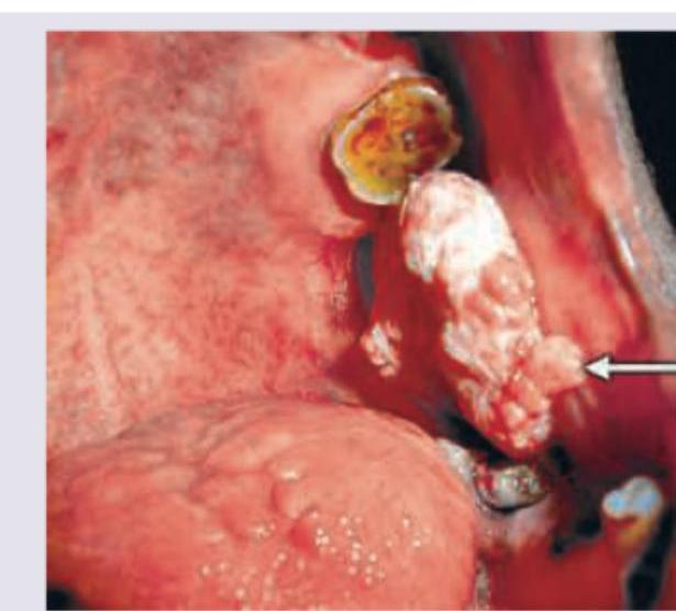
A patient presents with a gingival lesion as shown in the image. What is the most likely diagnosis?

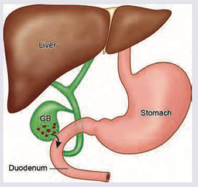
A patient with chronic cholelithiasis develops the complication shown in the image below. This leads to development of:

A 3-year-old child has presented with abdominal lump and peculiar appearance of eyes. All genes are responsible for development of this condition except:
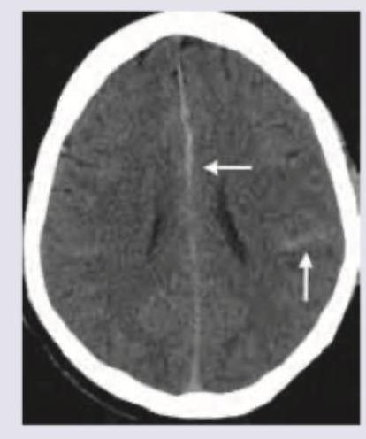
What is the correct diagnosis based on the image shown below?

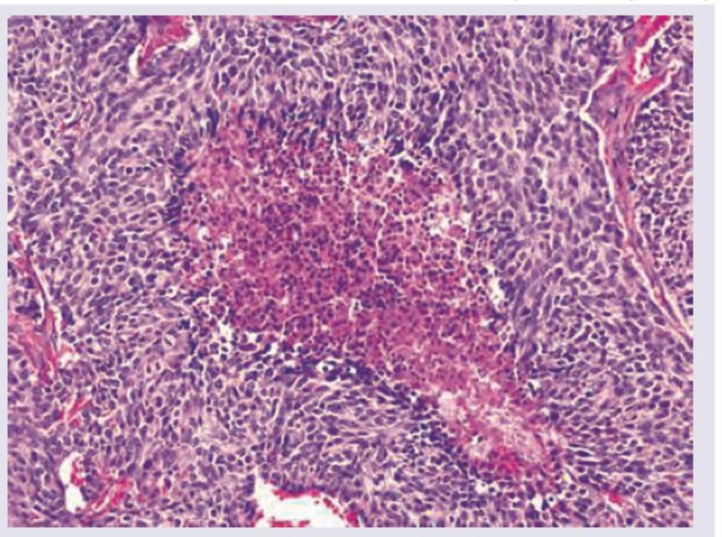
A 55-year-old male patient presented with a 4 month history of cough and hemoptysis. Bronchoscopy revealed an intrabronchial polyp. Biopsy from the polyp showed small cells with salt and pepper chromatin, with microscopic necrosis and 5 mitotic figures per 10 high power fields as shown below. Chromogranin staining was positive. What is the diagnosis and grade of the lesion?

Which of the following are poor prognostic factors in endometrial adenocarcinoma? I. Estrogen and progesterone receptor positivity II. HER-2/neu gene expression III. Histologic types papillary serous or clear cell carcinoma IV. Aneuploid tumours Select the correct answer using the code given below :
What is the most common type of tumour of Vermiform Appendix?
Which of the following are the malignancies associated with lymphoedema? I. Kaposi Sarcoma II. Squamous cell carcinoma III. Malignant melanoma IV. Leukaemia Select the correct answer using the code given below :
'Schiller-Duval body' is a characteristic histological feature of which one of the following cancers?
Masaoka staging is used for staging:
Explanation: ***Fibrous epulis*** - The image shows a **focal, exophytic, firm, nodular growth** on the gingiva, which is characteristic of a **fibrous epulis** [1]. These are common reactive lesions caused by **chronic irritation or trauma** [1]. - They are typically **pink, well-demarcated**, and may be pedunculated or sessile, composed primarily of **fibrous connective tissue** [1]. *Carcinoma alveolar margin* - Malignant lesions like **carcinoma** often present with irregular, ulcerated, fungating, or infiltrative borders, and may be associated with **bleeding, pain, or rapid growth** [4]. - While it can occur at the alveolar margin, the lesion in the image appears more organized and smooth-surfaced, which is less consistent with a typical malignant presentation. *Leukoplakia* - **Leukoplakia** is characterized by a **white patch or plaque** that cannot be rubbed off and is not diagnosable as any other disease [3]. - The lesion in the image is a clearly defined, **flesh-colored, nodular mass**, not a flat white patch. *Hyperplastic candidiasis* - **Hyperplastic candidiasis** typically presents as **persistent white plaques** that are adherent to the mucosa and may have a **nodular or granular appearance** [2]. - While it can be firm, the overall presentation in the image, particularly the color and distinct fibrous-like texture, is less suggestive of candidal infection. **References:** [1] Kumar V, Abbas AK, et al.. Robbins and Cotran Pathologic Basis of Disease. 9th ed. The Lung, pp. 735-736. [2] Kumar V, Abbas AK, et al.. Robbins and Cotran Pathologic Basis of Disease. 9th ed. The Lung, pp. 736-737. [3] Cross SS. Underwood's Pathology: A Clinical Approach. 6th ed. Common Clinical Problems From Alimentary System Disease, pp. 344-345. [4] Kumar V, Abbas AK, et al.. Robbins and Cotran Pathologic Basis of Disease. 9th ed. The Lung, pp. 739-741.
Explanation: ***Bouveret syndrome*** - The image shows gallstones in the gallbladder (GB) moving into the duodenum through a **cholecystoenteric fistula**, specifically into the stomach and duodenum, which is the mechanism leading to **Bouveret syndrome** [3]. - **Bouveret syndrome** is a rare form of gastric outlet obstruction caused by a large gallstone eroding through the gallbladder wall into the duodenum or stomach [3]. *Emphysematous cholecystitis* - This condition is characterized by the presence of **gas in the gallbladder wall or lumen** due to infection by gas-forming organisms, which is not depicted in the image. - It's a severe form of acute cholecystitis, often seen in diabetics or immunocompromised patients, and does not involve stone migration into the gut as shown. *Cholesterolosis* - **Cholesterolosis** (also known as "strawberry gallbladder") is a benign condition characterized by an accumulation of cholesterol esters in the macrophages of the gallbladder wall. - It does not involve the migration of gallstones into the gastrointestinal tract or the formation of fistulas and is not represented by the stones shown entering the duodenum. *Carcinoma of gallbladder* - **Gallbladder carcinoma** is a malignant tumor arising from the epithelial lining of the gallbladder [1]. - While chronic gallstones can be a risk factor for gallbladder cancer, the image specifically illustrates a mechanical complication of gallstone migration, not a neoplastic process [1][2]. **References:** [1] Kumar V, Abbas AK, et al.. Robbins and Cotran Pathologic Basis of Disease. 9th ed. Liver and Gallbladder, p. 886. [2] Kumar V, Abbas AK, et al.. Robbins and Cotran Pathologic Basis of Disease. 9th ed. Liver and Gallbladder, pp. 883-884. [3] Cross SS. Underwood's Pathology: A Clinical Approach. 6th ed. Common Clinical Problems From Liver And Biliary System Disease, pp. 404-405.
Explanation: ***K-ras gene*** - The K-ras gene is an **oncogene** commonly associated with various adult cancers, such as pancreatic and colorectal cancer, but it is **not typically implicated** in the development of Wilms tumor or retinoblastoma. - While K-ras mutations can drive cell proliferation, they are not a characteristic genetic alteration in the syndrome suggested by the child's presentation. *WT1* - The **WT1 gene** is a **tumor suppressor gene** whose inactivation is directly linked to the development of **Wilms tumor**, a common pediatric kidney cancer presenting as an abdominal lump [1], [2]. - Mutations in WT1 are also associated with **WAGR syndrome** (Wilms tumor, Aniridia, Genitourinary anomalies, and intellectual disability), which explains the "peculiar appearance of eyes" (aniridia) [1]. *P53* - The **P53 tumor suppressor gene** (also known as TP53) is a master regulator of the cell cycle and apoptosis [3]. - Mutations in p53 are associated with **Li-Fraumeni syndrome**, which increases the risk of various cancers, including Wilms tumor and osteosarcoma, although it's less specific to the eye abnormalities seen here. *Beta-catenin* - **Beta-catenin** (encoded by the CTNNB1 gene) is involved in various cellular processes, including cell adhesion and **Wnt signaling pathway** [3]. - Mutations in beta-catenin, particularly oncogenic activation, have been found in a subset of **Wilms tumors**, contributing to their development and progression. **References:** [1] Kumar V, Abbas AK, et al.. Robbins and Cotran Pathologic Basis of Disease. 9th ed. Diseases of Infancy and Childhood, pp. 487-488. [2] Cross SS. Underwood's Pathology: A Clinical Approach. 6th ed. (Basic Pathology) introduces the student to key general principles of pathology, both as a medical science and as a clinical activity with a vital role in patient care. Part 2 (Disease Mechanisms) provides fundamental knowledge about the cellular and molecular processes involved in diseases, providing the rationale for their treatment. Part 3 (Systematic Pathology) deals in detail with specific diseases, with emphasis on the clinically important aspects., pp. 211-212. [3] Kumar V, Abbas AK, et al.. Robbins and Cotran Pathologic Basis of Disease. 9th ed. Neoplasia, pp. 305-306.
Explanation: ***Dense MCA sign*** - The image displays a hyperdense (bright) appearance of the **Middle Cerebral Artery (MCA)**, particularly noticeble in the Sylvian fissure on the left side (indicated by the shorter arrow). This is highly suggestive of a **thrombus within the MCA lumen**, one of the earliest signs of an acute ischemic stroke on non-contrast CT [1]. - This finding is a strong indicator of **large vessel occlusion** and is crucial for guiding acute stroke management, such as the administration of thrombolytics or mechanical thrombectomy [1]. *Lacunar stroke* - Lacunar strokes are typically **small, deep infarcts** caused by occlusion of small penetrating arteries, which are not directly visible as a hyperdense vessel sign on non-contrast CT [3]. - The image shows a larger-scale vascular finding, not a small, isolated infarct characteristic of a lacunar stroke [3]. *Subarachnoid hemorrhage* - Subarachnoid hemorrhage (SAH) appears as **high-density blood** filling the subarachnoid spaces, fissures, and sulci [2]. - While the image shows some hyperdensity, it is specifically confined to a major arterial structure (MCA), not diffuse within the subarachnoid space as seen in SAH. *Intraventricular hemorrhage* - Intraventricular hemorrhage (IVH) is characterized by **hyperdense blood within the ventricular system** of the brain [2]. - The image does not show blood within the ventricles; the hyperdensity is clearly located along the course of a major cerebral artery. **References:** [1] Kumar V, Abbas AK, et al.. Robbins and Cotran Pathologic Basis of Disease. 9th ed. The Central Nervous System, pp. 1266-1268. [2] Cross SS. Underwood's Pathology: A Clinical Approach. 6th ed. Common Clinical Manifestations Of Central And Peripheral Nervous System Disease, pp. 706-707. [3] Kumar V, Abbas AK, et al.. Robbins and Cotran Pathologic Basis of Disease. 9th ed. The Central Nervous System, pp. 1269-1270.
Explanation: ***Atypical carcinoid grade 2*** - The presence of **salt and pepper chromatin**, **microscopic necrosis**, and **5 mitotic figures per 10 high power fields** are characteristic features of an **atypical carcinoid tumor** [1]. - **Positive chromogranin staining** confirms neuroendocrine differentiation, and the mitotic rate coupled with necrosis indicates a Grade 2 (atypical) carcinoid based on WHO classification [1]. *Carcinoid grade 1* - A typical carcinoid (Grade 1) would show **no necrosis** and a mitotic count of **less than 2 mitoses per 10 high power fields**, which contradicts the findings [1]. - While it features **salt and pepper chromatin** and positive neuroendocrine markers, the higher mitotic activity and necrosis exclude a typical carcinoid. *Small cell carcinoma grade IV* - **Small cell carcinoma** typically presents with extensive necrosis, very high mitotic activity (often >10 mitoses/10 HPF), and a more **scanty cytoplasm** than seen here, and often **crush artifact** [2]. - Although it is a high-grade neuroendocrine tumor, the described features (only 5 mitoses/10HPF, "salt and pepper chromatin" is less typical for SCLC which has more uniform nuclei, and distinct necrosis without widespread crush artifact) are more consistent with an atypical carcinoid [1], [2]. *Large cell neuroendocrine carcinoma grade IV* - **Large cell neuroendocrine carcinoma** (LCNEC) is characterized by **large cells** with prominent nucleoli, high mitotic counts (often >10-11 mitoses/10HPF), and extensive necrosis [2]. - The "small cells" and **salt and pepper chromatin** described in the biopsy are inconsistent with the large cell morphology of LCNEC. **References:** [1] Kumar V, Abbas AK, et al.. Robbins and Cotran Pathologic Basis of Disease. 9th ed. The Lung, pp. 725-727. [2] Cross SS. Underwood's Pathology: A Clinical Approach. 6th ed. Common Clinical Problems From Respiratory Tract Disease, pp. 337-338.
Explanation: ***II, III and IV*** - **HER-2/neu gene expression**, **papillary serous or clear cell histologic types**, and **aneuploid tumors** are all associated with a more aggressive disease course and worse outcomes in endometrial adenocarcinoma [1]. - These factors indicate less differentiated and often more resistant cancer, leading to higher recurrence rates and lower survival [1]. *I, III and IV* - This option incorrectly includes **estrogen and progesterone receptor positivity** as a poor prognostic factor, which is actually a favorable prognostic indicator. - **HER-2/neu gene expression** is a significant poor prognostic factor but is excluded from this option. *I, II and IV* - This option incorrectly includes **estrogen and progesterone receptor positivity** as a poor prognostic factor. - It also incorrectly excludes **histologic types papillary serous or clear cell carcinoma**, which are well-established poor prognostic factors [1]. *I, II and III* - This option incorrectly lists **estrogen and progesterone receptor positivity** as a poor prognostic factor. - It also incorrectly excludes **aneuploid tumors**, which are recognized indicators of poor prognosis. **References:** [1] Kumar V, Abbas AK, et al.. Robbins and Cotran Pathologic Basis of Disease. 9th ed. The Female Genital Tract, pp. 1017-1024.
Explanation: ***Carcinoid tumour*** - **Carcinoid tumors** (neuroendocrine tumors) are the **most common primary neoplasms of the appendix**, accounting for approximately 30-50% of all appendiceal tumors. [1] - They typically originate from the **enterochromaffin cells** in the appendiceal mucosa and are often discovered incidentally during appendectomy for suspected appendicitis. - Most appendiceal carcinoids are **small (<2 cm), benign, and located at the tip** of the appendix. [1] *Adenocarcinoma* - **Adenocarcinomas** are the second most common primary tumor of the appendix, representing about 10-20% of cases. - These **epithelial malignancies** include mucinous and non-mucinous subtypes and can present with symptoms mimicking acute appendicitis. - Mucinous adenocarcinomas may lead to **pseudomyxoma peritonei** if they rupture. *Germ cell tumour* - **Germ cell tumors** are exceptionally rare in the appendix and more commonly arise from the gonads (testes, ovaries) or midline structures. - These tumors originate from **pluripotent germ cells** and are not a significant consideration for appendiceal neoplasms. *Papillary cell tumour* - This term describes a **morphological growth pattern** (papillary architecture) rather than a specific primary tumor classification. - While some epithelial tumors may exhibit papillary features, this is **not a recognized primary tumor type** of the appendix. **References:** [1] Cross SS. Underwood's Pathology: A Clinical Approach. 6th ed. Common Clinical Problems From Alimentary System Disease, pp. 375-376.
Explanation: ***I, II and IV*** - **Kaposi sarcoma** is a well-documented malignancy that can develop in chronically lymphoedematous limbs, particularly in classic and endemic forms. - **Squamous cell carcinoma** can arise as a complication of chronic lymphoedema, developing in areas of long-standing skin changes and inflammation [1]. - **Leukaemia** is included here as it can cause lymphadenopathy and secondary lymphoedema, representing a bidirectional relationship where leukemic infiltration leads to lymphatic obstruction. - **Note:** The most classic malignancy associated with chronic lymphoedema is **angiosarcoma (Stewart-Treves syndrome)**, though it is not listed among the options. *II, III and IV* - While this includes **squamous cell carcinoma** (correct) [1], it incorrectly includes **malignant melanoma**. - **Malignant melanoma** has no established association with lymphoedema as a predisposing condition, though melanoma can cause lymphoedema through nodal metastases [2]. *I, III and IV* - This incorrectly includes **malignant melanoma** and omits **squamous cell carcinoma**. - **Squamous cell carcinoma** is a more clearly established malignancy that can arise in chronic lymphoedema [1]. *I, II and III* - This correctly includes **Kaposi sarcoma** and **squamous cell carcinoma** but incorrectly includes **malignant melanoma**. - **Malignant melanoma** does not have a recognized causal relationship with pre-existing lymphoedema. **References:** [1] Cross SS. Underwood's Pathology: A Clinical Approach. 6th ed. Disorders Involving Inflammatory And Haemopoietic Cells, pp. 643-644. [2] Cross SS. Underwood's Pathology: A Clinical Approach. 6th ed. (Basic Pathology) introduces the student to key general principles of pathology, both as a medical science and as a clinical activity with a vital role in patient care. Part 2 (Disease Mechanisms) provides fundamental knowledge about the cellular and molecular processes involved in diseases, providing the rationale for their treatment. Part 3 (Systematic Pathology) deals in detail with specific diseases, with emphasis on the clinically important aspects., pp. 234-235.
Explanation: ***Endodermal sinus tumour*** - **Schiller-Duval bodies** are pathognomonic histological structures found in **endodermal sinus tumours** (also known as yolk sac tumours). - These structures mimic the primitive glomerulus, consisting of a central capillary surrounded by tumour cells within a cyst-like space. *Non-gestational ovarian choriocarcinoma* - Characterized by the presence of **syncytiotrophoblast** and **cytotrophoblast** cells, often arranged in bilaminar structures [2]. - While it can produce **human chorionic gonadotropin (hCG)**, it does not typically feature Schiller-Duval bodies [2], [3]. *Dysgerminoma* - Composed of large, rounded, uniform cells with clear cytoplasm and prominent nuclei, often arranged in cords or nests separated by fibrous septa infiltrated by **lymphocytes**. - This tumour is analogous to testicular seminoma and does not contain Schiller-Duval bodies. *Sex cord stromal tumours* - A diverse group of tumours, including **granulosa cell tumours** and **Sertoli-Leydig cell tumours**, which originate from the ovarian stroma or sex cords [1]. - Histological features vary widely but generally involve granulosa cells, theca cells, Sertoli cells, or Leydig cells, and do not include Schiller-Duval bodies [1]. **References:** [1] Kumar V, Abbas AK, et al.. Robbins and Cotran Pathologic Basis of Disease. 9th ed. The Female Genital Tract, pp. 1037-1038. [2] Kumar V, Abbas AK, et al.. Robbins and Cotran Pathologic Basis of Disease. 9th ed. The Lower Urinary Tract and Male Genital System, p. 982. [3] Kumar V, Abbas AK, et al.. Robbins and Cotran Pathologic Basis of Disease. 9th ed. The Female Genital Tract, pp. 1035-1036.
Explanation: ***Thymoma*** - The **Masaoka staging system** is specifically designed for evaluating the extent of **thymomas**, a type of tumor originating from the thymus gland [1]. - This system assesses tumor invasion into surrounding structures, such as the mediastinal fat, pleura, pericardium, and great vessels, which is critical for determining prognosis and treatment [1],[2]. *Germ cell tumours* - **Germ cell tumors** are typically staged using systems specific to their primary site (e.g., testicular, ovarian, mediastinal) that often involve imaging, tumor markers (e.g., AFP, beta-hCG), and histopathological findings. - While germ cell tumors can occur in the mediastinum, the Masaoka system is not their primary staging method. *Neurogenic tumours* - **Neurogenic tumors** encompass a broad range of tumors arising from nervous tissue (e.g., neuroblastoma, schwannoma, ganglioneuroma) and are staged using various systems depending on the specific tumor type and location (e.g., INPC staging for neuroblastoma). - The Masaoka system is not applicable to these tumors. *Lymphoma* - **Lymphomas** are staged using the **Ann Arbor classification system** (or modified Lugano classification), which primarily considers the number and location of involved lymph node regions, as well as extranodal involvement. - This system is distinct from the Masaoka staging system, which is anatomically focused on the thymus and its surrounding structures. **References:** [1] Kumar V, Abbas AK, et al.. Robbins and Cotran Pathologic Basis of Disease. 9th ed. Diseases of White Blood Cells, Lymph Nodes, Spleen, and Thymus, pp. 634-635. [2] Cross SS. Underwood's Pathology: A Clinical Approach. 6th ed. Common Clinical Problems From Diseases Of The Urinary And Male Genital Tracts, pp. 572-574.
Nomenclature and Classification of Tumors
Practice Questions
Characteristics of Benign and Malignant Neoplasms
Practice Questions
Molecular Basis of Cancer
Practice Questions
Carcinogenesis and Carcinogens
Practice Questions
Tumor Progression and Metastasis
Practice Questions
Tumor Markers
Practice Questions
Paraneoplastic Syndromes
Practice Questions
Genetic Basis of Cancer
Practice Questions
Tumor Immunity
Practice Questions
Cancer Epidemiology and Prevention
Practice Questions
Get full access to all questions, explanations, and performance tracking.
Start For Free