Hereditary non-polyposis colorectal cancer (HNPCC), also known as Lynch syndrome, is caused by mutations in which type of genes?
HPV infection is most commonly associated with which type of cancer?
Which of the following is not related to disease progression?
Which of the following is true about p53?
A three-year-old boy has been observed by his parents to be increasingly clumsy for the past 6 months. On a physical examination, there is a cauliflower-like growth in the left eye, leukocoria, and an absent red reflex. The microscopic appearance of the specimen shows Flexner-Wintersteiner rosettes. Which gene is involved in the given condition?
Asbestos exposure may be associated with which of the following malignancies?
Most common cause of squamous cell carcinoma at the base of the tongue is:
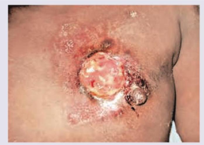
All of the following are true regarding this picture except: (Recent NEET Pattern 2016-17)

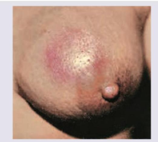
This appearance of mammary skin is due to:

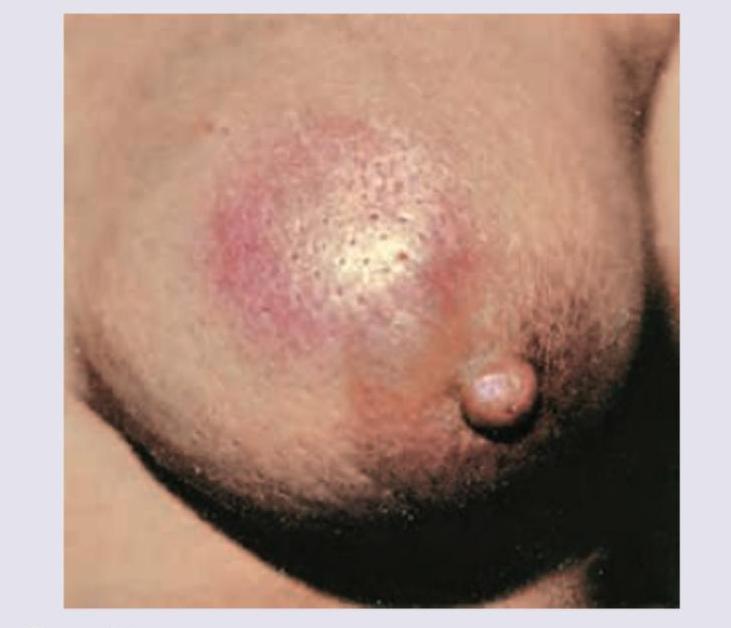
All are true about the condition shown in the image except:

Explanation: ***MMR*** - Hereditary non-polyposis colorectal cancer (**HNPCC**), also known as **Lynch syndrome**, is caused by germline mutations in **DNA Mismatch Repair (MMR)** genes [1]. - Defective MMR function, most commonly due to mutations in **MLH1**, **MSH2**, **MSH6**, or **PMS2**, leads to an accumulation of mutations throughout the genome, a state known as **microsatellite instability (MSI)**. *APC* - Mutations in the **APC** gene, a tumor suppressor, are the cause of **Familial Adenomatous Polyposis (FAP)**, a different hereditary colorectal cancer syndrome [1]. - FAP is characterized by the development of hundreds to thousands of colonic polyps, whereas HNPCC typically presents with fewer polyps that rapidly progress to carcinoma [1]. *MYC* - **MYC** is a **proto-oncogene** involved in cell cycle control and proliferation; its dysregulation contributes to the development of many cancers, but it is not the primary genetic cause of HNPCC. - It is not a DNA repair gene, but rather a transcription factor that, when overexpressed, promotes uncontrolled cell growth. *TERT1* - This likely refers to the **TERT** gene, which encodes **telomerase reverse transcriptase**, an enzyme essential for maintaining telomere length. - While reactivation of **telomerase** is a critical step for cellular immortalization in many cancers, inherited mutations in this gene are not the cause of Lynch syndrome. **References:** [1] Kumar V, Abbas AK, et al.. Robbins and Cotran Pathologic Basis of Disease. 9th ed. The Gastrointestinal Tract, pp. 817, 821-822.
Explanation: ***Oropharyngeal cancer*** - HPV (particularly HPV-16 and HPV-18) is strongly associated with oropharyngeal cancers, especially those affecting the **base of tongue and tonsils** [2], [3] - HPV-positive oropharyngeal cancers represent a distinct epidemiological entity with **better prognosis** compared to tobacco/alcohol-related cases - Among the options listed, oropharyngeal cancer is the **only HPV-associated malignancy** [3] - Note: Cervical cancer is the **most common HPV-related cancer overall** (not listed in options) [1], [3] *Incorrect: Esophageal cancer* - Primarily associated with **tobacco, alcohol, Barrett's esophagus, and chronic GERD** - Not significantly associated with HPV infection *Incorrect: Lung cancer* - Main risk factors include **smoking, radon exposure, asbestos, and air pollution** - No established HPV association *Incorrect: Stomach cancer* - Associated with **H. pylori infection, dietary factors (nitrosamines), and chronic gastritis** - Not linked to HPV infection **References:** [1] Kumar V, Abbas AK, et al.. Robbins and Cotran Pathologic Basis of Disease. 9th ed. The Female Genital Tract, pp. 1007-1008. [2] Kumar V, Abbas AK, et al.. Robbins and Cotran Pathologic Basis of Disease. 9th ed. The Lung, pp. 739-741. [3] Cross SS. Underwood's Pathology: A Clinical Approach. 6th ed. (Basic Pathology) introduces the student to key general principles of pathology, both as a medical science and as a clinical activity with a vital role in patient care. Part 2 (Disease Mechanisms) provides fundamental knowledge about the cellular and molecular processes involved in diseases, providing the rationale for their treatment. Part 3 (Systematic Pathology) deals in detail with specific diseases, with emphasis on the clinically important aspects., pp. 219-220.
Explanation: ***Duration*** - While a history of a long-standing lesion that changes is important for diagnosis, the absolute duration of its existence is not a primary prognostic factor used in staging skin cancers like melanoma. - Prognosis is determined by objective pathological features like depth and evidence of spread [3], not how long the patient reports having had the lesion. *Depth* - The **vertical depth of invasion** (e.g., **Breslow depth** for melanoma) is the single most important prognostic factor for primary cutaneous tumors [2]. - A greater depth directly correlates with an increased risk of **metastasis** and poorer survival rates, thus being a key measure of disease progression [4]. *Site* - The **病理部位 (anatomical location)** of the primary tumor is a known independent prognostic factor for melanoma. - Tumors located on the head, neck, trunk, hands, or feet often have a worse prognosis than those on the extremities. *Stage of Cancer* - The **stage** of cancer, determined by systems like **TNM** (Tumor, Node, Metastasis), is a comprehensive summary of the disease's extent [1]. - It is the definitive measure of disease progression, integrating primary tumor characteristics, lymph node involvement, and distant metastasis to guide treatment and predict outcome [1]. **References:** [1] Cross SS. Underwood's Pathology: A Clinical Approach. 6th ed. (Basic Pathology) introduces the student to key general principles of pathology, both as a medical science and as a clinical activity with a vital role in patient care. Part 2 (Disease Mechanisms) provides fundamental knowledge about the cellular and molecular processes involved in diseases, providing the rationale for their treatment. Part 3 (Systematic Pathology) deals in detail with specific diseases, with emphasis on the clinically important aspects., pp. 236-237. [2] Cross SS. Underwood's Pathology: A Clinical Approach. 6th ed. Disorders Involving Inflammatory And Haemopoietic Cells, pp. 650-651. [3] Cross SS. Underwood's Pathology: A Clinical Approach. 6th ed. (Basic Pathology) introduces the student to key general principles of pathology, both as a medical science and as a clinical activity with a vital role in patient care. Part 2 (Disease Mechanisms) provides fundamental knowledge about the cellular and molecular processes involved in diseases, providing the rationale for their treatment. Part 3 (Systematic Pathology) deals in detail with specific diseases, with emphasis on the clinically important aspects., pp. 36-37. [4] Kumar V, Abbas AK, et al.. Robbins and Cotran Pathologic Basis of Disease. 9th ed. The Skin, pp. 1151-1152.
Explanation: p53, known as the "**guardian of the genome**," has multiple functions beyond its classical role as a tumor suppressor [2]. While primarily known for **cell cycle arrest, DNA repair coordination, and apoptosis induction** [1], p53 also plays a role in **RNA metabolism and stabilization** through its RNA-binding properties and interaction with various RNA-binding proteins. This contributes to its regulation of gene expression at the post-transcriptional level. *Incorrect: It retains the telomerase enzyme in the nucleus* - **p53 actually REPRESSES telomerase activity** by downregulating hTERT (telomerase reverse transcriptase) expression - Loss of p53 function in cancer cells allows telomerase reactivation, contributing to cellular immortalization - This is the opposite of the stated function *Incorrect: It increases telomere length in the liver* - p53 does not increase telomere length; it inhibits telomerase activity - No specific tissue-specific telomere lengthening function in the liver *Incorrect: It synthesizes RNA* - p53 is a **transcription factor** that regulates gene expression but does not synthesize RNA itself [1] - RNA synthesis is performed by RNA polymerase enzymes [3] **References:** [1] Kumar V, Abbas AK, et al.. Robbins and Cotran Pathologic Basis of Disease. 9th ed. Neoplasia, pp. 302-304. [2] Cross SS. Underwood's Pathology: A Clinical Approach. 6th ed. (Basic Pathology) introduces the student to key general principles of pathology, both as a medical science and as a clinical activity with a vital role in patient care. Part 2 (Disease Mechanisms) provides fundamental knowledge about the cellular and molecular processes involved in diseases, providing the rationale for their treatment. Part 3 (Systematic Pathology) deals in detail with specific diseases, with emphasis on the clinically important aspects., pp. 226-227. [3] Kumar V, Abbas AK, et al.. Robbins and Cotran Pathologic Basis of Disease. 9th ed. With Illustrations By, pp. 30-31.
Explanation: ***Rb gene***- The constellation of leukocoria, absent red reflex, and the microscopic finding of **Flexner-Wintersteiner rosettes** are pathognomonic for **retinoblastoma** [1, 3]. - The **Rb gene** (Retinoblastoma gene) is a critical **tumor suppressor gene** located on chromosome 13q14, and its inactivation leads to the development of this malignancy [1, 2]. *ATP 7B gene* - This gene is responsible for **Wilson disease**, an autosomal recessive disorder causing excessive **copper accumulation** in the liver, brain, and cornea (Kayser-Fleischer rings). - It is not associated with primary malignant retinal tumors like retinoblastoma. *Rhodopsin gene* - Mutations in the **rhodopsin gene** are the most common cause of **autosomal dominant retinitis pigmentosa**, which presents with progressive night blindness and visual field loss. - This condition involves retinal degeneration and does not cause a mass-forming neoplasm with leukocoria. *FBN 1 gene* - The **FBN1 gene** codes for **Fibrillin-1** and is mutated in **Marfan syndrome**, a connective tissue disorder. - While Marfan syndrome often causes ocular issues like **ectopia lentis** (lens dislocation), it does not cause retinoblastoma. **References:** [1] Cross SS. Underwood's Pathology: A Clinical Approach. 6th ed. Common Clinical Manifestations Of Central And Peripheral Nervous System Disease, pp. 737-738. [2] Kumar V, Abbas AK, et al.. Robbins and Cotran Pathologic Basis of Disease. 9th ed. Neoplasia, p. 300. [3] Kumar V, Abbas AK, et al.. Robbins and Cotran Pathologic Basis of Disease. 9th ed. The Eye, p. 1342.
Explanation: ***Mesothelioma***- The definitive and most recognized malignancy strongly linked to inhalation of **asbestos fibers** is **malignant mesothelioma**, primarily affecting the pleura or less commonly the peritoneum [1], [2].- Exposure typically involves a long latent period, often 20 to 50 years, after the initial exposure to asbestos minerals like **chrysotile** or **amphibole** [3].*Acute myeloid leukemia*- **Acute myeloid leukemia (AML)** is often linked to exposure to **benzene** or therapeutic **alkylating agents**, but not typically associated with asbestos inhalation [2].- AML is a malignancy of the **bone marrow** affecting myeloid precursors, distinct from the fibrotic and solid tumors caused by asbestos.*Hepatic angiosarcoma*- **Hepatic angiosarcoma** is a rare vascular tumor of the liver typically associated with chemical carcinogens such as **vinyl chloride monomer**, **arsenic**, or **thorium dioxide (Thorotrast)**.- There is no established primary association between asbestos exposure and the development of hepatic angiosarcoma.*Skin carcinoma*- **Skin carcinoma** (e.g., basal or squamous cell carcinoma) is overwhelmingly linked to chronic exposure to **ultraviolet (UV) radiation**.- While asbestos is a potent carcinogen associated with lung and pleural malignancies, it is not a recognized major risk factor for typical cutaneous carcinomas. **References:** [1] Cross SS. Underwood's Pathology: A Clinical Approach. 6th ed. (Basic Pathology) introduces the student to key general principles of pathology, both as a medical science and as a clinical activity with a vital role in patient care. Part 2 (Disease Mechanisms) provides fundamental knowledge about the cellular and molecular processes involved in diseases, providing the rationale for their treatment. Part 3 (Systematic Pathology) deals in detail with specific diseases, with emphasis on the clinically important aspects., pp. 221-222. [2] Kumar V, Abbas AK, et al.. Robbins and Cotran Pathologic Basis of Disease. 9th ed. Neoplasia, p. 286. [3] Cross SS. Underwood's Pathology: A Clinical Approach. 6th ed. Common Clinical Problems From Respiratory Tract Disease, pp. 339-340.
Explanation: ***HPV*** - **Human Papillomavirus**, particularly **HPV-16**, is the predominant etiological factor for **squamous cell carcinoma (SCC) of the oropharynx**, which includes the base of the tongue [1]. - **HPV-positive SCC** has rapidly increased in incidence and constitutes the majority of these cancers in non-smokers and non-drinkers [1]. *EBV* - **Epstein-Barr Virus (EBV)** is strongly associated with **Nasopharyngeal Carcinoma (NPC)**, endemic Burkitt's lymphoma, and post-transplant lymphoproliferative disorder [2]. - It is not considered the primary or most common oncogenic agent for **base of tongue SCC** [2]. *HCV* - **Hepatitis C Virus (HCV)** is the chief cause of viral **Hepatocellular Carcinoma (HCC)** and is also linked to certain **B-cell non-Hodgkin's lymphomas**. - There is no established role for HCV in the pathogenesis or high prevalence of **squamous cell carcinoma of the head and neck**. *CMV* - **Cytomegalovirus (CMV)** is a herpesvirus typically associated with opportunistic infections (e.g., retinitis, colitis) in **immunocompromised patients**. - While studies have investigated its oncogenic potential, CMV is not a significant or common causative agent for **oral or pharyngeal SCC**. **References:** [1] Kumar V, Abbas AK, et al.. Robbins and Cotran Pathologic Basis of Disease. 9th ed. The Lung, pp. 738-741. [2] Kumar V, Abbas AK, et al.. Robbins and Cotran Pathologic Basis of Disease. 9th ed. Head and Neck, pp. 744-745.
Explanation: ***Usually bilateral*** - Paget's disease of the nipple, as depicted in the image (lesions on the nipple with surrounding dermatitis-like changes), is almost always an **unilateral condition**. - Its bilateral occurrence is exceedingly rare and would suggest a different or coincidental pathology. - **This is the FALSE statement** - Paget's disease is typically unilateral, not bilateral. *Associated with underlying invasive carcinoma of breast* - **Paget's disease of the nipple** is histologically characterized by the presence of **Paget cells** (adenocarcinoma cells) in the epidermis of the nipple and areola [2]. - In a vast majority of cases (90-95%), it is associated with an underlying **ductal carcinoma in situ (DCIS)** or an **invasive ductal carcinoma** originating from the lactiferous ducts [1], [2]. *Itching is a common symptom* - **Itching (pruritus)** is a **prominent and common symptom** of Paget's disease of the nipple, along with burning, pain, and crusting. - The eczematous changes often lead to significant discomfort and itching. *Treatment is simple mastectomy with axillary lymph node dissection* - The standard treatment for Paget's disease of the nipple, especially when associated with an underlying invasive carcinoma, is **surgical excision**. - A **simple mastectomy** removes the entire breast and is often combined with **axillary lymph node dissection** or sentinel lymph node biopsy to assess for nodal involvement [1]. **References:** [1] Cross SS. Underwood's Pathology: A Clinical Approach. 6th ed. Common Clinical Problems From Liver And Biliary System Disease, pp. 456-457. [2] Kumar V, Abbas AK, et al.. Robbins and Cotran Pathologic Basis of Disease. 9th ed. The Breast, pp. 1061-1062.
Explanation: ***Lymphatic penetration*** - The image illustrates **peau d'orange**, a classic sign of **inflammatory breast cancer**, caused by tumor cells blocking lymphatic drainage in the skin of the breast [1]. - This blockage leads to **edema** and accentuation of hair follicles, creating an orange peel-like appearance [1]. *Intra-epithelial cancer* - **Intra-epithelial cancer** (e.g., Paget disease of the nipple) primarily affects the epidermis and presents as a scaly, eczematous rash, not the diffuse skin thickening and pitting seen here [2]. - While it can be associated with an underlying invasive cancer, the visual presentation in the image is not typical for a purely intra-epithelial process [2]. *Sub-epidermal cancer* - **Sub-epidermal cancer** refers to malignancies located beneath the epidermis, often presenting as a palpable mass or nodule [1]. - It would not typically cause the widespread **skin edema** and **lymphatic obstruction** characteristic of peau d'orange, which affects a broader area of the skin due to involvement of dermal lymphatics [1]. *Vascular embolization* - **Vascular embolization** involves the blockage of blood vessels, which can lead to ischemia, necrosis, or localized discoloration. - It does not explain the characteristic **lymphatic obstruction** and **edema** that causes the peau d'orange appearance in the mammary skin [1]. **References:** [1] Cross SS. Underwood's Pathology: A Clinical Approach. 6th ed. Common Clinical Problems From Liver And Biliary System Disease, pp. 453-454. [2] Kumar V, Abbas AK, et al.. Robbins and Cotran Pathologic Basis of Disease. 9th ed. The Breast, pp. 1061-1062.
Explanation: ***Overexpression of HER 2/neu shows poor response to adriamycin*** - This statement is **FALSE** and represents the correct answer. **HER2-positive breast cancers** actually show **GOOD response** to anthracycline-based chemotherapy including adriamycin (doxorubicin) [1]. - Multiple clinical trials have demonstrated that **HER2-overexpressing tumors are chemosensitive** to anthracyclines, with improved response rates compared to HER2-negative tumors [1]. - Modern treatment combines adriamycin with **HER2-targeted therapy** (trastuzumab) for optimal outcomes in HER2+ inflammatory breast cancer [1]. *Occurs due to lymphatics destruction* - This is **TRUE** for inflammatory breast cancer. The pathognomonic feature involves **dermal lymphatic invasion** by malignant cells causing obstruction. - This lymphatic blockage leads to the characteristic clinical presentation including **peau d'orange appearance** and rapid onset of breast erythema and edema. *Peau d'orange* - This is **TRUE** and represents a classic sign of inflammatory breast cancer. The **"orange peel" appearance** results from skin edema with tethering at hair follicles. - Caused by **dermal lymphatic obstruction** by tumor emboli, leading to lymphedema that accentuates the skin pores. *Van Nuys prognostic index* - This statement is **TRUE** as written. The **Van Nuys Prognostic Index (VNPI)** is a scoring system specifically developed for **ductal carcinoma in situ (DCIS)**, not for inflammatory breast cancer. - Inflammatory breast cancer is an **aggressive invasive carcinoma** (stage IIIB minimum) and uses different prognostic systems including TNM staging and molecular markers, making VNPI inappropriate for this condition. **References:** [1] Kumar V, Abbas AK, et al.. Robbins and Cotran Pathologic Basis of Disease. 9th ed. The Breast, p. 1066.
Nomenclature and Classification of Tumors
Practice Questions
Characteristics of Benign and Malignant Neoplasms
Practice Questions
Molecular Basis of Cancer
Practice Questions
Carcinogenesis and Carcinogens
Practice Questions
Tumor Progression and Metastasis
Practice Questions
Tumor Markers
Practice Questions
Paraneoplastic Syndromes
Practice Questions
Genetic Basis of Cancer
Practice Questions
Tumor Immunity
Practice Questions
Cancer Epidemiology and Prevention
Practice Questions
Get full access to all questions, explanations, and performance tracking.
Start For Free