Which of the following is a triple negative breast cancer?
The most common malignancy of the oral cavity is:
Which of the following is not a tumor marker?
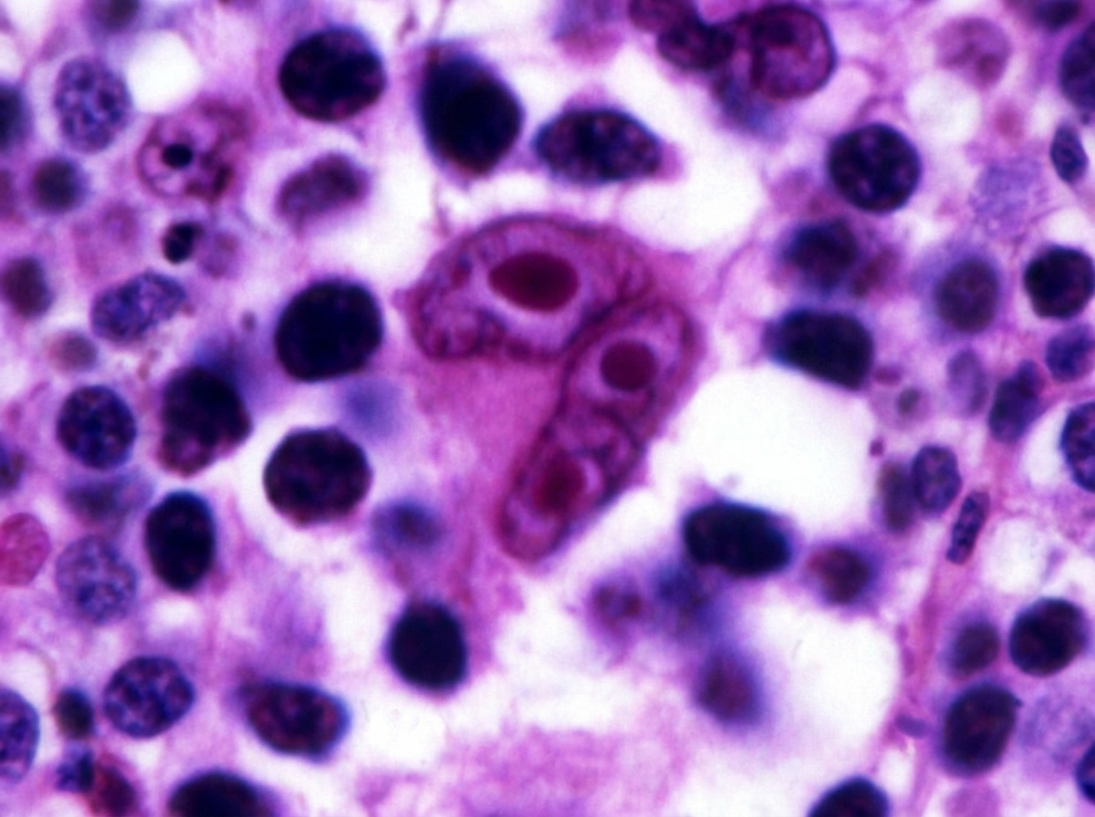
Epstein-Barr virus (EBV) is associated with which type of cancer?
A patient was incidentally diagnosed with a lump during a routine health checkup. Following ultrasound and guided biopsy, histopathological examination revealed a diagnosis. The patient was initiated on chemotherapy that included a drug obtained from a specific plant. What is the site of action of this drug?

What type of fibroadenosis is most likely to undergo malignant change?
Which of the following is not a benign tumor?
Paget's disease of the nipple is stained positive for:
Gleason's staging is used in which of the following malignancies?
RET gene mutation is associated with which malignancy?
Explanation: **Triple-Negative Breast Cancer (TNBC)** refers to tumors that lack expression of Estrogen Receptor (ER), Progesterone Receptor (PR), and Human Epidermal Growth Factor Receptor 2 (HER2) [1]. These are typically aggressive, but certain rare subtypes have a more indolent course [1]. 1. **Why Secretory Cell Cancer is correct:** Secretory carcinoma of the breast is a rare, distinct subtype characterized by a specific **t(12;15) translocation**, creating the *ETV6-NTRK3* fusion gene. It characteristically presents as a **triple-negative** tumor. Despite being TNBC, it has an excellent prognosis, especially in children and young adults, unlike the typical aggressive "basal-like" TNBCs. 2. **Analysis of Incorrect Options:** * **Colloid (Mucinous) Carcinoma:** These are typically **ER/PR positive** and HER2 negative. They occur in older women and have a favorable prognosis due to their slow growth. * **Acinic Cell Carcinoma:** While rare in the breast and often triple-negative, it is not the classic "textbook" association for TNBC in this context compared to Secretory carcinoma. (Note: It is more common in salivary glands). * **Mucinous Carcinoma:** (Same as Colloid) These are luminal-type cancers (ER+) characterized by abundant extracellular mucin. **High-Yield Clinical Pearls for NEET-PG:** * **Most common TNBC:** The "Basal-like" subtype (associated with BRCA1 mutations) [1]. * **Morphology of Secretory Cancer:** Shows intracellular and extracellular secretions that stain positive with **PAS (Periodic Acid-Schiff)**. * **Prognostic Paradox:** While most TNBCs are high-grade (Grade 3), **Secretory** and **Adenoid Cystic Carcinomas** of the breast are TNBCs with an **excellent prognosis** [1]. * **Targeted Therapy:** The *NTRK* fusion makes these tumors potentially responsive to TRK inhibitors like Larotrectinib. **References:** [1] Kumar V, Abbas AK, et al.. Robbins and Cotran Pathologic Basis of Disease. 9th ed. The Breast, pp. 1060-1069.
Explanation: **Explanation:** **Squamous Cell Carcinoma (SCC)** is the most common malignancy of the oral cavity, accounting for approximately **90-95%** of all oral cancers. The oral cavity is lined by stratified squamous epithelium; chronic exposure to carcinogens—most notably **tobacco** (smoking and smokeless) and **alcohol**—leads to a progression from dysplasia to invasive carcinoma [1]. In recent years, **Human Papillomavirus (HPV-16)** has also been identified as a significant risk factor, particularly for oropharyngeal sites [1]. **Analysis of Incorrect Options:** * **Basal Cell Carcinoma (BCC):** While BCC is the most common skin cancer, it rarely affects the oral mucosa. It typically occurs on sun-exposed skin, such as the upper face. * **Transitional Cell Carcinoma:** This malignancy arises from the urothelium and is characteristic of the urinary tract (bladder, ureters). It is not found in the oral cavity. * **Melanoma:** Oral mucosal melanoma is extremely rare (less than 1% of oral malignancies). Although highly aggressive, it is far less common than SCC. **High-Yield Clinical Pearls for NEET-PG:** * **Most common site:** The lateral border of the tongue and the floor of the mouth are the most frequent locations for oral SCC [1]. * **Precursor lesions:** Leukoplakia (white patch) and Erythroplakia (red patch) are significant premalignant conditions; Erythroplakia carries a much higher risk of malignant transformation [1]. * **Field Cancerization:** This concept explains why patients with one oral SCC are at high risk for developing secondary primary tumors due to diffuse carcinogen exposure across the mucosal surface. * **Lymphatic Spread:** Oral SCC typically metastasizes first to the submental and deep cervical lymph nodes. **References:** [1] Kumar V, Abbas AK, et al.. Robbins and Cotran Pathologic Basis of Disease. 9th ed. The Lung, pp. 738-741.
Explanation: **Explanation:** The correct answer is **Human leucocyte antigen A2 (HLA-A2)**. Tumor markers are substances (proteins, enzymes, or hormones) produced by cancer cells or by the body in response to cancer, which can be detected in blood, urine, or tissues [1]. * **Why HLA-A2 is the correct answer:** HLA-A2 is a Major Histocompatibility Complex (MHC) Class I molecule. Its primary role is to present peptides to T-cells for immune recognition [4]. While HLA expression may be altered in cancer (downregulated to evade the immune system), it is a normal constituent of cell surfaces used for tissue typing and is **not** used as a diagnostic or prognostic marker for malignancy. * **Analysis of Incorrect Options:** * **CEA (Carcinoembryonic Antigen):** An oncofetal antigen primarily used to monitor recurrence in **Colorectal Carcinoma** [2]. It is also elevated in pancreatic, gastric, and breast cancers [3]. * **Tyrosinase:** A specific enzyme involved in melanin synthesis. It is a highly specific **tissue-based tumor marker** used in immunohistochemistry (IHC) to identify **Malignant Melanoma**. * **AFP (Alpha-Fetoprotein):** An oncofetal antigen used as a marker for **Hepatocellular Carcinoma (HCC)** and **Non-seminomatous germ cell tumors (Yolk sac tumor)** [2]. **NEET-PG High-Yield Pearls:** * **PSA:** Most specific marker for Prostate Cancer screening/monitoring [2]. * **CA-125:** Marker for Ovarian Cancer (Serous cystadenocarcinoma). * **CA 19-9:** Marker for Pancreatic and Cholangiocarcinoma. * **Calcitonin:** Marker for Medullary Carcinoma of the Thyroid. * **S-100:** Broad marker for Melanoma, Schwannoma, and Langerhans Cell Histiocytosis. **References:** [1] Cross SS. Underwood's Pathology: A Clinical Approach. 6th ed. (Basic Pathology) introduces the student to key general principles of pathology, both as a medical science and as a clinical activity with a vital role in patient care. Part 2 (Disease Mechanisms) provides fundamental knowledge about the cellular and molecular processes involved in diseases, providing the rationale for their treatment. Part 3 (Systematic Pathology) deals in detail with specific diseases, with emphasis on the clinically important aspects., pp. 254-255. [2] Kumar V, Abbas AK, et al.. Robbins and Cotran Pathologic Basis of Disease. 9th ed. Neoplasia, p. 346. [3] Kumar V, Abbas AK, et al.. Robbins and Cotran Pathologic Basis of Disease. 9th ed. Neoplasia, pp. 319-320. [4] Kumar V, Abbas AK, et al.. Robbins and Cotran Pathologic Basis of Disease. 9th ed. Neoplasia, pp. 318-319.
Explanation: **Explanation:** **1. Why Nasopharyngeal Carcinoma (NPC) is correct:** Epstein-Barr Virus (EBV), a member of the Herpesvirus family (HHV-4), is a potent oncogenic virus [1]. It infects B cells and epithelial cells by binding to the **CD21 (CR2) receptor**. In the nasopharynx, EBV infection leads to the expression of **LMP-1 (Latent Membrane Protein-1)**, which mimics CD40 signaling and activates the NF-κB and JAK/STAT pathways. This promotes cell survival and proliferation, specifically leading to the **undifferentiated (Type 3) variant** of Nasopharyngeal Carcinoma, which is highly prevalent in Southern China and parts of Africa [2]. **2. Why other options are incorrect:** * **Epidermodysplasia verruciformis:** This is a rare genetic susceptibility to **Human Papillomavirus (HPV)** [1], specifically types 5 and 8, which can lead to squamous cell carcinomas. * **Kaposi Sarcoma:** This is caused by **Human Herpesvirus-8 (HHV-8)**, not EBV [1]. It is a vascular tumor typically seen in immunocompromised patients (AIDS-defining illness). **3. NEET-PG High-Yield Clinical Pearls:** * **EBV-Associated Malignancies:** Remember the mnemonic **"B-N-H-L"**: **B**urkitt Lymphoma (starry-sky appearance, t(8;14)), **N**asopharyngeal Carcinoma, **H**odgkin Lymphoma (Mixed cellularity type), and **L**ymphomas in immunocompromised patients (CNS Lymphoma) [1]. * **Diagnostic Marker:** Elevated titers of **IgA antibodies** against EBV viral capsid antigen (VCA) are used for screening and monitoring recurrence in NPC. * **Other EBV associations:** Infectious Mononucleosis (Glandular fever) and Oral Hairy Leukoplakia (in HIV patients). **References:** [1] Cross SS. Underwood's Pathology: A Clinical Approach. 6th ed. (Basic Pathology) introduces the student to key general principles of pathology, both as a medical science and as a clinical activity with a vital role in patient care. Part 2 (Disease Mechanisms) provides fundamental knowledge about the cellular and molecular processes involved in diseases, providing the rationale for their treatment. Part 3 (Systematic Pathology) deals in detail with specific diseases, with emphasis on the clinically important aspects., pp. 219-221. [2] Kumar V, Abbas AK, et al.. Robbins and Cotran Pathologic Basis of Disease. 9th ed. Head and Neck, pp. 744-745.
Explanation: ***Tubulin (mitotic inhibitor)*** - Plant-derived chemotherapeutic agents like **vinca alkaloids** (vincristine, vinblastine from *Vinca rosea*) and **taxanes** (paclitaxel from *Taxus brevifolia*) target **tubulin** and disrupt **microtubule** formation. - These drugs prevent proper **mitotic spindle** formation, leading to cell cycle arrest in **M-phase** and subsequent **apoptosis** in rapidly dividing cancer cells. *DNA alkylation* - **Alkylating agents** like **cyclophosphamide** and **nitrogen mustards** form **covalent bonds** with DNA bases, causing cross-linking. - These are not typically plant-derived drugs and work through direct **DNA damage** rather than affecting cellular structures. *Topoisomerase II inhibition* - **Etoposide** and **doxorubicin** inhibit **topoisomerase II**, preventing DNA **supercoiling** and causing DNA breaks. - While some topoisomerase inhibitors are plant-derived, the question specifically hints at drugs affecting **cellular division machinery** rather than DNA enzymes. *DNA intercalation* - **Intercalating agents** like **actinomycin D** insert between **DNA base pairs**, distorting the double helix structure. - These drugs primarily affect **transcription** and **replication** processes rather than the **mitotic apparatus** targeted by plant-derived anti-mitotic agents.
Explanation: **Explanation:** The question addresses the spectrum of **Fibrocystic Changes (FCC)** of the breast and their relative risk for developing invasive carcinoma. **Why Epitheliosis is correct:** **Epitheliosis**, also known as **Ductal Hyperplasia**, refers to an increase in the number of epithelial cell layers (more than the normal two layers) lining the ducts or acini [2]. It is categorized into "usual" and "atypical" hyperplasia. While mild hyperplasia carries little risk, **Atypical Hyperplasia** (epitheliosis with architectural or cytologic atypia) is a significant pre-malignant condition, carrying a **4 to 5-fold increased risk** of malignant transformation [1]. If a family history is present, this risk increases further. **Analysis of Incorrect Options:** * **A. Adenosis:** This refers to an increase in the number of acini per lobule [2]. While it represents physiological activity, it is a non-proliferative change with no significant increase in cancer risk. * **C. Sclerosing Adenosis:** This involves both acinar proliferation and stromal fibrosis, often mimicking carcinoma clinically and mammographically [1],[2]. However, it is considered a proliferative lesion *without* atypia, carrying only a minimal risk (1.5 to 2 times). * **D. Cystic Change:** The formation of fluid-filled cysts (often with apocrine metaplasia) is the most common feature of FCC. These are non-proliferative lesions and do not increase the risk of malignancy [2]. **NEET-PG High-Yield Pearls:** * **Non-proliferative (No risk):** Cysts, Adenosis, Fibrosis, Apocrine metaplasia [2]. * **Proliferative without atypia (Mild risk 1.5-2x):** Sclerosing adenosis, Radial scar, Duct papilloma [1]. * **Proliferative with atypia (High risk 4-5x):** Atypical Ductal Hyperplasia (ADH) and Atypical Lobular Hyperplasia (ALH) [1]. * **Carcinoma in situ (Highest risk 8-10x):** LCIS and DCIS [1]. **References:** [1] Kumar V, Abbas AK, et al.. Robbins and Cotran Pathologic Basis of Disease. 9th ed. The Breast, pp. 1052-1056. [2] Cross SS. Underwood's Pathology: A Clinical Approach. 6th ed. Common Clinical Problems From Liver And Biliary System Disease, pp. 446-447.
Explanation: **Explanation:** In medical nomenclature, the suffix **"-oma"** typically denotes a benign tumor [1]. However, there are several high-yield exceptions where the suffix "-oma" is used to describe highly malignant neoplasms. **Hepatoma** (specifically Hepatocellular Carcinoma) is the correct answer because it is a **malignant** primary tumor of the liver [3]. Despite its name, it does not follow the standard naming convention for benign growths. **Analysis of Incorrect Options:** * **Lipoma (A):** A very common benign tumor of adipose (fat) tissue [2]. * **Leiomyoma (B):** A benign tumor of smooth muscle, most commonly found in the uterus (often referred to as "fibroids") [2]. * **Chondroma (C):** A benign tumor of cartilage cells (chondrocytes). **Clinical Pearls & High-Yield Facts for NEET-PG:** To excel in Neoplasia questions, remember the "Malignant Omas"—tumors that sound benign but are actually malignant: 1. **Hepatoma** (Hepatocellular Carcinoma) 2. **Melanoma** (Malignancy of melanocytes) 3. **Seminoma** (Malignancy of testicular germ cells) 4. **Lymphoma** (Malignancy of lymphoid tissue) 5. **Mesothelioma** (Malignancy of the mesothelium, often linked to asbestos) 6. **Multiple Myeloma** (Malignancy of plasma cells) **Note on Nomenclature:** While most benign tumors end in "-oma," malignant tumors of epithelial origin are called **Carcinomas**, and those of mesenchymal origin are called **Sarcomas** [1]. **References:** [1] Cross SS. Underwood's Pathology: A Clinical Approach. 6th ed. (Basic Pathology) introduces the student to key general principles of pathology, both as a medical science and as a clinical activity with a vital role in patient care. Part 2 (Disease Mechanisms) provides fundamental knowledge about the cellular and molecular processes involved in diseases, providing the rationale for their treatment. Part 3 (Systematic Pathology) deals in detail with specific diseases, with emphasis on the clinically important aspects., pp. 208-209. [2] Kumar V, Abbas AK, et al.. Robbins and Cotran Pathologic Basis of Disease. 9th ed. Neoplasia, pp. 276-278. [3] Cross SS. Underwood's Pathology: A Clinical Approach. 6th ed. Common Clinical Problems From Liver And Biliary System Disease, pp. 398-399.
Explanation: **Explanation:** **Paget’s disease of the nipple** is a condition where malignant glandular cells (Paget cells) migrate from an underlying ductal carcinoma in situ (DCIS) or invasive carcinoma into the squamous epithelium of the nipple and areola [1], [2]. **Why CEA is the correct answer:** Paget cells are **adenocarcinoma** cells. Therefore, they express markers associated with glandular differentiation. **Carcinoembryonic Antigen (CEA)** is a classic oncofetal antigen expressed by various adenocarcinomas, including those of the breast. In Paget’s disease, CEA staining helps highlight these malignant cells within the epidermis, distinguishing them from surrounding keratinocytes. Other positive markers include **Mucicarmine**, **PAS (diastase resistant)**, and **Cytokeratin 7 (CK7)**. **Why other options are incorrect:** * **S-100:** This is a marker for cells of neural crest origin, such as **Melanocytes**. S-100 is used to diagnose **Malignant Melanoma**, which is the primary differential diagnosis for Paget’s disease. Paget cells are S-100 negative, whereas Melanoma cells are S-100 positive. * **Both of the above:** Incorrect because Paget’s disease is epithelial/glandular in origin, not melanocytic. **High-Yield Clinical Pearls for NEET-PG:** * **Clinical Presentation:** Presents as a chronic, eczematous, or crusting lesion of the nipple that does not heal with topical steroids [1]. * **Pathognomonic Feature:** Presence of large, pale, vacuolated cells (Paget cells) arranged singly or in small clusters within the epidermis [2]. * **Key Differential:** **Superficial Spreading Melanoma** (S-100+, HMB-45+, CEA-) and **Bowen’s Disease** (p63+). * **Underlying Malignancy:** Almost 100% of cases are associated with an underlying breast carcinoma (usually DCIS) [1], [2]. * **HER2/neu:** Paget cells frequently show overexpression of the HER2/neu protein [1]. **References:** [1] Kumar V, Abbas AK, et al.. Robbins and Cotran Pathologic Basis of Disease. 9th ed. The Breast, pp. 1061-1062. [2] Cross SS. Underwood's Pathology: A Clinical Approach. 6th ed. Common Clinical Problems From Liver And Biliary System Disease, pp. 456-457.
Explanation: **Explanation:** **Gleason’s Grading System** is the gold standard for determining the prognosis of **Prostate Adenocarcinoma** [1]. Unlike many other cancers where grading is based on cellular atypia, the Gleason system is based entirely on the **architectural patterns** of the tumor glands under low-power magnification. 1. **Why Option A is Correct:** The Gleason system assigns a grade from 1 (well-differentiated) to 5 (poorly differentiated/anaplastic) based on the glandular arrangement. Because prostate cancer is often multifocal and heterogeneous, a **Gleason Score** is calculated by adding the primary (most dominant) pattern and the secondary (second most dominant) pattern (e.g., 3+4=7) [1]. A higher score indicates a more aggressive tumor and a poorer prognosis [1]. 2. **Why Other Options are Incorrect:** * **Carcinoma of the Bladder:** Typically graded using the **WHO/ISUP classification** (Low grade vs. High grade) based on cytological features and architectural complexity. * **Carcinoma of the Pancreas:** Graded based on the degree of glandular differentiation (Well, Moderate, or Poorly differentiated). * **Carcinoma of the Colon:** Primarily staged using the **TNM system** and graded based on the percentage of gland formation (e.g., Grade 1 is >95% gland formation). **High-Yield Clinical Pearls for NEET-PG:** * **Modified Gleason System:** The lowest score assigned in modern practice is typically 6 (3+3), as patterns 1 and 2 are rarely diagnosed on needle biopsies. * **Grade Groups:** To simplify clinical decision-making, Gleason scores are now categorized into **Grade Groups 1 to 5** (e.g., Score ≤6 is Group 1; Score 10 is Group 5). * **Prostate Specific Antigen (PSA):** While Gleason score determines the grade, PSA levels and TNM staging are used alongside it to determine the overall clinical stage [1]. **References:** [1] Kumar V, Abbas AK, et al.. Robbins and Cotran Pathologic Basis of Disease. 9th ed. The Lower Urinary Tract and Male Genital System, pp. 989-994.
Explanation: **Explanation:** The **RET (REarranged during Transformation)** gene is a proto-oncogene located on chromosome 10 that encodes a receptor tyrosine kinase. Mutations in this gene lead to constitutive activation of signaling pathways, promoting uncontrolled cell growth [1]. 1. **Why Medullary Carcinoma of the Thyroid (MTC) is correct:** RET mutations are the hallmark of MTC. Approximately 95-100% of hereditary MTC (associated with **MEN 2A and 2B**) and about 50% of sporadic MTC cases harbor RET mutations [1]. In MEN 2 syndromes, germline mutations lead to C-cell hyperplasia, the precursor to MTC [2]. 2. **Why other options are incorrect:** * **Pheochromocytoma:** While pheochromocytoma *is* associated with RET mutations (as part of MEN 2A/2B), it is not the primary diagnostic association compared to MTC [1]. MTC is the most consistent feature of MEN 2. * **Lymphoma:** Associated with mutations/translocations like *c-MYC* (Burkitt), *BCL-2* (Follicular), or *BCL-6*. * **Renal Cell Carcinoma:** Primarily associated with *VHL* gene mutations (Clear cell RCC) or *MET* proto-oncogene (Papillary RCC). **High-Yield Clinical Pearls for NEET-PG:** * **MEN 2A (Sipple Syndrome):** MTC + Pheochromocytoma + Parathyroid Hyperplasia [1]. * **MEN 2B (Williams-Pollock Syndrome):** MTC + Pheochromocytoma + Mucosal Neuromas + Marfanoid habitus [2]. * **Prophylactic Thyroidectomy:** Recommended for children carrying germline RET mutations because MTC penetrance is nearly 100%. * **Papillary Thyroid Carcinoma:** Associated with **RET/PTC rearrangements** (different from the point mutations seen in MTC) [1]. **References:** [1] Kumar V, Abbas AK, et al.. Robbins and Cotran Pathologic Basis of Disease. 9th ed. The Endocrine System, pp. 1097-1098. [2] Kumar V, Abbas AK, et al.. Robbins and Cotran Pathologic Basis of Disease. 9th ed. The Endocrine System, pp. 1102-1103.
Nomenclature and Classification of Tumors
Practice Questions
Characteristics of Benign and Malignant Neoplasms
Practice Questions
Molecular Basis of Cancer
Practice Questions
Carcinogenesis and Carcinogens
Practice Questions
Tumor Progression and Metastasis
Practice Questions
Tumor Markers
Practice Questions
Paraneoplastic Syndromes
Practice Questions
Genetic Basis of Cancer
Practice Questions
Tumor Immunity
Practice Questions
Cancer Epidemiology and Prevention
Practice Questions
Get full access to all questions, explanations, and performance tracking.
Start For Free