In a patient with non-Hodgkin B cell lymphoma, a nuclear gene is actively transcribed to mRNA and transported to the cytoplasm, where a protein is translated. This process is associated with up-regulation of BCL2. In a control group without lymphoma, translation of this mRNA does not occur. How is the silencing of this active gene's mRNA most likely to occur?
Which type of cancer is most commonly associated with radiation exposure?
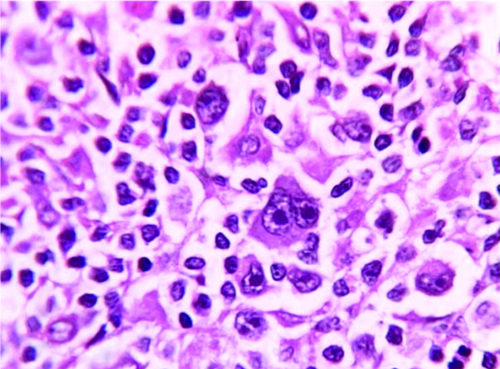
Histopathological examination reveals the diagnosis as:

Which type of breast carcinoma is characterized by a pushing border?
In Wilms tumor, which of the following leads to emergence of resistance to chemotherapy?
Endodermal sinus tumor is associated with which of the following?
Which of the following immunohistochemical markers is positive in neuroendocrine tumors?
S100 is a marker of which of the following?
What is the most common extragonadal site for germ cell tumors?
A patient complains of epigastric pain that fails to respond to antacids. Endoscopy demonstrates an ulcerated mass on the greater curvature of the stomach. Genetic studies on the tumor demonstrate an altered DCC gene. Which of the following tumor suppressor genes is found on the same chromosome as DCC?
Explanation: ### Explanation The question describes a scenario where a gene is actively transcribed into mRNA, but its translation into protein is inhibited in healthy individuals. This is a classic description of **post-transcriptional gene silencing** mediated by **microRNA (miRNA)** [1], [3]. **1. Why "Binding to microRNA (miRNA)" is correct:** MicroRNAs are small, non-coding single-stranded RNA molecules (approx. 22 nucleotides). They function by base-pairing with complementary sequences within the 3' untranslated region (UTR) of target messenger RNA (mRNA) [3]. * If there is **perfect complementarity**, the mRNA is cleaved and degraded [1]. * If there is **imperfect complementarity** (more common in humans), it leads to **translational repression** [3]. In this case, the BCL2 gene is transcribed, but miRNA prevents its translation in healthy cells. In B-cell lymphomas (like Follicular Lymphoma), this regulation is lost or bypassed (often via the t(14;18) translocation), leading to BCL2 protein overexpression and evasion of apoptosis [2], [4]. **2. Why other options are incorrect:** * **Absence of tRNA:** tRNA is essential for all protein synthesis; its absence would be lethal to the cell and is not a mechanism for gene-specific regulation. * **Methylation of DNA:** This is an **epigenetic silencing** mechanism that occurs at the transcriptional level [5]. If DNA were methylated, the gene would not be "actively transcribed to mRNA" in the first place [1]. * **Mutation of mRNA:** While mutations can occur in DNA, mRNA itself is not typically "mutated" to silence a gene; rather, it is regulated or degraded. **3. Clinical Pearls for NEET-PG:** * **OncomiRs:** miRNAs that contribute to cancer by downregulating tumor suppressor genes or upregulating oncogenes (by losing their inhibitory function) [1]. * **BCL2 Function:** It is an **anti-apoptotic** protein located on the outer mitochondrial membrane. It prevents the release of Cytochrome C [2]. * **Follicular Lymphoma:** Characterized by **t(14;18)**, which places the *BCL2* gene under the influence of the IgH promoter, leading to massive protein overproduction [2], [4]. **References:** [1] Cross SS. Underwood's Pathology: A Clinical Approach. 6th ed. (Basic Pathology) introduces the student to key general principles of pathology, both as a medical science and as a clinical activity with a vital role in patient care. Part 2 (Disease Mechanisms) provides fundamental knowledge about the cellular and molecular processes involved in diseases, providing the rationale for their treatment. Part 3 (Systematic Pathology) deals in detail with specific diseases, with emphasis on the clinically important aspects., pp. 230-231. [2] Kumar V, Abbas AK, et al.. Robbins and Cotran Pathologic Basis of Disease. 9th ed. Neoplasia, pp. 310-311. [3] Kumar V, Abbas AK, et al.. Robbins and Cotran Pathologic Basis of Disease. 9th ed. With Illustrations By, pp. 16-17. [4] Kumar V, Abbas AK, et al.. Robbins and Cotran Pathologic Basis of Disease. 9th ed. Diseases of White Blood Cells, Lymph Nodes, Spleen, and Thymus, pp. 602-604. [5] Kumar V, Abbas AK, et al.. Robbins and Cotran Pathologic Basis of Disease. 9th ed. With Illustrations By, pp. 15-16.
Explanation: **Explanation:** Radiation-induced carcinogenesis is a well-documented phenomenon where ionizing radiation causes DNA damage (double-strand breaks), leading to mutations [3]. While radiation can induce various malignancies [1], **Leukemia** (specifically Acute Myeloid Leukemia and Chronic Myeloid Leukemia) is the most common and earliest malignancy to appear following significant radiation exposure. * **Why Leukemia is Correct:** Hematopoietic cells are highly proliferative and sensitive to radiation [4]. Leukemia has a relatively short **latent period** (typically 5–10 years) compared to solid tumors, making it the most frequent "early" malignancy observed in survivors of atomic blasts or therapeutic radiation. * **Why other options are incorrect:** * **Thyroid Carcinoma:** This is the most common radiation-induced **solid tumor**, especially in children (e.g., post-Chernobyl) [2]. However, across all age groups and timeframes, leukemia remains more frequent. * **Bronchogenic Carcinoma:** Associated with mining (Radon gas) and tobacco, but not the most common overall radiation-linked cancer. * **Breast Cancer:** While radiation to the chest (e.g., for Hodgkin lymphoma) increases risk, it is less common than leukemia [4]. **High-Yield Clinical Pearls for NEET-PG:** * **Shortest Latent Period:** Leukemia (5–10 years). * **Longest Latent Period:** Solid tumors like Skin and Bone cancers (up to 20+ years). * **Most Radiosensitive Tissue:** Lymphocytes and Bone marrow [3]. * **Most Radioresistant Tissue:** Nerve cells and Muscle. * **Hierarchy of Risk:** Leukemia > Thyroid (in children) > Breast > Lung > Salivary glands [4]. **References:** [1] Cross SS. Underwood's Pathology: A Clinical Approach. 6th ed. (Basic Pathology) introduces the student to key general principles of pathology, both as a medical science and as a clinical activity with a vital role in patient care. Part 2 (Disease Mechanisms) provides fundamental knowledge about the cellular and molecular processes involved in diseases, providing the rationale for their treatment. Part 3 (Systematic Pathology) deals in detail with specific diseases, with emphasis on the clinically important aspects., pp. 220-221. [2] Kumar V, Abbas AK, et al.. Robbins and Cotran Pathologic Basis of Disease. 9th ed. The Endocrine System, pp. 1098-1099. [3] Kumar V, Abbas AK, et al.. Robbins and Cotran Pathologic Basis of Disease. 9th ed. Central Nervous System Synapse, pp. 438-439. [4] Cross SS. Underwood's Pathology: A Clinical Approach. 6th ed. (Basic Pathology) introduces the student to key general principles of pathology, both as a medical science and as a clinical activity with a vital role in patient care. Part 2 (Disease Mechanisms) provides fundamental knowledge about the cellular and molecular processes involved in diseases, providing the rationale for their treatment. Part 3 (Systematic Pathology) deals in detail with specific diseases, with emphasis on the clinically important aspects., pp. 111-112.
Explanation: ***Hodgkin lymphoma*** - Characterized by the presence of **Reed-Sternberg cells** with **owl-eye nucleoli** and **binucleated giant cells**. - The classic histopathological appearance includes a **mixed inflammatory infiltrate** surrounding the characteristic malignant cells. *Ewing sarcoma* - Shows **small round blue cells** with **uniform nuclear morphology** and **high nuclear-to-cytoplasmic ratio**. - Typically positive for **CD99** immunostain and lacks the characteristic giant cells seen in lymphomas. *Kaposi Sarcoma* - Demonstrates **spindle cells** arranged in **fascicles** with **slit-like vascular spaces**. - Associated with **HHV-8 infection** and shows **extravasated red blood cells** with **hemosiderin deposits**. *Burkitt lymphoma* - Exhibits a **starry-sky pattern** due to **scattered tingible body macrophages** among uniform lymphoid cells. - Composed of **monomorphic medium-sized B cells** with **multiple nucleoli** and high mitotic activity.
Explanation: ### Explanation **Correct Answer: D. Medullary carcinoma** **Understanding the Concept:** Medullary carcinoma of the breast is a distinct subtype of invasive carcinoma characterized by a **well-circumscribed, "pushing" (non-infiltrative) border** [1]. Unlike most breast cancers that invade the surrounding stroma in a jagged, irregular fashion, medullary carcinoma grows as a solid mass that displaces rather than infiltrates adjacent tissue. Microscopically, it is defined by a "triad": 1. Solid sheets of large pleomorphic cells with high mitotic rates [1]. 2. A prominent **lymphoplasmacytic infiltrate** (host immune response) [1]. 3. A non-invasive, pushing margin. Despite its high-grade histological appearance, it often carries a **better prognosis** than standard invasive ductal carcinoma [1]. **Why other options are incorrect:** * **Invasive Ductal Carcinoma (NOS):** This is the most common type and typically presents with **stellate or irregular borders** due to significant desmoplasia (fibrosis), giving it a "gritty" feel on sectioning. * **Invasive Lobular Carcinoma:** Characterized by a **diffuse, single-file (Indian file) pattern** of small cells. It is notorious for being "insidious" and often lacks a distinct mass or border, making it difficult to detect on mammography. * **Mucinous (Colloid) Carcinoma:** While it can be well-circumscribed, it is characterized by "lakes of extracellular mucin" and does not classically define the "pushing border" terminology used for medullary types. **High-Yield Pearls for NEET-PG:** * **Genetic Association:** Medullary carcinoma is frequently associated with **BRCA1 mutations**. * **Triple Negative:** Most medullary carcinomas are **ER, PR, and HER2/neu negative**, yet they maintain a favorable prognosis. * **Differential Diagnosis:** On imaging, the pushing border can mimic a benign **fibroadenoma**; therefore, core biopsy is essential. **References:** [1] Cross SS. Underwood's Pathology: A Clinical Approach. 6th ed. Common Clinical Problems From Liver And Biliary System Disease, pp. 455-456.
Explanation: **Explanation:** **Correct Answer: C. Anaplasia** In Wilms tumor (Nephroblastoma), the presence of **anaplasia** is the single most important histological predictor of prognosis [2]. Anaplasia is defined by the presence of giant, hyperchromatic, pleomorphic nuclei and abnormal multipolar mitoses [1]. Its clinical significance lies in its strong association with **resistance to standard chemotherapy** and a higher likelihood of tumor recurrence [4]. While Wilms tumor generally has an excellent cure rate, the "unfavorable histology" (anaplastic variant) requires more aggressive treatment protocols. **Analysis of Incorrect Options:** * **A. Nephrogenic rests:** These are focus of abnormally persistent embryonic cells that are considered **precursor lesions** for Wilms tumor [2]. While they increase the risk of developing a contralateral tumor (bilaterality), they do not directly cause chemoresistance. * **B. Monoblastic morphology:** This is not a standard histological feature of Wilms tumor. Classic Wilms tumor is **triphasic**, consisting of blastemal, stromal, and epithelial cells [2]. * **D. Capsular infiltration:** This relates to the **staging** of the tumor (Stage II) rather than its biological response to chemotherapy. While it indicates local spread, it does not inherently change the chemosensitivity of the cells. **High-Yield Clinical Pearls for NEET-PG:** * **Triphasic Pattern:** Blastema (small blue cells), Stroma (fibrocytic/myxoid), and Epithelium (tubules/glomeruli) [2]. * **Genetic Associations:** WAGR syndrome (WT1 mutation, 11p13), Denys-Drash syndrome (WT1), and Beckwith-Wiedemann syndrome (WT2, 11p15.5) [3]. * **Most common site of metastasis:** Lungs. * **Key Marker:** Anaplasia is linked to mutations in the **TP53** tumor suppressor gene. **References:** [1] Kumar V, Abbas AK, et al.. Robbins and Cotran Pathologic Basis of Disease. 9th ed. Neoplasia, p. 278. [2] Kumar V, Abbas AK, et al.. Robbins and Cotran Pathologic Basis of Disease. 9th ed. Diseases of Infancy and Childhood, pp. 488-490. [3] Kumar V, Abbas AK, et al.. Robbins and Cotran Pathologic Basis of Disease. 9th ed. Diseases of Infancy and Childhood, pp. 487-488. [4] Cross SS. Underwood's Pathology: A Clinical Approach. 6th ed. (Basic Pathology) introduces the student to key general principles of pathology, both as a medical science and as a clinical activity with a vital role in patient care. Part 2 (Disease Mechanisms) provides fundamental knowledge about the cellular and molecular processes involved in diseases, providing the rationale for their treatment. Part 3 (Systematic Pathology) deals in detail with specific diseases, with emphasis on the clinically important aspects., pp. 212-213.
Explanation: **Explanation:** **Endodermal Sinus Tumor (Yolk Sac Tumor)** is the most common germ cell tumor in children [1]. The hallmark histological feature of this tumor is the **Schiller-Duval body**. 1. **Why Option A is Correct:** A Schiller-Duval body consists of a central capillary surrounded by visceral and parietal layers of neoplastic cells within a space, resembling a primitive glomerulus. This structure is pathognomonic for Yolk Sac Tumors. Additionally, these tumors characteristically produce **Alpha-Fetoprotein (AFP)**, which serves as a vital serum marker for diagnosis and monitoring. 2. **Why Other Options are Incorrect:** * **B. Multinucleate giant cells:** These are typically seen in granulomatous inflammation (e.g., Tuberculosis) or specific tumors like Choriocarcinoma (Syncytiotrophoblasts) and Giant Cell Tumor of the bone [2]. * **C. R-S cells (Reed-Sternberg cells):** These are the diagnostic "owl-eye" cells found in **Hodgkin Lymphoma**, not germ cell tumors. * **D. Plasma cells:** These are mature B-lymphocytes seen in chronic inflammation and **Multiple Myeloma** (Fried-egg appearance/Clock-face chromatin). **High-Yield Clinical Pearls for NEET-PG:** * **Serum Marker:** Elevated **AFP** is the most significant biochemical finding. * **Histology:** Look for **intracytoplasmic hyaline droplets** (PAS-positive) in addition to Schiller-Duval bodies. * **Age Group:** It is the most common testicular tumor in infants and young children (under 3 years) [1]. * **Ovarian counterpart:** In females, it presents as a rapidly growing, painful pelvic mass, usually in young women. **References:** [1] Kumar V, Abbas AK, et al.. Robbins and Cotran Pathologic Basis of Disease. 9th ed. The Lower Urinary Tract and Male Genital System, pp. 979-980. [2] Kumar V, Abbas AK, et al.. Robbins and Cotran Pathologic Basis of Disease. 9th ed. The Lower Urinary Tract and Male Genital System, pp. 980-982.
Explanation: **Explanation:** **Neuroendocrine tumors (NETs)** arise from cells of the endocrine and nervous systems. To confirm a neuroendocrine origin, pathologists look for markers associated with secretory vesicles and neural differentiation. * **Why Synaptophysin is correct:** **Synaptophysin** is a glycoprotein found in the membranes of synaptic vesicles in neurons and neurosecretory granules of neuroendocrine cells. It is considered the most specific and reliable pan-neuroendocrine marker. Along with **Chromogranin A** (found in the matrix of these granules) and **CD56 (NCAM)**, it forms the standard IHC panel for diagnosing NETs (e.g., Carcinoid, Small cell carcinoma) [2]. **Analysis of Incorrect Options:** * **A. Cytokeratin:** This is a marker for **epithelial differentiation**. While many NETs (like Small cell lung cancer) can be CK positive, it is not specific to neuroendocrine cells and is used primarily to identify carcinomas. * **C. Calretinin:** This is a calcium-binding protein used as a primary marker for **Mesothelioma**. It is also seen in sex cord-stromal tumors (e.g., Granulosa cell tumors). * **D. GFAP (Glial Fibrillary Acidic Protein):** This is the specific marker for **Glial cells** (Astrocytes). It is used to diagnose CNS tumors like Astrocytomas and Ependymomas. **High-Yield NEET-PG Pearls:** * **Chromogranin A** is the most specific serum marker for monitoring NETs. * **Neuron-Specific Enolase (NSE)** is another neuroendocrine marker but is less specific than Synaptophysin. * **Salt and Pepper Chromatin:** The classic nuclear morphology seen in neuroendocrine tumors on histopathology [1]. * **Homer-Wright Rosettes:** Often seen in primitive neuroectodermal tumors (e.g., Neuroblastoma). **References:** [1] Kumar V, Abbas AK, et al.. Robbins and Cotran Pathologic Basis of Disease. 9th ed. The Gastrointestinal Tract, pp. 781-782. [2] Cross SS. Underwood's Pathology: A Clinical Approach. 6th ed. Common Clinical Problems From Respiratory Tract Disease, pp. 337-338.
Explanation: **Explanation:** **S100** is a low-molecular-weight calcium-binding protein originally isolated from the brain. It is a highly sensitive, though not highly specific, immunohistochemical (IHC) marker used to identify cells derived from the **neural crest** and certain other lineages. 1. **Melanoma (Option A):** Melanocytes are neural crest-derived cells. S100 is the most sensitive marker for malignant melanoma (approaching 100% sensitivity), making it the primary screening tool, although markers like HMB-45 and Melan-A are more specific. 2. **Schwannoma (Option B):** Schwann cells are also neural crest-derived [2]. S100 shows strong and diffuse cytoplasmic and nuclear positivity in nerve sheath tumors like Schwannomas and Neurofibromas [3]. 3. **Histiocytoma (Option C):** S100 is expressed in specific subsets of histiocytic cells, most notably **Langerhans cells** [1]. It is a diagnostic marker for Langerhans Cell Histiocytosis (LCH) and Rosai-Dorfman disease. Since S100 is positive in all three conditions, **Option D (All of the above)** is the correct answer. **High-Yield Clinical Pearls for NEET-PG:** * **Neural Crest Origin:** Remember the mnemonic "S100 = **S**eventeen (Chr 17), **S**chwannoma, **S**kin (Melanoma), **S**ustentacular cells." * **Other S100+ Tissues:** Chondrocytes (Chondrosarcoma), Adipocytes (Liposarcoma), Salivary gland myoepithelial cells, and Astrocytes (Gliomas). * **Differential Diagnosis:** While S100 is excellent for *ruling out* melanoma (high negative predictive value), its lack of specificity means it must be used as part of a panel. * **Langerhans Cell Histiocytosis (LCH):** Characteristic IHC profile is **S100(+), CD1a(+), and CD207 (Langerin)(+).** [1] **References:** [1] Kumar V, Abbas AK, et al.. Robbins and Cotran Pathologic Basis of Disease. 9th ed. Diseases of White Blood Cells, Lymph Nodes, Spleen, and Thymus, pp. 629-630. [2] Kumar V, Abbas AK, et al.. Robbins and Cotran Pathologic Basis of Disease. 9th ed. Peripheral Nerves and Skeletal Muscles, pp. 1249-1250. [3] Cross SS. Underwood's Pathology: A Clinical Approach. 6th ed. Common Clinical Manifestations Of Central And Peripheral Nervous System Disease, pp. 727-728.
Explanation: **Explanation:** Germ cell tumors (GCTs) typically arise in the gonads (testes and ovaries). However, during embryogenesis, primordial germ cells migrate from the yolk sac endoderm to the gonadal ridges. If these cells fail to reach the gonads or migrate abnormally along the midline of the body, they can give rise to **Extragonadal Germ Cell Tumors (EGGCTs).** **1. Why Mediastinum is Correct:** In **adults**, the **mediastinum** (specifically the anterior mediastinum) is the most common site for extragonadal germ cell tumors. They represent approximately 50-70% of all EGGCTs. The most common histological subtype in this location is the mature teratoma. **2. Analysis of Incorrect Options:** * **Sacrococcygeal region (Option B):** This is the most common site for extragonadal GCTs in **infants and children**, but not in the general population or adults [3]. * **Retroperitoneum (Option A):** This is the second most common site in adults. However, a primary retroperitoneal GCT must be distinguished from a metastatic deposit from an occult testicular primary (Burned-out tumor) [2]. * **Pineal gland (Option C):** This is a classic midline site for intracranial GCTs (Germinomas), but it is less frequent than the mediastinal location [1]. **3. NEET-PG High-Yield Pearls:** * **Most common EGGCT in children:** Sacrococcygeal teratoma (more common in females) [3]. * **Most common EGGCT in adults:** Mediastinal GCT (more common in males). * **Klinefelter Syndrome (47, XXY):** Strongly associated with an increased risk of mediastinal germ cell tumors. * **Tumor Marker:** Non-seminomatous GCTs in the mediastinum often show elevated **AFP** (Yolk sac component) or **β-hCG** (Choriocarcinoma component). * **Hematologic Association:** Mediastinal non-seminomatous GCTs are uniquely associated with the development of hematologic malignancies like **Acute Myeloid Leukemia (AML).** **References:** [1] Kumar V, Abbas AK, et al.. Robbins and Cotran Pathologic Basis of Disease. 9th ed. The Endocrine System, pp. 1140-1141. [2] Cross SS. Underwood's Pathology: A Clinical Approach. 6th ed. Common Clinical Problems From Diseases Of The Urinary And Male Genital Tracts, pp. 510-512. [3] Kumar V, Abbas AK, et al.. Robbins and Cotran Pathologic Basis of Disease. 9th ed. Diseases of Infancy and Childhood, pp. 482-483.
Explanation: **Explanation:** The question tests your knowledge of tumor suppressor gene (TSG) chromosomal locations. The **DCC gene** (Deleted in Colorectal Carcinoma) is located on **Chromosome 18q**. **1. Why DPC is correct:** The **DPC4 gene** (Deleted in Pancreatic Carcinoma, also known as **SMAD4**) is also located on **Chromosome 18q**. Both genes are frequently mutated or deleted in gastrointestinal malignancies. While DCC is classically associated with colorectal cancer (and occasionally gastric cancer, as seen in this clinical vignette), DPC4/SMAD4 is a critical marker for pancreatic adenocarcinoma and juvenile polyposis syndrome. **2. Why other options are incorrect:** * **BRCA-1:** Located on **Chromosome 17q**. It is associated with hereditary breast and ovarian cancer. * **NF-1 (Neurofibromin):** Located on **Chromosome 17q**. Mutation leads to Neurofibromatosis Type 1 (von Recklinghausen disease). * **NF-2 (Merlin):** Located on **Chromosome 22q**. Mutation leads to Neurofibromatosis Type 2 (bilateral acoustic neuromas). **Clinical Pearls for NEET-PG:** * **Mnemonic for Chromosome 18:** Remember "**18** is **D**eleted in **D**PC and **D**CC" (The 3 Ds). * **SMAD4/DPC4** is a signaling transducer for the TGF-β pathway; its loss leads to unchecked cellular proliferation. * **Chromosome 17** is a high-yield "hotspot" containing **TP53** (17p), **NF-1** (17q), and **BRCA-1** (17q). * **Chromosome 13** contains **RB1** and **BRCA-2**. * **Chromosome 5** contains **APC** (Adenomatous Polyposis Coli). **Clinical Presentation Note:** Gastric adenocarcinomas may present as an ulcerated tumor [1], with common symptoms including dyspepsia and epigastric pain [1]. **References:** [1] Kumar V, Abbas AK, et al.. Robbins and Cotran Pathologic Basis of Disease. 9th ed. The Gastrointestinal Tract, pp. 779-781.
Nomenclature and Classification of Tumors
Practice Questions
Characteristics of Benign and Malignant Neoplasms
Practice Questions
Molecular Basis of Cancer
Practice Questions
Carcinogenesis and Carcinogens
Practice Questions
Tumor Progression and Metastasis
Practice Questions
Tumor Markers
Practice Questions
Paraneoplastic Syndromes
Practice Questions
Genetic Basis of Cancer
Practice Questions
Tumor Immunity
Practice Questions
Cancer Epidemiology and Prevention
Practice Questions
Get full access to all questions, explanations, and performance tracking.
Start For Free