
Neoplasia — MCQs

On this page
Masaoka staging is used for staging:
The term "Gompertzian curve" is related to which one of the following ?
Which of the following are correct regarding Li-Fraumeni syndrome? 1. It has autosomal dominant inheritance and is associated with P53 gene. 2. It has autosomal recessive inheritance and is associated with P53 gene. 3. It is associated with an increased risk of sarcomas and leukaemia. 4. It is associated with an increased risk of brain tumours and osteosarcomas. Select the answer using the code given below.
The commonest variety of peritoneal metastasis is
Which of the following statements regarding papillary thyroid cancer is correct? 1. It is the most common malignant tumour of thyroid gland. 2. It is more common in young females. 3. It has propensity for haematogenous spread. 4. Distant metastases are uncommon.
The histological grade best correlates with the prognosis in which one of the following malignancies?
Which one of the following is NOT associated with BRCA1/BRCA2 genes?
In which of the following is the term low and high grade squamous intraepithelial neoplasia used?
Which of the following pairs is not correctly matched?
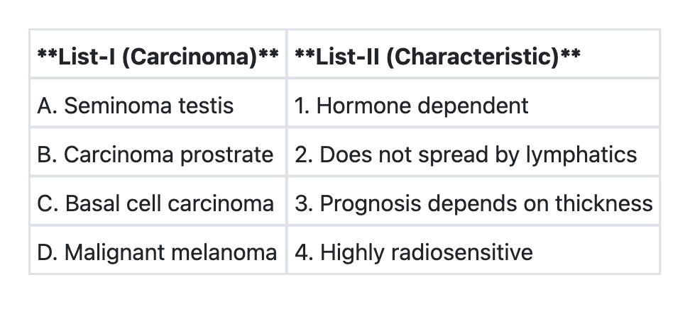
Match List-I with List-II and select the correct answer using the code given below the Lists:
Practice by Chapter
Nomenclature and Classification of Tumors
Practice Questions
Characteristics of Benign and Malignant Neoplasms
Practice Questions
Molecular Basis of Cancer
Practice Questions
Carcinogenesis and Carcinogens
Practice Questions
Tumor Progression and Metastasis
Practice Questions
Tumor Markers
Practice Questions
Paraneoplastic Syndromes
Practice Questions
Genetic Basis of Cancer
Practice Questions
Tumor Immunity
Practice Questions
Cancer Epidemiology and Prevention
Practice Questions
Want unlimited practice?
Get full access to all questions, explanations, and performance tracking.
Scan to download app