An 18 year old boy had a closed lower limb injury while riding his motorbike. He was brought to hospital where on examination he had severe pain which increased on passive movement of affected limb with distal sensory disturbances. What is the probable diagnosis?
In a fracture of shaft of longbone, the component which contributes least in fracture healing is:
Which one of the following statements about Compartment Syndrome is NOT correct?
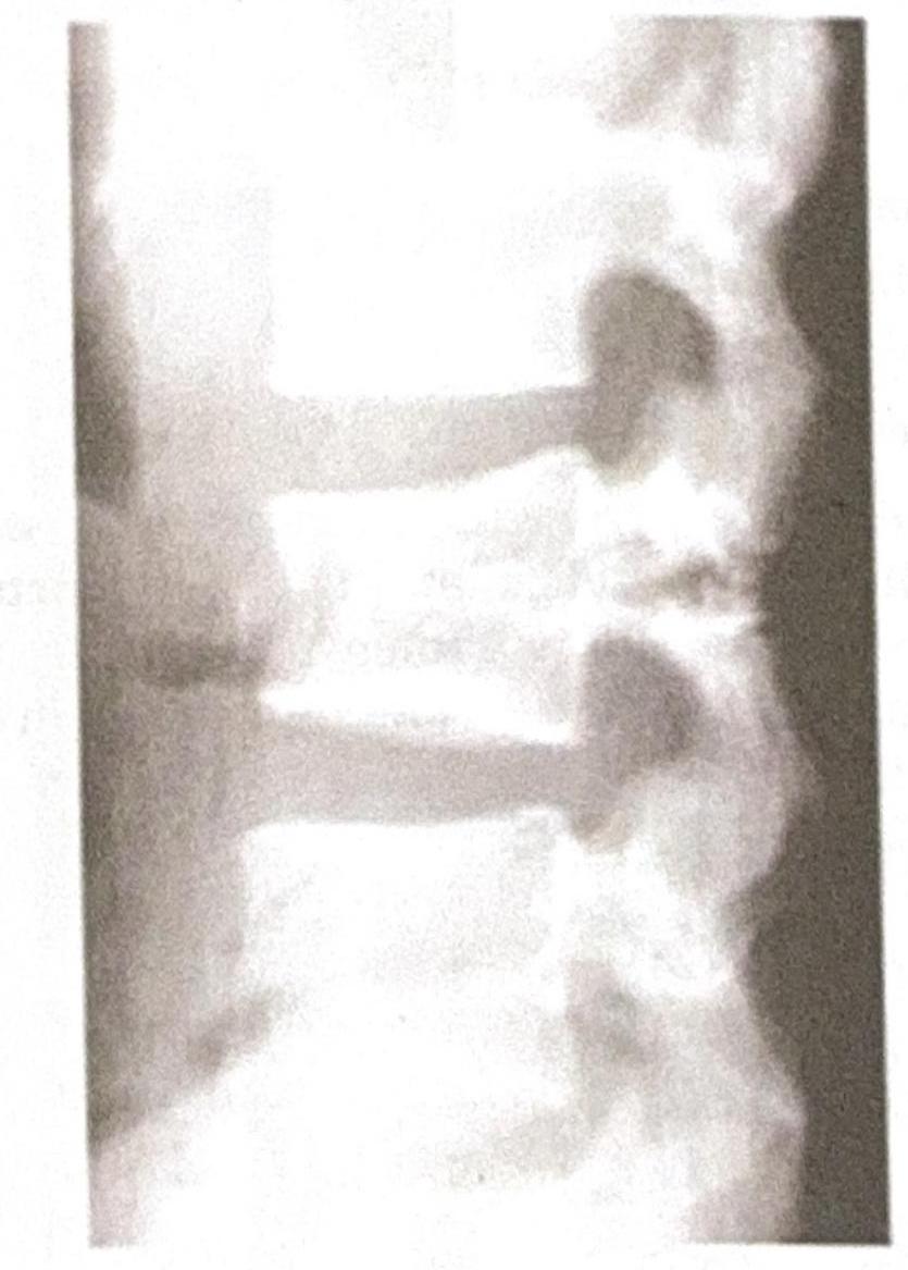
A 35-year-old male presents to the emergency department following a high-speed motor vehicle accident. He complains of severe lower back pain but denies any loss of consciousness or abdominal pain. A lateral X-ray of the lumbar spine is obtained, as shown in the image. The image reveals a horizontal fracture through the vertebral body, extending through the posterior elements. Based on the clinical presentation and imaging findings, what is the most likely diagnosis?

A patient fell off a bicycle and now complains of pain around the hip, with shortening of the affected limb. The hip is held in a position of flexion, adduction, and internal rotation. What is the most likely diagnosis?
Following a road traffic accident, a patient develops type IIIa compound tibial fracture. Arrange the following external fixators in decreasing order of their stability (highest to lowest) 1. Ilizarov fixator 2. Uniplanar with a single rod 3. Uniplanar with double rod 4. Biplanar frame/Ring with a cylindrical rod
Following a femoral shaft fracture, your consultant asks you to provide tibia traction. Which of the following will you request from the nurse? 1. Thomas splint 2. K-wire 3. Steinmann pin 4. Denham's pin 5. Bohler's stirrup 6. Bohler Braun splint
A 30-year-old male presents with pain and limited movement in his shoulder following a fall. X-ray reveals an anterior dislocation of the glenohumeral joint. Which of the following structures is most likely to be damaged in this injury?
Most common complication of extra capsular fracture of neck of femur is:
Which nerve is commonly damaged in fracture of neck of fibula?
Explanation: ***Compartment syndrome*** - This diagnosis is strongly suggested by the presence of **severe pain out of proportion to injury**, pain with **passive stretch** of muscles, **paresthesia** (sensory disturbances), and a history of **closed lower limb injury**. These are classic signs of increased pressure within a confined fascial compartment. - The elevated pressure compromises **perfusion**, leading to muscle and nerve ischemia. Prompt recognition and treatment (fasciotomy) are crucial to prevent permanent damage. *Deep Vein thrombosis* - While DVT can cause lower limb pain and swelling, it is typically associated with **venous stasis**, endothelial injury, and hypercoagulability, not immediate severe pain with passive movement and sensory deficits following acute trauma. - The pain in DVT is usually described as a **dull ache** and not typically out of proportion to what would be expected, nor does it typically present with acute sensory loss without major vascular compromise. *Degloving Injury* - A degloving injury involves the **separation of skin and subcutaneous tissue** from deeper structures, often with significant visible skin avulsion. - While it can cause severe pain, the key diagnostic features in this scenario (closed injury, pain on passive movement, and distal sensory disturbances) are more classic for compartment syndrome rather than a primary external soft tissue avulsion. *High Pressure Injection injury* - This type of injury results from the **introduction of foreign material** under high pressure into tissues, often leading to rapid swelling, pain, and tissue necrosis. - It usually has a distinct history of exposure to high-pressure equipment (e.g., paint gun, grease gun), which is not mentioned here, and while severe, the precise constellation of symptoms points away from this specific mechanism.
Explanation: ***Endosteum*** - The **endosteum** is a thin vascular membrane that lines the inner surface of the bony tissue forming the medullary cavity of long bones. - While it has osteogenic potential, its contribution to callus formation and overall fracture healing is **less significant** compared to the richly vascularized periosteum and the surrounding musculature. *Blood vessels* - **Blood vessels** are crucial for delivering essential nutrients, oxygen, and cells (like **osteoblasts** and **osteoclasts**) to the fracture site, which are fundamental for callus formation and bone remodeling. - Their disruption can significantly impair healing, making them a **major contributor** to the process. *Periosteum* - The **periosteum** is a dense, fibrous membrane covering the outer surface of bones, except at articular surfaces, and is rich in **osteogenic cells** and blood vessels. - It plays a **dominant role** in forming the external callus, especially in long bone fractures, due to its **cambial layer** containing progenitor cells. *Matrix* - The **bone matrix** (both organic and inorganic components) is the framework upon which new bone is laid down during fracture healing. - While it provides structural support, the matrix itself does not actively *contribute* to the cellular processes of healing; rather, it is the **product of these processes** involving cells like osteoblasts.
Explanation: ***Pain is on active movement but not on passive movement of muscles*** - This statement is incorrect because pain in compartment syndrome is characteristically **out of proportion to the injury** and is **exacerbated by passive stretching of the muscles** within the affected compartment. - While active movement can cause pain, the hallmark sign related to pain is its intensification with passive stretching due to increased pressure. *Fasciotomy is the treatment of choice* - **Fasciotomy** is indeed the definitive surgical treatment for compartment syndrome to relieve pressure and prevent irreversible tissue damage. - It involves incising the fascia to decompress the affected muscle compartment. *It is commonest in a closed fracture* - Compartment syndrome most frequently occurs after a **closed fracture**, particularly in the tibia and forearm, because the intact fascial compartments restrict expansion, leading to increased pressure. - The swelling and hemorrhage associated with the fracture are contained, causing pressure to rise rapidly. *Volkmann's Ischaemic contracture is a late complication* - **Volkmann's ischemic contracture** is a severe and debilitating late complication of unresolved or undertreated compartment syndrome, primarily affecting the forearm muscles. - It results from prolonged ischemia, causing muscle necrosis, fibrosis, and subsequent shortening and contracture.
Explanation: ***Chance fracture*** - A **chance fracture** is characterized by a **horizontal fracture** through the entire vertebral body and posterior elements, including the neural arch, typically caused by a **flexion-distraction mechanism** in accidents like those involving seatbelts (lap belt only), consistent with the high-speed motor vehicle accident scenario. - The image distinctly shows a fracture line traversing the vertebral body and extending into the posterior elements, which is the hallmark of this type of injury. *Burst fracture* - A **burst fracture** involves a comminuted fracture of the vertebral body with **retropulsion of bone fragments** into the spinal canal due to axial loading, which is not clearly depicted here. - While it can result from high-impact trauma, the characteristic horizontal disruption of both anterior and posterior segments points away from a solely compressive mechanism. *Compression fracture* - A **compression fracture** primarily involves the **anterior wedging** or collapse of the vertebral body, resulting from only axial compression forces without significant involvement of the posterior elements. - The presented image shows a fracture extending through the posterior elements, which is not typical for a simple compression fracture. *Spondylolisthesis* - **Spondylolisthesis** is the **anterior slippage of one vertebral body over another**, often due to pars interarticularis defects (spondylolysis) or degenerative changes. - This condition involves vertebral displacement, not a fresh fracture line across the body and posterior elements as seen in the image.
Explanation: **Posterior dislocation** - **Posterior hip dislocations** typically occur after high-energy trauma (e.g., falls from height, motor vehicle accidents) and present with the affected limb in a classic position of **flexion, adduction, and internal rotation**. - **Shortening of the limb** is also a hallmark sign, often due to the femoral head displacing posteriorly and superiorly. *Intertrochanteric fracture (IT fracture)* - **Intertrochanteric fractures** usually present with the affected limb in **external rotation** and shortening, which is contrary to the internal rotation described in the case. - While pain is present, the specific rotational deformity helps differentiate it from a hip dislocation. *Transcervical fracture* - **Transcervical fractures** (femoral neck fractures) also typically present with the leg in **external rotation** and shortening. - These fractures are common in older adults and often associated with less severe trauma or falls. *Anterior dislocation* - **Anterior hip dislocations** are less common and typically present with the affected limb in a position of **flexion, abduction, and external rotation**. - This presentation is directly opposite to the adduction and internal rotation described in the question.
Explanation: ***1>4>3>2*** - **Ilizarov fixator** utilizes multiple wires **under tension** and rings, providing the most **biologically stable** and rigid fixation due to its distributed force across the bone. - **Biplanar frames/Rings with a cylindrical rod** offer high stability by providing pin fixation in **two different planes**, significantly resisting bending and torsional forces. - **Uniplanar with double rod** provides better stability than a single rod by increasing the **moment of inertia** and reducing deflection under axial and bending loads. - **Uniplanar with a single rod** is the least stable due to its limited resistance to **torsional** and **bending forces** as pin placement is restricted to a single plane.
Explanation: ***3,5,6*** - For **tibia traction** in a femoral shaft fracture, you would need a **Steinmann pin** for skeletal traction, a **Bohler's stirrup** to apply the traction force, and a **Bohler-Braun splint** to support the limb. - The **Steinmann pin** is inserted into the proximal tibia, the **Bohler's stirrup** attaches to the pin, and the **Bohler-Braun splint** provides a fixed structure for the traction system. *1,2,3,4,5,6* - This option incorrectly includes items not specifically used for applying **tibia traction** (e.g., K-wire is for internal fixation, Thomas splint is for early femur fracture management but not specifically for tibia traction application). - While some components might be used in general fracture management, not all are directly involved in setting up tibia traction as requested. *3,4,5* - This option correctly includes the **Steinmann pin** and **Bohler's stirrup** but incorrectly replaces the **Bohler-Braun splint** with a **Denham's pin**. - A **Denham's pin** is an alternative to a Steinmann pin for skeletal traction, but a **Bohler-Braun splint** is crucial for supporting the limb in this setup, which is missing here. *1,2,4* - This option includes a **Thomas splint** (used for femur fracture support, not tibia traction application), a **K-wire** (used for internal fixation, not traction), and a **Denham's pin** (an alternative to Steinmann pin, but lacks the necessary support and traction application equipment). - These items are not suitable for setting up comprehensive **tibia traction** for a femoral shaft fracture.
Explanation: ***Anterior inferior glenohumeral ligament*** - This ligament is a primary static stabilizer against **anterior dislocation** of the shoulder; thus, it is frequently stretched or torn during such an event. - Damage to this ligament is often associated with a **Bankart lesion**, which is an injury to the anterior inferior labrum that can lead to recurrent dislocations. *Long head of biceps tendon* - While the **long head of the biceps tendon** can be injured in shoulder trauma, it is more commonly associated with chronic overuse or superior labral tears (**SLAP lesions**), rather than primary anterior dislocation. - Injuries to this tendon might occur as a secondary complication but are not the most likely primary soft tissue damage in an acute anterior dislocation. *Acromioclavicular ligament* - The **acromioclavicular ligament** stabilizes the **acromioclavicular (AC) joint**, which is distinct from the glenohumeral joint. - Injuries to this ligament typically result from direct trauma to the top of the shoulder, causing AC joint separation, not glenohumeral dislocation. *Supraspinatus tendon* - The **supraspinatus tendon** is part of the rotator cuff and is most commonly injured in impingement syndrome or rotator cuff tears, which can result from falls but are not the primary structure damaged in an **anterior glenohumeral dislocation**. - Its role is mainly in abduction of the arm, and while it can be involved in large tears associated with advanced age, it is not the initial or most common structure to fail in this specific injury. *Coracoclavicular ligament* - The **coracoclavicular ligament** is composed of the conoid and trapezoid ligaments, which are crucial for the stability of the **acromioclavicular (AC) joint**. - Injury to this ligament is indicative of a more severe AC joint separation (usually **type III or higher**) and is not the primary structure damaged in a glenohumeral dislocation.
Explanation: ***Non union*** - **Non-union** is a common complication in extracapsular femoral neck fractures due to the **disrupted blood supply** and mechanical forces across the fracture site. - The **fracture fragments** may fail to heal properly, leading to persistent pain, instability, and functional impairment. *Malunion* - **Malunion** occurs when the fracture heals in an **unacceptable anatomical position**, causing deformity or altered biomechanics. - While it can be a complication, **non-union** is generally more prevalent and problematic in extracapsular femoral neck fractures. *Ischemic necrosis* - **Ischemic necrosis** (or avascular necrosis) is less common in extracapsular femoral neck fractures compared to intracapsular fractures. - This is because the **extracapsular location** often spares the crucial blood supply to the femoral head, which is frequently compromised in intracapsular injuries. *Pulmonary complications* - **Pulmonary complications** (e.g., pneumonia, pulmonary embolism) are significant risks in elderly patients with hip fractures due to prolonged immobility and surgery. - However, direct fracture-related complications like **non-union** are distinct and represent issues specifically with bone healing.
Explanation: ***Common peroneal*** - The **common peroneal nerve** (also known as the **common fibular nerve**) wraps superficially around the **neck of the fibula**, making it highly vulnerable to injury in fractures of this region. - Damage to this nerve typically results in **foot drop** and sensory loss over the dorsum of the foot and lateral leg, due to impaired dorsiflexion and eversion. *Tibial* - The **tibial nerve** lies in the posterior compartment of the leg and is generally well-protected, making it less susceptible to injury from a fibular neck fracture. - Injury to the tibial nerve would primarily affect plantarflexion of the foot and sensation to the sole. *Superficial peroneal* - The **superficial peroneal nerve** is a branch of the common peroneal nerve that descends along the lateral compartment of the leg. - While it originates from the common peroneal, a direct fracture of the fibular neck is more likely to injure the main common peroneal trunk rather than just this specific branch, leading to a broader deficit. *Deep peroneal* - The **deep peroneal nerve** is another branch of the common peroneal nerve that runs through the anterior compartment of the leg. - Similar to the superficial peroneal nerve, a fracture at the fibular neck is more likely to affect the main **common peroneal nerve** directly.
Principles of Fracture Management
Practice Questions
Upper Limb Fractures
Practice Questions
Lower Limb Fractures
Practice Questions
Spinal Trauma
Practice Questions
Pelvic and Acetabular Fractures
Practice Questions
Open Fractures
Practice Questions
Fractures in Children
Practice Questions
Fracture Complications
Practice Questions
Nonunion and Malunion
Practice Questions
Polytrauma Management
Practice Questions
Joint Dislocations
Practice Questions
Soft Tissue Injuries
Practice Questions
Get full access to all questions, explanations, and performance tracking.
Start For Free