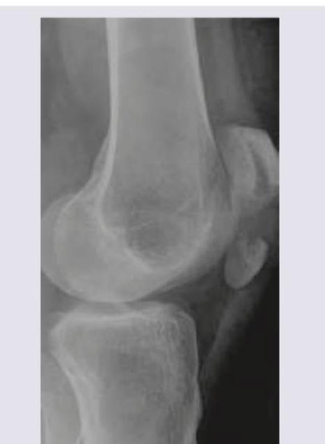
Which is the suggested treatment of the lesion shown in the X-ray?

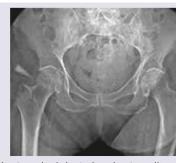
An elderly patient slipped in the bathroom and sustained injury over the hip joint. X- Ray is shown below. Her attitude of leg will be?

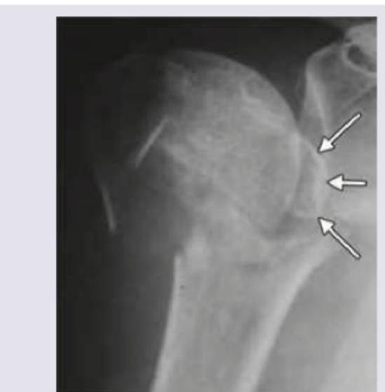
What type of fracture is shown in X-ray of left shoulder?

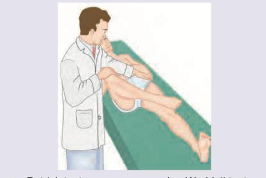
What is the test being performed in the patient?

Consider the following : 1. Pain relief 2. Prevention of infection 3. Anaesthesia 4. Restoration of anatomy Which of the features given above are priorities for fracture treatment?
The commonest complication of fracture of clavicle is :
The nerve most likely to get injured in patients with fracture of upper end of radius is :
Which one of the following fractures is most often complicated by fat embolism ?
Avascular necrosis may develop in the following fractures except
What is the most common injury sustained due to fall on outstretched hand by a person aged 65 years?
Explanation: ***Tension band wiring*** - The X-ray image shows a **transverse patellar fracture** with some displacement, which is well-suited for tension band wiring. - This technique converts tensile forces on the anterior surface of the patella into compressive forces at the fracture site during knee flexion. *Intramedullary nail* - **Intramedullary nailing** is primarily used for **long bone fractures** (e.g., femur, tibia, humerus) and is not appropriate for patellar fractures. - This method is designed to stabilize diaphyseal or metaphyseal fractures in load-bearing long bones by placing a rod within the medullary canal. *Patellectomy* - **Patellectomy** (surgical removal of the patella) is typically reserved for **severely comminuted** or irreparable patellar fractures, or in cases of infection, which is not indicated by the X-ray. - This procedure can lead to significant functional impairment, including reduced quadriceps strength and increased tibiofemoral joint stress. *Above knee cast* - While an **above-knee cast** can provide - An above-knee cast does not provide **sufficient reduction and compression** for displaced patellar fractures, which are subjected to significant tensile forces. - Conservative management is generally reserved for **non-displaced or minimally displaced** patellar fractures where the extensor mechanism remains intact.
Explanation: ***Shortened, abducted and externally rotated*** - The X-ray image reveals an **intertrochanteric fracture** of the right hip, characterized by a fracture line between the greater and lesser trochanters. - Due to the pull of strong muscles acting on the fractured fragments (e.g., iliopsoas, gluteal muscles), the limb typically assumes a position of **shortening, abduction, and external rotation**. *Shortened and abducted* - While **shortening** and **abduction** are present in intertrochanteric fractures, this answer is incomplete as it misses the crucial component of **external rotation**. - The powerful **external rotators** and the **gravity** acting on the unstable distal fragment contribute significantly to the external rotation. *Lengthened and internally rotated* - This attitude is characteristic of a **posterior hip dislocation**, where the femoral head is driven posteriorly and superiorly, typically leading to limb lengthening and internal rotation. - This is opposite to the typical presentation of an intertrochanteric fracture as seen in the X-ray. *Flexed and adducted and internally rotated* - A flexed, adducted, and internally rotated position is also seen in **posterior hip dislocations**, which is not consistent with the X-ray findings of an intertrochanteric hip fracture. - Hip fractures usually result in some degree of **external rotation** due to the muscle forces.
Explanation: ***Ideberg classification grade 4*** - The X-ray shows a **scapular fracture**, specifically involving the glenoid. Ideberg classification is used for **glenoid fractures**. - An Ideberg grade 4 fracture involves a **transverse fracture below the scapular notch**, separating the load-bearing part of the glenoid from the rest of the scapula, which appears consistent with the image. *Neer classification grade 4* - The **Neer classification** system is used for **proximal humerus fractures**, not scapular fractures. - A grade 4 Neer fracture involves **four part displacement** of the humeral head, greater tuberosity, lesser tuberosity, and humeral shaft. *Garden classification grade 3* - The **Garden classification** system is specifically used for **femoral neck fractures**, which are fractures of the hip. - A Garden grade 3 fracture represents a **complete, displaced fracture of the femoral neck**, distinct from the shoulder injury shown. *Schatzker classification grade 5* - The **Schatzker classification** system is used for **tibial plateau fractures**, which are fractures of the knee joint. - A Schatzker grade 5 fracture involves a **bicondylar fracture** of the tibial plateau, which is severe and impacts the knee, not the shoulder.
Explanation: ***Hoover test*** - The image depicts the **Hoover test**, where the examiner places one hand under the asymptomatic heel while the patient attempts to lift the symptomatic leg. The hand under the asymptomatic heel should feel downward pressure if the patient is genuinely trying to lift the affected leg. - This test is used to detect **malingering** or non-organic weakness in the lower limbs, often in cases of suspected sciatica or lumbar radiculopathy. *Patrick test* - The **Patrick test**, also known as the **FABER test** (Flexion, Abduction, External Rotation), assesses the hip joint and sacroiliac joint. - It involves placing the foot of the symptomatic leg on the opposite knee and allowing the knee to fall towards the examination table, which is not what is shown in the image. *Waddell test* - The **Waddell signs** are a group of physical signs used to identify non-organic (psychological) components in low back pain. - These signs include superficial tenderness, non-anatomic tenderness, simulation tests, distraction tests, and regional disturbances, none of which are individually represented by the specific maneuver in the image. *McMurray test* - The **McMurray test** is used to assess for **meniscal tears** in the knee. - It involves flexing and extending the knee while applying varus or valgus stress and internal or external rotation to elicit a click or pain, which is markedly different from the maneuver shown.
Explanation: **1, 3 and 4** - **Priorities in fracture treatment** always include alleviating pain, which can be severe and debilitating. - **Restoration of normal anatomy** is crucial for proper healing and optimal function of the fractured limb. - **Anesthesia** is often required to facilitate reduction and fixation of a fracture, as well as to manage pain during the procedure. *2, 3 and 4* - While **anesthesia** and **restoration of anatomy** are priorities, **prevention of infection** is primarily a concern for **open fractures** or surgical interventions. - **Pain relief** is a fundamental and immediate concern in all fracture cases, which is omitted in this option. *1, 2 and 3* - **Pain relief** and **anesthesia** are critical, and **prevention of infection** is important, but this option neglects the essential goal of **restoring anatomical alignment**. - **Restoring anatomy** directly impacts the long-term functional outcome and is a major goal of fracture management. *1, 2 and 4* - This option correctly identifies **pain relief**, **prevention of infection**, and **restoration of anatomy** as important. - However, it overlooks the immediate necessity of **anesthesia** to effectively manage pain during treatment procedures and allow for fracture reduction.
Explanation: ***malunion*** - **Malunion** is the most frequent complication following a clavicle fracture, meaning the bone heals in an anatomically incorrect or deformed position. - This often results in a palpable bump or cosmetic deformity, and can occasionally cause functional impairment. *non union* - **Non-union** occurs when the fracture fails to heal completely, leaving a persistent gap between the bone fragments. - While possible, it is less common than malunion in clavicle fractures, especially with appropriate management. *avascular necrosis* - **Avascular necrosis** is rare in clavicle fractures because the clavicle has a rich blood supply. - It typically affects bones with precarious blood supply, such as the femoral head or scaphoid. *Neurovascular injury* - **Neurovascular injury** involving the subclavian vessels or brachial plexus is a serious but relatively rare complication of clavicle fractures. - While possible, especially with displaced fractures, it is not the most common adverse outcome.
Explanation: ***Posterior interosseous*** - The **posterior interosseous nerve** (deep branch of radial nerve) winds around the **neck of radius** through the **radial tunnel** between the heads of the supinator muscle, making it most vulnerable to injury with upper radial fractures. - Injury results in weakness of **finger and thumb extensors** with no sensory loss, as it is a purely **motor nerve** branch. *Ulnar* - The **ulnar nerve** passes behind the **medial epicondyle** of the humerus, making it vulnerable to elbow injuries but not typically affected by upper radial fractures. - Injury leads to **claw hand deformity** and sensory loss in the **medial hand**, which is anatomically distant from the radius. *Median* - The **median nerve** travels through the **cubital fossa** and is anatomically protected from direct trauma by upper radial fractures. - More commonly injured by **distal radial fractures** (Colles' fracture) causing **carpal tunnel compression**, not upper end fractures. *Radial* - The **radial nerve trunk** courses around the **spiral groove** of the humerus, making it susceptible to **humeral shaft fractures**, not upper radial fractures. - Injury causes **wrist drop** and sensory loss over the **posterior forearm**, but the main trunk is anatomically distant from the upper radius.
Explanation: ***Shaft of femur*** - **Long bone fractures**, especially those involving the **femur**, are classic causes of **fat embolism syndrome (FES)** due to the large amount of fatty marrow released into the circulation. - The **intraosseous pressure** increases at the fracture site, pushing fat globules into the bloodstream, which then travel to the lungs and other organs. *Shaft of tibia* - While the tibia is also a long bone and can cause fat emboli, it contains less marrow than the femur, making **tibial shaft fractures** less frequently associated with severe **fat embolism syndrome** compared to femoral fractures. - The **mechanical disruption** is generally less extensive than in a femoral fracture, reducing the volume of fatty material released. *Supra condyler humerus* - This fracture involves the **distal humerus**, which is a long bone, but its **marrow content is significantly less** than that of the femur. - While any long bone fracture can theoretically cause a **fat embolism**, the risk is much lower for **supracondylar humerus fractures** due to the smaller amount of fatty marrow. *Lumbar vertebrae* - Vertebral fractures, particularly those in the lumbar region, are primarily associated with the release of **bone marrow cells** and potentially spinal cord injury, but not typically with significant **fat embolism syndrome**. - The **marrow in vertebrae** is predominantly hematopoietic and much less fatty compared to the large medullary cavities of long bones like the femur.
Explanation: ***Fracture of the calcaneum*** - The **calcaneum** (heel bone) has a rich and robust blood supply from multiple arteries, making it highly resistant to avascular necrosis (AVN) even after significant fractures. - While calcaneal fractures can lead to other complications like **subtalar arthritis** or wound issues, AVN is exceedingly rare due to its excellent vascularity. *Fracture of the talus* - The **talus** has a precarious blood supply, primarily from branches off the dorsalis pedis artery, peroneal artery, and posterior tibial artery, which enter at specific non-articular areas. - Fractures, especially those involving the **talus neck**, can disrupt these vital vascular channels, frequently leading to **avascular necrosis** of the talar body. *Subcapital fracture of the femoral neck* - **Subcapital fractures** occur within the hip joint capsule and often disrupt the **retinacular arteries** (medial and lateral circumflex femoral arteries), which are the main blood supply to the femoral head. - This interruption of blood flow to the femoral head is a very common cause of **avascular necrosis**, particularly in displaced fractures. *Fracture of the scaphoid* - The **scaphoid bone** has a unique blood supply where arteries typically enter the distal pole and travel proximally. - A fracture, especially in the **waist** or **proximal pole**, can easily disrupt this retrograde blood flow, leading to a high incidence of **avascular necrosis** in the proximal fragment.
Explanation: ***Colles' fracture*** - A **Colles' fracture** is a common injury in older adults, particularly post-menopausal women, due to **osteoporosis** weakening the bones. - It involves a **distal radius fracture** with dorsal displacement of the distal fragment, occurring typically from a fall onto an **outstretched hand**. *Shoulder dislocation* - While shoulder dislocations can occur from falls, they are less common than wrist fractures in the elderly following a fall on an outstretched hand, especially given the prevalence of **osteoporosis**. - A shoulder dislocation typically involves the **humeral head** coming out of the glenoid fossa. *Fracture of metacarpals* - Metacarpal fractures can result from direct trauma to the hand or a fall, but they are generally less frequent than **distal radius fractures** in the elderly after a fall on an outstretched hand. - These fractures involve the bones within the hand itself and are often caused by **punching injuries** or direct impact. *Supracondylar fracture* - **Supracondylar fractures** are more common in children due to their bone structure and are typically associated with falls on an outstretched hand. - In adults, particularly the elderly, this type of fracture is much less common than a **Colles' fracture** following such a mechanism.
Principles of Fracture Management
Practice Questions
Upper Limb Fractures
Practice Questions
Lower Limb Fractures
Practice Questions
Spinal Trauma
Practice Questions
Pelvic and Acetabular Fractures
Practice Questions
Open Fractures
Practice Questions
Fractures in Children
Practice Questions
Fracture Complications
Practice Questions
Nonunion and Malunion
Practice Questions
Polytrauma Management
Practice Questions
Joint Dislocations
Practice Questions
Soft Tissue Injuries
Practice Questions
Get full access to all questions, explanations, and performance tracking.
Start For Free