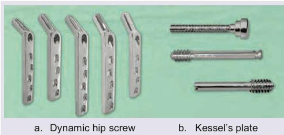
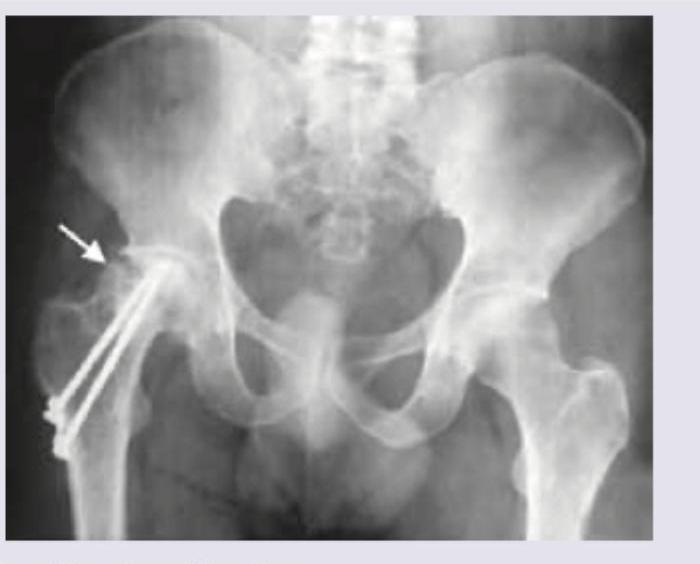
What does the given image show?

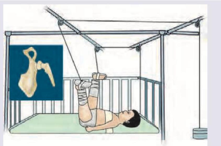
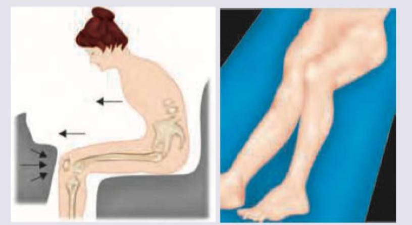
Identify the traction show in the image:

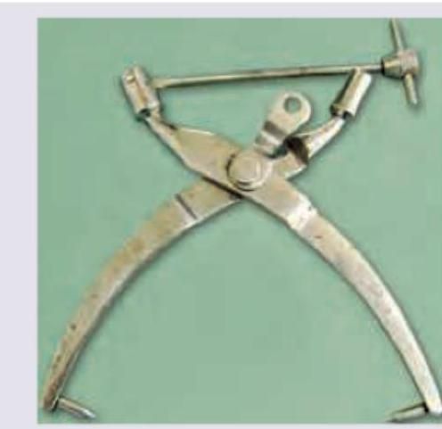
Identify the instrument:

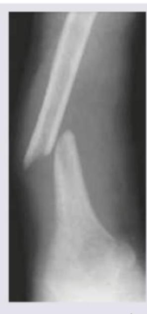
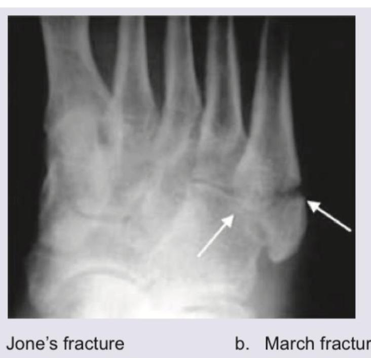
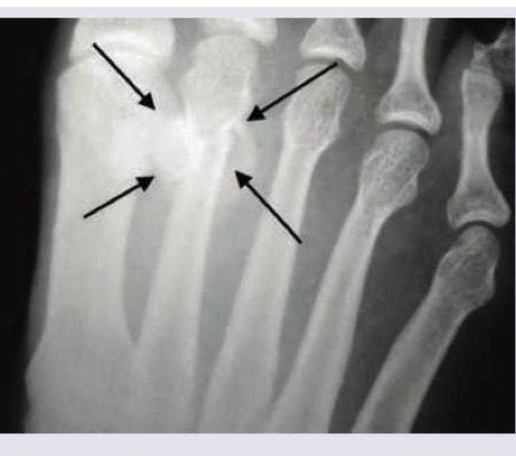
Identify the fracture shown in the image below:

Which of the following classifications is used to assess the fracture shown below?

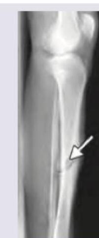
The X-ray was taken 8 weeks after sustaining fracture. What is the diagnosis?

Which of the following is most commonly seen after the accident shown?

Comment on the diagnosis:

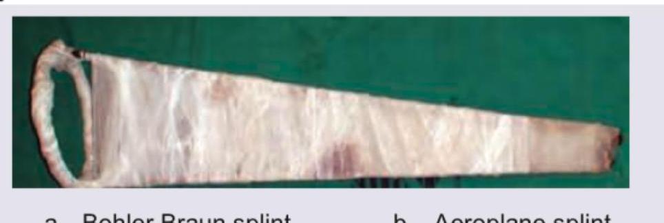
What is the name of the splint shown here?

Comment on the diagnosis:

Explanation: ***Kessel's plate*** - The image clearly labels the items on the right side as "b. Kessel's plate," which is a type of **osteosynthesis plate** used for fixing bone fractures. - Kessel's plates are designed to provide **stable fixation** for specific types of fractures, often in the distal femur or tibia. *Dynamic hip screw* - The image labels the items on the left side as "a. Dynamic hip screw," which is a different orthopedic implant than the one in question. - A dynamic hip screw (DHS) is primarily used for the fixation of **intertrochanteric hip fractures** and has a distinct design with a sliding screw. *Amp rasp* - An Amp rasp is a surgical tool used in **hip arthroplasty** to prepare the femoral canal for the stem of a hip prosthesis. - The items shown in the image are **implants** for fixation, not surgical rasps. *Bipolar hip prosthesis* - A bipolar hip prosthesis is a type of **hip replacement implant** used in hemiarthroplasty, consisting of two articulating components. - The image displays **fixation plates and screws**, not a joint replacement prosthesis.
Explanation: ***$90-90$ traction*** - In this traction, hips and knees are bent at **$90$-degree angles**, and the lower leg is suspended with a traction device, often used for **femur fractures** in young children. - This position helps in aligning bone fragments and providing stable fixation. *Gallow's traction* - **Gallow's traction** involves suspending both legs vertically, with the hips flexed to **$90$ degrees**, and the child's buttocks slightly off the bed. - It is typically used for **femoral shaft fractures** in infants weighing less than **$12$ kg**. *Russell traction* - **Russell traction** involves a sling under the knee and weights applied in longitudinal and vertical directions. - This setup creates a **horizontal force** to align **femoral shaft fractures**. *Smith's traction* - **Smith's traction** is a type of skeletal traction for femoral shaft fractures, usually involving a **pin inserted into the distal femur** or proximal tibia. - This method is characterized by a direct pull on the bone for more effective reduction and stabilization.
Explanation: ***Crutchfield tongs*** - These tongs are used for **skeletal traction** in cases of **cervical spine injuries**, particularly cervical dislocations. - They are specifically designed for insertion into the temporal bones of the skull to apply continuous traction, as seen with the pointed ends and adjustable bar. *Boyle-Davis gag* - A Boyle-Davis gag is a type of **mouth gag** used to hold the mouth open during oral and pharyngeal surgeries, and it does not resemble the instrument shown. - It usually features a tongue depressor and often a method for suction, which are absent here. *Decapitator* - A decapitator is a historical surgical instrument used for **fetal decapitation** during difficult childbirth, and it has a very different morphology, usually involving a sharp cutting or crushing mechanism. - Its design is entirely distinct from the traction device pictured. *Jewett nail* - A Jewett nail is an **intramedullary nail** used in orthopedic surgery to fix **intertrochanteric hip fractures**. - It is an internal fixation device, not an external traction tong, and bears no resemblance to the instrument shown.
Explanation: ***Fracture tibia*** - The image displays a **long bone fracture** in the lower leg, characterized by a visible fracture line (indicated by the arrow) through the shaft. - The upper part of the image clearly shows the **knee joint**, confirming this as a view of the tibia (shin bone). *Fracture radius* - The radius is a bone in the **forearm**, not the lower leg. - The image clearly depicts the **lower extremity** with a knee joint visible. *Colle's fracture* - A Colles' fracture is a specific type of fracture of the **distal radius** (near the wrist), which is in the forearm. - The fracture shown is in the **tibia**, a bone of the lower leg, and the location is not characteristic of a Colles' fracture. *Supracondylar fracture humerus* - The humerus is the **upper arm bone**, and a supracondylar fracture occurs near the elbow joint. - The image shows a fracture in the **lower leg**, specifically the tibia, not the upper arm.
Explanation: ***Pipkins classification*** - The image shows a **femoral head fracture**, indicated by the arrow, with internal fixation. The Pipkins classification system specifically categorizes fractures of the **femoral head**. - This system helps guide treatment and prognostication for these particular hip injuries, differentiating based on location and associated injuries. *Gustilo classification* - The Gustilo classification is used for **open fractures**, assessing the extent of soft tissue damage and contamination. - The image displays a post-operative X-ray of a surgically fixed fracture, and the classification is not applicable here as it pertains to the initial presentation of an open fracture. *Salter and Harris classification* - The Salter-Harris classification system is used for **growth plate (physeal) fractures** in children. - The image shows a fracture in an adult bone (as evidenced by the fused growth plates and the morphology of the bone), making this classification irrelevant. *Weber classification* - The Weber classification is used for **ankle fractures**, specifically those involving the distal fibula. - The fracture shown in the image is of the **femoral head**, not the ankle, hence the Weber classification is not appropriate.
Explanation: ***Nonunion*** - The X-ray image shows a clear **gap** between the two bone fragments, and the bone ends appear **sclerotic** and rounded, indicating that the bone has failed to heal after 8 weeks. - Nonunion is the failure for a fracture to heal after a reasonable period, often defined by the absence of signs of healing for 3-6 months depending on the bone and site, but after 8 weeks with clearly visible sclerotic fragment ends suggests this condition. *Malunion* - **Malunion** occurs when a fracture heals, but in a **deformed** or anatomically incorrect position, which is not evident in the provided image as the fragments are still separate. - The primary problem here is the *absence* of healing, not the healing in a misaligned fashion. *Myositis ossificans* - **Myositis ossificans** is the formation of **heterotopic bone** within muscle or soft tissue, typically following trauma, and would appear as calcification *outside* the bone shaft. - The image clearly depicts unhealed bone fragments, not new bone formation within surrounding soft tissues. *Avascular necrosis* - **Avascular necrosis** (AVN) is the death of bone tissue due to interruption of blood supply, often leading to **collapse of the bone**, which is usually seen in particular bones like the femoral head or scaphoid. - While AVN can complicate fractures, the image primarily shows a lack of bony bridging rather than changes indicative of bone death and collapse.
Explanation: ***Posterior hip dislocation*** - The image depicts a classic mechanism of injury in a dashboard injury, where the **knee strikes the dashboard**, driving the femur posteriorly. - This force, especially when the hip is flexed and adducted, can eject the femoral head **posteriorly out of the acetabulum**. *Anterior hip dislocation* - This type of dislocation typically occurs with the hip in **flexion, abduction, and external rotation**, a mechanism not suggested by the diagram. - It is much **less common** than posterior hip dislocation in dashboard injuries. *Posterior tibial dislocation* - While a dashboard injury can affect the knee, a direct posterior force on the tibia itself is more likely to cause a **tibial plateau fracture** or **ligamentous injury** (e.g., PCL tear) than a complete tibiofemoral dislocation in this context. - A posterior tibial dislocation results from severe hyperextension or a direct blow to the anterior tibia when the knee is flexed, which is a different vector of force than the one illustrated. *Anterior tibial dislocation* - This injury typically results from a **hyperextension injury** of the knee or a force applied to the posterior aspect of the proximal tibia. - The image shows a force specifically directed towards the *anterior* aspect of the knee (dashboard impact), making posterior displacement of the femur relative to the pelvis more likely.
Explanation: ***Jones fracture*** - The image indicates a fracture located at the **proximal metaphyseal-diaphyseal junction of the fifth metatarsal**, which is characteristic of a **Jones fracture**. - This fracture involves the **base of the fifth metatarsal** and is often associated with a higher risk of nonunion due to limited blood supply. *March fracture* - A **March fracture** is a type of stress fracture, typically affecting the **shaft of the second, third, or fourth metatarsals**, often seen in military recruits or those who engage in prolonged walking or running. - It results from repetitive stress rather than an acute injury, and its location is distinct from the proximal fifth metatarsal. *Shepherd's fracture* - A **Shepherd's fracture** refers to an avulsion fracture of the **posterolateral tubercle of the talus**, also known as an os trigonum fracture. - This fracture is located in the ankle region, distinct from the metatarsals. *Cotton's fracture* - A **Cotton's fracture** is a trimalleolar fracture of the ankle, involving the **medial malleolus**, **lateral malleolus**, and the **posterior malleolus** of the tibia. - This is a complex ankle injury, entirely unrelated to fractures of the metatarsals.
Explanation: ***Bohler Braun splint*** - The image displays a **Bohler Braun splint**, characterized by its **trapezoidal shape** and often used for lower extremity fractures. - It features a frame designed to provide **traction and support**, with a U-shaped or ring component at one end and a tapering support surface. *Aeroplane splint* - An aeroplane splint positions the arm in **abduction** (away from the body) and often **external rotation**, resembling an airplane wing. - It is typically used for shoulder and upper arm injuries, which is distinct from the lower limb support seen in the image. *Von Rosen splint* - The Von Rosen splint is a **hip abduction brace** used in infants for congenital hip dislocation. - It positions the hips in **flexion and abduction**, which is entirely different from the long, tapering splint shown. *Thomas splint* - A Thomas splint is a **traction splint** used for femoral shaft fractures, characterized by a large ring that fits around the groin and long sidebars. - While it provides traction like the Bohler Braun splint, its design, particularly the **prominent thigh ring**, is distinctly different from the broad, tapering shape in the image.
Explanation: ***March fracture*** - The image indicates a **stress fracture** of a metatarsal bone, which is characteristic of a **March fracture**. - This type of fracture commonly affects the **2nd or 3rd metatarsal** and is often seen in individuals who engage in strenuous physical activity. *June's fracture* - This is not a recognized eponym for a specific type of fracture in the foot. - The term "June's fracture" does not correspond to any known medical diagnosis related to metatarsal stress fractures. *Shepherd's fracture* - A Shepherd's fracture refers to a **fracture of the posterior process of the talus**. - It is distinct from a metatarsal stress fracture and typically results from forced plantarflexion. *Cotton's fracture* - A Cotton's fracture is a **trimalleolar ankle fracture**, involving the medial, lateral, and posterior malleoli of the ankle. - This is a complex ankle injury, unrelated to a metatarsal stress fracture.
Principles of Fracture Management
Practice Questions
Upper Limb Fractures
Practice Questions
Lower Limb Fractures
Practice Questions
Spinal Trauma
Practice Questions
Pelvic and Acetabular Fractures
Practice Questions
Open Fractures
Practice Questions
Fractures in Children
Practice Questions
Fracture Complications
Practice Questions
Nonunion and Malunion
Practice Questions
Polytrauma Management
Practice Questions
Joint Dislocations
Practice Questions
Soft Tissue Injuries
Practice Questions
Get full access to all questions, explanations, and performance tracking.
Start For Free