A 23 year old male epileptic patient presented with pain in right shoulder region. Examination revealed that right upper limb was abducted and externally rotated and the movements could not be performed. Which of the following is the most likely diagnosis?
The diagnosis in a patient who is unable to perform internal and external rotation of the hip is?
A patient is referred to a higher center with the diagnosis of fracture. On examination, the forearm pulses were not palpable. An upper arm X-ray was done, which is given below. Which artery is most likely to be injured in this condition?
An 80-year-old female patient with a history of slip and fall in the bathroom was brought to the casualty. She is having pain in the left hip and is unable to walk. She has no history of osteoarthritis. On examination, the left lower limb is shortened and externally rotated. X-ray findings are shown below. How will you manage this patient?
A 60-year-old female patient complains of pain and swelling in the left wrist following a fall on an outstretched hand. On examination, dinner fork deformity can be noticed. What is the most likely displacement seen in this patient?
A 65-year-old osteoporotic female falls on her outstretched hand and presents with severe wrist pain and deformity. X-ray shows a distal radius fracture with dorsal angulation of 25°, radial shortening of 8mm, and intra-articular extension with 3mm step-off at the radiocarpal joint. Ulnar styloid is also fractured. Which of the following treatment options provides the best functional outcome and lowest complication rate in this patient?
A teenager presents to the emergency department with wrist pain after falling off his skateboard. He has snuff-box tenderness. Which bone is likely fractured?
What is the preferred treatment for an inter-trochanteric fracture in a 72-year-old female?
A 25-year-old man presents to the emergency department following a motorbike accident and is found to have a closed midshaft fracture of the left tibia. Six hours later, he develops severe leg pain that is disproportionate to the injury and worsens with passive dorsiflexion of the foot. The pain is not relieved by analgesics. On examination, dorsalis pedis and posterior tibial pulses are present, but there is no sensation over the first dorsal webspace. What is the most appropriate next step in management?
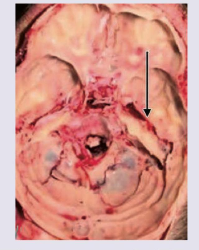
Identify the fracture given in the image given below:

Explanation: ***Subglenoid dislocation of shoulder*** - The presentation of the upper limb held in **abduction** and **external rotation** is the hallmark clinical finding of an **anterior shoulder dislocation**, of which the **subglenoid type** is the most frequent variant. - Subglenoid dislocation accounts for approximately **60-75% of anterior dislocations** and occurs when the humeral head displaces anteriorly and inferiorly to rest below the glenoid fossa. - Although the patient has a history of **epilepsy** (a common cause of posterior dislocation during seizures), the current physical examination findings definitively point to an **anterior presentation**. *Incorrect: Posterior dislocation of shoulder* - **Posterior dislocation** is most commonly associated with events causing unopposed muscle contraction, such as **seizures**, **electric shock**, and **electroconvulsive therapy**. - However, the typical clinical presentation of a posterior dislocation is the arm held in **adduction** and **internal rotation**, directly contradicting the observed **external rotation** in this case. - Posterior dislocations represent only **2-4% of all shoulder dislocations**. *Incorrect: Luxation erecta* - This is an unstable **inferior shoulder dislocation** where the arm is fixed in a position of **extreme abduction** (pointing straight overhead, typically >110-160 degrees). - The humeral head is displaced inferiorly with the humeral shaft positioned vertically. - While it involves abduction, the specific combination of **abduction and external rotation** without explicit maximal elevation fits better with the common anterior (subglenoid) dislocation. *Incorrect: Intrathoracic dislocation of shoulder* - This is an **extremely rare** and severe type of shoulder dislocation resulting from massive trauma, where the humeral head penetrates the chest cavity. - It is not typically associated with muscle contractions from seizures and presents with **dramatic symptoms** including respiratory compromise and hemodynamic instability. - This diagnosis would require high-energy trauma and is inconsistent with the clinical presentation.
Explanation: ***Dislocation of hip***- Inability to perform **internal and external rotation** (both active and passive) is a hallmark sign of a **dislocated joint**, indicating mechanical blockage due to the displacement of the **femoral head** from the acetabulum.- Hip dislocations (especially posterior) present with severe pain and a **fixed deformity** (usually internal rotation, adduction, and mild flexion), which mechanically prevents any rotary movement.*Femur head fracture*- While a **femur head fracture** causes severe pain and guarded movement, it typically allows some rotation, provided the displacement is not severe enough to cause mechanical locking within the joint.- Differentiating features usually include shortening and **external rotation** of the limb (in displaced fractures), but complete mechanical block of all rotation is less specific than in frank dislocation.*Acetabular fractures*- These fractures cause gross instability and pain, but motion may still be present unless the fracture fragments are severely displaced or impinging on the **femoral head**.- The primary focus of a symptomatic acetabular fracture is significant pain, often exacerbated by axial loading, rather than a total mechanical block of rotation.*Pelvis fracture*- **Pelvis fractures** (especially stable types) cause severe pain and limit weight-bearing, but the hip joint often retains some range of motion, particularly passive rotation, if the acetabulum is intact.- Unstable pelvic ring injuries (e.g., Malgaigne fracture) are defined by instability of the bony ring and potential for hemorrhage, not by complete mechanical inability to rotate the hip joint itself.
Explanation: ***Brachial artery*** - The **brachial artery** runs in the anterior compartment of the arm, in close proximity to the humeral shaft. A mid-shaft humeral fracture, as shown in the X-ray, can directly injure or compress this vessel. - The clinical finding of impalpable **radial** and **ulnar pulses** strongly suggests a vascular injury proximal to the elbow, pointing directly to the brachial artery, which is the main arterial supply to the forearm. *Radial artery* - The **radial artery** is a terminal branch of the brachial artery located in the forearm. The fracture is in the humerus (upper arm), making a direct injury to the radial artery unlikely. - An absent radial pulse in this context is a *consequence* of the proximal brachial artery injury, not the primary site of damage. *Ulnar artery* - The **ulnar artery**, like the radial artery, is a major artery of the forearm that arises from the bifurcation of the brachial artery in the cubital fossa. It is not located near the humeral shaft fracture. - Injury to the ulnar artery alone would typically spare the radial pulse; the absence of both pulses points to a more proximal vascular compromise. *Anterior interosseous artery* - The **anterior interosseous artery** is a deep branch of the ulnar artery in the forearm. It is anatomically well-protected and distant from the site of the humeral fracture. - This artery is most commonly injured in association with complex forearm fractures, not humeral shaft fractures.
Explanation: ***Hemiarthroplasty*** - This is the treatment of choice for a **displaced intracapsular femoral neck fracture** in an elderly patient (typically >75 years) due to the high risk of **avascular necrosis (AVN)** and **non-union** if treated with internal fixation. - It involves replacing the femoral head with a prosthesis, which allows for **early mobilization** and weight-bearing, significantly reducing the risk of complications associated with immobility in geriatric patients, such as DVT and pneumonia. *Internal fixation with cancellous screws* - This approach is reserved for **undisplaced femoral neck fractures** or for displaced fractures in younger, more physiologically fit patients (<65 years) where preserving the native femoral head is a priority. - In an 80-year-old with a displaced fracture and likely poor bone quality, the risk of fixation failure, **non-union**, or subsequent **AVN** is unacceptably high, often requiring a second surgery. *Meyer's operation* - This is a **muscle-pedicle bone graft** procedure, typically using the quadratus femoris, designed to improve the blood supply to the femoral head. - It is not a primary treatment for an acute fracture but is sometimes used as an adjunct to internal fixation in younger patients to prevent AVN, or as a treatment for early-stage AVN itself. *McMurray's osteotomy* - This is a type of **intertrochanteric osteotomy** historically used to treat **non-union** of femoral neck fractures by converting shear forces at the fracture site into compressive forces. - It is not indicated for the primary management of an acute femoral neck fracture in an elderly patient and has been largely superseded by modern arthroplasty techniques.
Explanation: ***Distal and dorsal displacement of radius*** This patient presents with a **Colles' fracture**, the most common distal radius fracture in elderly patients following a fall on an outstretched hand (FOOSH injury). **Key Features of Colles' Fracture:** - Fracture of the **distal radius** (within 2.5 cm of radiocarpal joint) - **Dorsal (posterior) displacement** of the distal fragment - **Dorsal angulation** of the distal fragment - **Radial displacement and shortening** - Creates the characteristic **"dinner fork deformity"** when viewed from the side - The intact proximal radius fragment remains in normal position **Why the dinner fork deformity occurs:** The dorsal displacement and angulation of the distal radius fragment causes the wrist to have a bayonet-like appearance on lateral view, resembling the curve of a dinner fork. *Incorrect - Proximal displacement options:* In Colles' fracture, it is the **distal fragment** that displaces, not the proximal fragment. The proximal radius remains attached to the elbow and stays in its normal anatomical position. *Incorrect - Ventral (volar/palmar) displacement:* Volar displacement would be seen in **Smith's fracture** (reverse Colles'), which is much less common and occurs from a fall on the back of the hand. This would produce a "garden spade deformity," not a dinner fork deformity. *Incorrect - Ulnar displacement:* The ulna is not the primary bone involved in dinner fork deformity. While ulnar styloid fractures may occur concurrently in 50-60% of cases, the characteristic deformity results from distal radius displacement.
Explanation: ***Open Reduction and Internal Fixation (ORIF)*** - The fracture characteristics (dorsal angulation >20°, radial shortening >5mm, significant intra-articular step-off >2mm) classify this as an **unstable and complex fracture**, typically managed surgically in active, osteoporotic patients. - **Volar locking plate fixation** (a form of ORIF) provides rigid fixation, allowing for early mobilization, superior maintenance of reduction, and the best functional outcome for intra-articular, unstable distal radius fractures. *Closed Reduction and Cast Immobilization* - This non-operative method is suitable only for **stable, extra-articular fractures** with minimal displacement or for low-demand patients. - The current fracture's instability, intra-articular extension, and significant comminution/displacement make successful closed reduction and maintenance of alignment highly unlikely, leading to a high risk of **secondary displacement** and poor outcome. *External Fixation alone* - Primarily used for highly comminuted, severe open fractures, or when soft tissue injury precludes internal fixation (pilon or highly comminuted fractures). - Offers limited ability to restore the articular congruity and often requires supplementary **K-wire fixation** to control the fragments, which may not be as stable as a plate. *Percutaneous Pinning (K-wires) Following Closed Reduction* - Suitable for **extra-articular or simple intra-articular fractures** in which reduction is easily maintained and stability is good. - In this case, the significant dorsal angulation (25°), radial shortening (8mm), and the **3mm intra-articular step-off** require direct visualization (ORIF) and strong internal fixation (plate) to restore the articular surface and provide adequate stability.
Explanation: ***Scaphoid*** - **Snuff-box tenderness** is the classic and most reliable clinical sign indicating a scaphoid fracture, usually sustained after a **fall onto an outstretched hand (FOOSH)**, common in young adults and teenagers. - The **scaphoid** is the most frequently fractured carpal bone (approx. 60%) and requires careful immobilization and follow-up due to the high risk of **avascular necrosis** from its retrograde blood supply. ***Trapezoid*** - Fractures of the trapezoid are extremely rare (less than 1% of carpal fractures) because of its stable position wedged between the **second metacarpal** and other carpals. - Tenderness for an isolated trapezoid fracture would be localized to the dorsal aspect of the wrist, more centrally, not specifically within the anatomical snuff box which is bounded by the **extensor tendons** of the thumb. ***Capitate*** - The capitate is the largest carpal bone, located in the center of the wrist, articulating with the **third metacarpal**; it is the second most commonly fractured carpal bone after the scaphoid. - Tenderness would be localized more centrally over the dorsal wrist, proximal to the third metacarpal base, and is not associated with classic **snuff-box tenderness**. ***Trapezium*** - The trapezium carpal bone articulates primarily with the **first metacarpal (thumb)**, forming the highly mobile carpometacarpal joint. - A fracture typically results in tenderness localized to the base of the thumb or the **thenar eminence**, rather than the radial dorsal wrist corresponding to the anatomical snuff box.
Explanation: ***Hemiarthroplasty / Dynamic Hip Screw (DHS)*** - **DHS** is the standard treatment for stable (Type I and II) inter-trochanteric fractures, providing controlled collapse and compression at the fracture site. - For unstable fractures (Type III and IV), especially in elderly patients with poor bone quality, **Intramedullary (IM) nailing** is often preferred over DHS due to superior biomechanical stability, though the combination option provided suggests the widely applicable stabilization principles for this age group. ***Intramedullary nailing*** - **IM nailing** is generally the preferred choice for unstable inter-trochanteric fractures (e.g., reverse oblique pattern or severe comminution) as it resists varus collapse more effectively than DHS. - While highly effective, it is not the *only* preferred treatment, and DHS remains primary for stable patterns, making the combined option more comprehensive for standard fracture care in the elderly. ***Open Reduction and Internal Fixation (ORIF) with plating*** - ORIF with plate fixation (other than DHS) is rarely used for inter-trochanteric fractures today, as it involves extensive soft tissue stripping and offers inferior biomechanical stability compared to compression screws (DHS) or nails. - This technique is typically reserved for highly unusual fracture patterns or as a salvage procedure, not as the primary 'preferred' method. ***Boot and bar*** - **Traction (boot and bar)** is historical and obsolete for treating hip fractures, including inter-trochanteric fractures. - Modern management mandates operative fixation as soon as the patient is medically optimized to allow early mobilization, reduce pain, and prevent complications like **deep vein thrombosis (DVT)** and pneumonia.
Explanation: ***Immediate fasciotomy*** - The clinical presentation of severe, disproportionate pain, pain on passive stretching, and altered sensation (**paresthesia**) in the setting of a major fracture is highly indicative of **acute compartment syndrome**. - **Fasciotomy** is the definitive, urgent treatment to decompress the muscle compartments and prevent irreversible tissue necrosis and nerve damage. *Elevate the limb and observe* - Elevating the limb may further decrease the **arterial-venous pressure gradient**, potentially worsening an already compromised perfusion. - Observation is inappropriate because **acute compartment syndrome** requires immediate surgical intervention to prevent serious, permanent deficits or loss of function. *Administer opioid analgesics and continue observation* - Analgesics, even strong opioids, will only mask the classic symptom (**disproportionate pain**) but will not address the underlying pathology (increased intracompartmental pressure). - Continued observation will lead to progression of **ischemia**, resulting in irreversible muscle and nerve damage after approximately 6-8 hours. *Apply cast and follow up* - Applying a cast in the setting of suspected **compartment syndrome** is contraindicated, as the rigid cast can contribute to or exacerbate the compartment pressure. - Following up later is dangerous and negligent, as this condition is a surgical emergency requiring intervention within the **golden hour(s)** to salvage limb function.
Explanation: ***Gutter*** - A **gutter fracture** involves a linear fracture with depression of the adjacent bone fragment, often forming a "gutter" or trough-like deformity, as indicated by the arrow in the image. - This type of fracture is typically caused by a **blunt impact** to the skull that causes focal indentation. *Pond* - A **pond fracture** is a type of depressed skull fracture seen in infants, characterized by a smooth, bowl-like depression without sharp edges or fragmentation, resembling an indentation from a thumb. - It results from **low velocity impact** and the skull's plasticity in infants, which is not what is seen here. *Hinge* - A **hinge fracture** is a type of basilar skull fracture that involves the skull base, often extending bilaterally through structures like the sphenoid bone, creating a "hinge" effect. - This fracture pattern usually results from **severe trauma** and is not depicted in this image, which shows a localized depression. *Comminuted* - A **comminuted fracture** is characterized by the bone breaking into three or more fragments at the site of injury. - While there is bone fragmentation in the image, the primary descriptive feature highlighted by the arrow is the **depressed trough** rather than multiple distinct pieces.
Principles of Fracture Management
Practice Questions
Upper Limb Fractures
Practice Questions
Lower Limb Fractures
Practice Questions
Spinal Trauma
Practice Questions
Pelvic and Acetabular Fractures
Practice Questions
Open Fractures
Practice Questions
Fractures in Children
Practice Questions
Fracture Complications
Practice Questions
Nonunion and Malunion
Practice Questions
Polytrauma Management
Practice Questions
Joint Dislocations
Practice Questions
Soft Tissue Injuries
Practice Questions
Get full access to all questions, explanations, and performance tracking.
Start For Free