
Trauma — MCQs

On this page
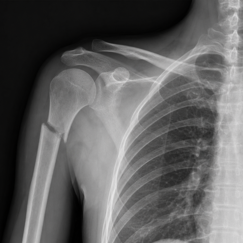
X-ray below shows which of the following fractures?
A 40-year-old male with a history of road traffic accident (RTA) and multiple long bone fractures develops tachypnea, periumbilical rashes, and has urinary fat globules. What is the most likely diagnosis?
Uncomplicated shoulder dislocation most commonly occurs in which direction?
A 56-year-old woman visits the emergency department after falling on wet pavement. Radiographic examination reveals osteoporosis and a Colles' fracture. Which of the following carpal bones are often fractured or dislocated with a Colles' fracture?
A child develops severe forearm pain after a supracondylar fracture reduction and POP cast application. The child describes the pain as worse than the fracture pain and has limited finger movement. Volkmann's ischemic contracture is suspected. Which muscle is most commonly affected in Volkmann's ischemia?
A 30-year-old biker sustained a compound fracture of the leg following a road traffic accident. The fracture has been classified as Gustilo-Anderson type IIIB. Which of the following best represents this injury classification?
Fracture of the neck of the humerus is common in:
In fracture shaft of femur, which nail is commonly used for ORIF?
Open book and bucket handle injuries are seen in which anatomical region?
The Judet and Lectournal classification is used for which type of fracture?
Practice by Chapter
Principles of Fracture Management
Practice Questions
Upper Limb Fractures
Practice Questions
Lower Limb Fractures
Practice Questions
Spinal Trauma
Practice Questions
Pelvic and Acetabular Fractures
Practice Questions
Open Fractures
Practice Questions
Fractures in Children
Practice Questions
Fracture Complications
Practice Questions
Nonunion and Malunion
Practice Questions
Polytrauma Management
Practice Questions
Joint Dislocations
Practice Questions
Soft Tissue Injuries
Practice Questions
Want unlimited practice?
Get full access to all questions, explanations, and performance tracking.
Scan to download app