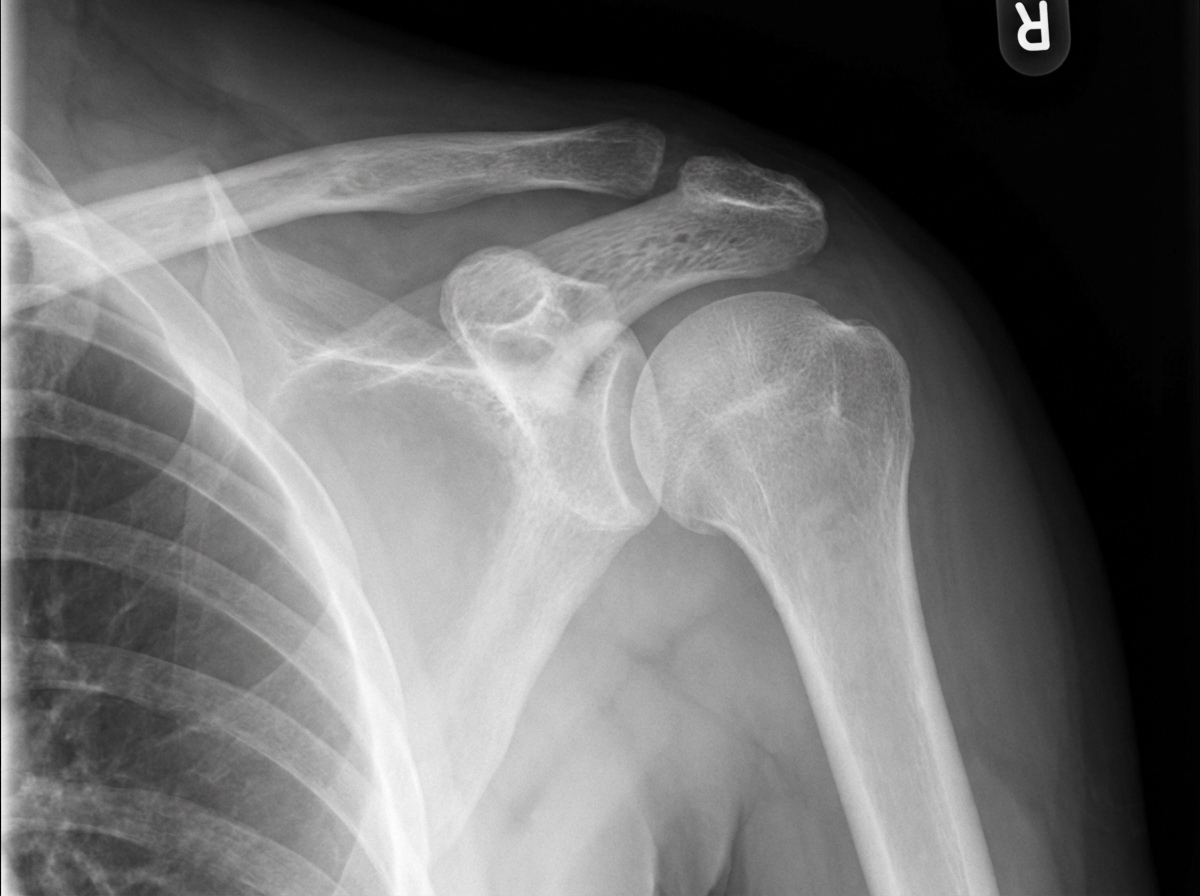
Which of the following statements regarding this diagnosis is true?

A 37-year-old female patient has a fracture of the clavicle. The junction of the inner and middle third of the bone exhibits overriding of the medial and lateral fragments. The arm is rotated medially, but it is not rotated laterally. Which of the following complications is most likely to occur secondary to the fractured clavicle?
Which of the following is NOT a recognized feature of myositis ossificans?
Which of the following statements regarding supracondylar fracture of the humerus in children is true?
What is a fracture of the 5th metatarsal called?
A student presents with a shoulder injury sustained while playing football. On comparing the symmetry of his two shoulders, a marked elevation of the distal end of the clavicle with respect to the acromion is noted on the injured side. X-ray examination reveals a grade III shoulder separation. Which ligament must be torn for this injury to have occurred?
What is the most common dangerous complication of posterior dislocation of the knee?
Fracture of the neck of the fifth metacarpal bone occurs in which type of fracture?
What is Jones fracture?
A 21-year-old male with a fracture of the surgical neck of the humerus presents with a regimental badge sign and difficulty in abduction. What is the most likely nerve injury?
Explanation: ***Recurrence is a common complication*** - **Anterior shoulder dislocation** has a high recurrence rate, especially in **young adults under 30 years**, where recurrence can exceed 80-90%. - **Recurrent dislocations** occur due to damage to the **glenoid labrum** (Bankart lesion) and capsular structures during the initial injury. *Common in children and rare in adults* - Anterior shoulder dislocation is **most common in adults**, particularly in the **20-40 year age group**. - In children, shoulder dislocations are relatively **rare** due to the greater elasticity of their joint capsules and ligaments. *May be overlooked as clinically occult* - Anterior shoulder dislocation presents with **obvious clinical signs** including loss of normal shoulder contour and severe pain. - The **squared-off shoulder** appearance and inability to move the arm make this injury readily apparent, not occult. *Is not associated with other bony or cartilaginous injury* - Anterior shoulder dislocation is commonly associated with **Hill-Sachs lesion** (compression fracture of the humeral head). - Also frequently involves **Bankart lesion** (tear of the anterior glenoid labrum) and potential **greater tuberosity fractures**.
Explanation: **Explanation:** The clavicle is the most commonly fractured bone in the body, with the majority of fractures occurring at the junction of the **medial two-thirds and lateral one-third** (the weakest point). **1. Why Option B is Correct:** The clavicle lies in close anatomical proximity to the **subclavian vein**, which passes directly behind the middle third of the bone, separated only by the subclavius muscle. In displaced fractures, particularly with overriding fragments, the sharp edges of the bone can cause direct trauma to the vessel wall or lead to compression. This stasis and endothelial injury (Virchow’s Triad) can result in **subclavian vein thrombosis**. If the thrombus dislodges, it travels through the right heart into the pulmonary circulation, causing a **pulmonary embolism**. **2. Why the Other Options are Incorrect:** * **Option A:** The **brachiocephalic vein** is located deeper in the mediastinum, posterior to the sternoclavicular joint. While a posterior dislocation of the sternoclavicular joint might threaten it, a mid-shaft clavicle fracture is unlikely to reach this vessel. * **Option C:** While the subclavian artery is also posterior to the clavicle, an arterial thrombus would travel **distally** toward the arm (causing digital ischemia), not retrogradely into the ascending aorta. * **Option D:** The **lower trunk** (C8-T1) of the brachial plexus is more vulnerable to clavicular trauma than the upper trunk, as it lies directly behind the middle third of the bone. **Clinical Pearls for NEET-PG:** * **Deformity:** In mid-shaft fractures, the **medial fragment** is pulled **upward** by the Sternocleidomastoid, while the **lateral fragment** drops **downward** due to the weight of the arm. * **Most common site:** Junction of middle and lateral thirds. * **Management:** Most are treated conservatively with a **Figure-of-8 bandage** or a triangular sling. Surgery is indicated for skin tenting, neurovascular injury, or non-union.
Explanation: **Explanation:** **Myositis Ossificans (Traumatic Myositis Ossificans)** is a condition characterized by heterotopic ossification within a muscle following trauma. It is essentially a metaplastic process where mesenchymal cells differentiate into bone-forming cells. **Why Option C is the correct answer (The False Statement):** Contrary to the statement, myositis ossificans is **more common in children and young adults**. This is due to their more active periosteum and higher osteogenic potential. In children, even minor repetitive trauma or a single significant injury (like a supracondylar fracture) can trigger exuberant bone formation. **Analysis of Incorrect Options (True Statements):** * **Option A:** It is indeed a **post-traumatic ossification**. It typically occurs following a hematoma within the muscle (commonly the brachialis or quadriceps) that undergoes calcification and subsequent ossification. * **Option B:** The **elbow joint** is the most common site, specifically following posterior dislocations or supracondylar fractures. Forceful passive stretching or vigorous massage of a stiff elbow post-injury significantly increases the risk. * **Option D:** **Skiagraphy (X-ray)** is the definitive diagnostic tool. It reveals a characteristic "zonal phenomenon" where the lesion has a mature peripheral radiopaque rim of bone and a radiolucent center, distinguishing it from osteosarcoma. **NEET-PG High-Yield Pearls:** * **Most common muscle involved:** Brachialis (elbow) and Quadriceps femoris (thigh). * **Clinical Warning:** Never massage a recently injured joint (especially the elbow), as it promotes the development of myositis ossificans. * **Management:** Initial treatment is **rest and immobilization**. Surgery is only indicated after the bone has "matured" (usually 6–12 months), evidenced by well-defined margins on X-ray and a negative bone scan. Early surgery leads to high recurrence rates.
Explanation: **Explanation:** Supracondylar fracture of the humerus is the most common elbow fracture in children (peak age 5–8 years). It is considered an **orthopaedic emergency** due to its proximity to vital neurovascular structures. **1. Why Option A is correct:** Admission following reduction is mandatory for **monitoring neurovascular status**. The primary concern is the development of **Volkmann’s Ischemia**, which can lead to Volkmann’s Ischemic Contracture (VIC). Post-reduction swelling or tight casting can compromise the brachial artery or cause Compartment Syndrome. Hourly observation for the "5 Ps" (Pain, Pallor, Pulselessness, Paresthesia, Paralysis) is essential for at least 24 hours. **2. Why the other options are incorrect:** * **Option B:** It is typically caused by a **fall on an outstretched hand (FOOSH)** with the elbow in extension (Extension type, >95%). A fall on the point of the elbow usually results in the rarer flexion-type fracture. * **Option C:** Most cases are managed by **Closed Reduction and Internal Fixation (CRIF)** using percutaneous K-wires. Open reduction is reserved only for failed closed attempts, vascular compromise, or compound injuries. * **Option D:** These are usually **closed fractures**. While the sharp proximal fragment can pierce the brachialis muscle and skin (puckering sign), frank compound injuries are not the norm. **High-Yield Clinical Pearls for NEET-PG:** * **Most common nerve injured:** Anterior Interosseous Nerve (AIN) – branch of the Median nerve (test by asking the child to make an "OK" sign). * **Most common deformity:** Cubitus Varus (Gun-stock deformity) due to malunion (remodeling does not correct rotation). * **Gartland Classification:** Used to grade severity (Type I: Undisplaced; Type II: Displaced with intact posterior cortex; Type III: Completely displaced).
Explanation: **Explanation:** The correct answer is **Jones fracture**. This refers to a transverse fracture at the **base of the 5th metatarsal**, specifically at the metaphyseal-diaphyseal junction (Zone 2). This area is clinically significant because it is a "watershed area" with a precarious blood supply, making these fractures prone to delayed union or non-union. **Analysis of Options:** * **Jones Fracture (Correct):** Occurs about 1.5 to 3 cm distal to the tuberosity. It must be distinguished from a **Pseudo-Jones fracture** (Avulsion fracture of the styloid process/tuberosity), which is more common and heals more easily. * **Malgaigne's Fracture:** A vertical shear injury of the pelvis involving double vertical fractures (fracture of the pubic rami anteriorly and disruption of the SI joint or ilium posteriorly). * **Cotton's Fracture:** A trimalleolar fracture of the ankle involving the medial malleolus, lateral malleolus, and the posterior malleolus (posterior lip of the tibia). * **Pott's Fracture:** A generic term for bimalleolar ankle fractures caused by eversion-external rotation injuries. **NEET-PG High-Yield Pearls:** 1. **Mechanism:** Jones fracture usually occurs due to a forceful adduction of the forefoot with the ankle in plantar flexion. 2. **Classification:** The **Lawrence and Botte classification** divides 5th metatarsal base fractures into three zones: Zone 1 (Pseudo-Jones), Zone 2 (Jones), and Zone 3 (Stress fracture of the proximal shaft). 3. **Management:** Jones fractures often require non-weight-bearing casts for 6–8 weeks or internal fixation (intramedullary screw) in athletes due to the high risk of non-union.
Explanation: **Explanation:** The clinical presentation of a "marked elevation of the distal end of the clavicle" (Step-off deformity) following trauma is characteristic of an **Acromioclavicular (AC) joint dislocation**, commonly referred to as a "shoulder separation." **1. Why Coracoclavicular (CC) is correct:** The stability of the AC joint depends on two sets of ligaments: the **Acromioclavicular ligaments** (horizontal stability) and the **Coracoclavicular ligaments** (vertical stability). The CC ligament complex consists of the **Conoid** and **Trapezoid** ligaments. In a **Grade III injury**, both the AC and CC ligaments are completely ruptured. Because the CC ligaments normally anchor the clavicle down to the coracoid process, their tear allows the clavicle to be pulled superiorly by the trapezius muscle, resulting in the visible elevation noted in the question. **2. Why other options are incorrect:** * **Coracoacromial:** This ligament connects two parts of the same bone (scapula). It forms the coracoacromial arch but does not stabilize the clavicle. * **Costoclavicular:** This ligament anchors the medial (sternal) end of the clavicle to the first rib. It is not involved in distal shoulder separations. * **Superior glenohumeral:** This is a component of the shoulder joint capsule (glenohumeral joint) and provides stability against inferior translation of the humeral head, not the clavicle. **Clinical Pearls for NEET-PG:** * **Rockwood Classification:** Grade I (Sprain), Grade II (AC torn, CC intact), Grade III (Both AC and CC torn; 25-100% displacement). * **Piano Key Sign:** A classic physical exam finding where the elevated distal clavicle can be depressed but springs back up. * **Management:** Grade I-III are typically managed conservatively (sling/rehab), while Grade IV-VI usually require surgical intervention.
Explanation: ### Explanation **1. Why Popliteal Artery Injury is Correct:** Knee dislocation is a surgical emergency. The **popliteal artery** is the most vulnerable structure because it is tethered firmly at two points: the adductor hiatus (superiorly) and the tendinous arch of the soleus muscle (inferiorly). When the tibia displaces posteriorly relative to the femur, the artery is stretched or sheared against the posterior edge of the tibial plateau. This often results in an **intimal tear**, which can lead to delayed thrombosis and limb-threatening ischemia. While anterior dislocations can also cause injury, posterior dislocations carry the highest risk of complete arterial transection. **2. Why the Other Options are Incorrect:** * **Sciatic Nerve Injury:** The sciatic nerve bifurcates into the tibial and common peroneal nerves well above the knee joint. While the **Common Peroneal Nerve** is frequently injured in knee dislocations (especially posterolateral), the main trunk of the sciatic nerve is not typically involved. * **Ischemia of the Lower Leg Compartment:** This is a *consequence* (Compartment Syndrome) of the vascular injury or the trauma itself, rather than the primary anatomical complication. * **Femoral Artery Injury:** The femoral artery becomes the popliteal artery as it passes through the adductor hiatus. The injury occurs distal to this point, specifically within the popliteal fossa. **3. High-Yield Clinical Pearls for NEET-PG:** * **"The Rule of 1/3rd":** Approximately 1/3rd of knee dislocations involve a popliteal artery injury. * **Hard Signs:** If distal pulses are absent, immediate surgical exploration is required. * **ABI (Ankle-Brachial Index):** An ABI < 0.9 is a sensitive indicator for arterial injury; if found, a CT Angiogram is the gold standard for diagnosis. * **Nerve Involvement:** The **Common Peroneal Nerve** is the most common *nerve* injured, but popliteal artery injury is the most *dangerous* complication. * **Spontaneous Reduction:** Many knee dislocations reduce spontaneously before reaching the ER; always maintain a high index of suspicion for vascular injury even if the joint appears stable.
Explanation: **Explanation** **Correct Option: D. Boxer's fracture** A **Boxer’s fracture** is a fracture of the **neck of the fifth metacarpal**. It typically occurs when a person strikes a hard object with a closed fist. The mechanism involves an axial load transmitted through the shaft of the metacarpal, leading to volar (palmar) angulation of the distal fragment. While the name suggests professional boxing, it is more commonly seen in unskilled individuals who punch with an improper technique, impacting the object with the ulnar side of the hand rather than the second and third metacarpals. **Incorrect Options:** * **A. Hangman’s Fracture:** This refers to a traumatic spondylolisthesis of the **axis (C2)**, specifically a bilateral fracture through the pars interarticularis, usually caused by hyperextension of the neck. * **B. Jefferson’s Fracture:** This is a burst fracture of the **atlas (C1)**, involving both the anterior and posterior arches. It is typically caused by an axial load to the top of the head (e.g., diving into a shallow pool). * **C. Greenstick Fracture:** This is an **incomplete fracture** seen in children (pediatric population) where one side of the bone breaks while the other side merely bends, due to the higher collagen-to-mineral ratio in young bones. **High-Yield Clinical Pearls for NEET-PG:** * **Acceptable Angulation:** Up to 40° of volar angulation is often acceptable in the 5th metacarpal due to the mobility of the CMC joint; however, any **rotational deformity** (fingers overlapping on flexion) requires surgical correction. * **Splinting:** Managed with an **Ulnar Gutter Splint** in the "intrinsic plus" position. * **Barroom Fracture:** Occasionally, a fracture of the 4th or 5th metacarpal *shaft* is referred to as a Barroom fracture, though "Boxer's" specifically targets the neck.
Explanation: **Explanation:** **Jones Fracture** is a transverse fracture at the base of the **fifth metatarsal**, specifically occurring at the **metaphyseal-diaphyseal junction** (Zone 2). It is often caused by a forceful inversion of the foot. While the question identifies it as an avulsion fracture, it is clinically important to distinguish it from a "Pseudo-Jones" fracture. * **Why Option A is correct:** The base of the fifth metatarsal is a high-yield site for fractures. A true Jones fracture occurs about 1.5–3 cm distal to the tuberosity. It is notorious for **potential non-union** due to the "watershed area" (poor blood supply) at this specific junction. **Analysis of Incorrect Options:** * **Option B:** A bimalleolar fracture (involving medial and lateral malleoli) is known as a **Pott’s fracture**. * **Option C:** A burst fracture of the C1 vertebra (atlas) is known as a **Jefferson fracture**, typically caused by axial loading on the head. * **Option D:** An avulsion fracture of the medial femoral condyle (at the site of the MCL attachment) is known as a **Pellegrini-Stieda lesion** (often seen as calcification on X-ray). **NEET-PG High-Yield Pearls:** 1. **Pseudo-Jones (Dancer’s Fracture):** This is a true avulsion fracture of the **tuberosity** (Zone 1) by the **Peroneus brevis** tendon or plantar fascia. It has a better prognosis than a Jones fracture. 2. **Stress Fracture:** Occurs in Zone 3 (proximal diaphysis), common in athletes. 3. **Management:** Jones fractures often require non-weight-bearing casts or internal fixation (intramedullary screw) due to the high risk of non-union.
Explanation: ### **Explanation** The correct answer is **Axillary nerve injury**. **1. Why Axillary Nerve Injury is Correct:** The axillary nerve (C5, C6) winds around the **surgical neck of the humerus** within the quadrangular space. Fractures at this site or anterior dislocations of the shoulder commonly result in its injury. * **Regimental Badge Sign:** The axillary nerve provides sensory innervation to the skin over the lower half of the deltoid muscle via the upper lateral cutaneous nerve of the arm. Loss of sensation in this specific area is known as the "Regimental Badge Sign." * **Difficulty in Abduction:** The nerve supplies the **deltoid** (the primary abductor of the arm after 15°) and the teres minor. Paralysis of the deltoid leads to significant weakness in abduction. **2. Why Other Options are Incorrect:** * **Ulnar Nerve Injury:** Typically occurs with fractures of the medial epicondyle of the humerus. It presents with "claw hand" and sensory loss in the medial 1.5 fingers, not abduction deficits. * **Klumpke's Paralysis:** This is a lower brachial plexus injury (C8-T1) usually caused by hyper-abduction of the arm. It results in a total claw hand and sensory loss along the ulnar aspect of the forearm and hand. * **Supraclavicular Nerve Injury:** These are cutaneous nerves from the cervical plexus (C3, C4) supplying the skin over the shoulder and clavicle. Injury would not cause motor weakness in abduction. **3. NEET-PG High-Yield Pearls:** * **Most common nerve injured in shoulder dislocation:** Axillary nerve. * **Most common nerve injured in mid-shaft humerus fracture:** Radial nerve (presents with wrist drop). * **Deltoid Atrophy:** Chronic axillary nerve palsy leads to the loss of the rounded contour of the shoulder, making it appear "flat." * **Abduction sequence:** 0–15° (Supraspinatus), 15–90° (Deltoid), >90° (Serratus anterior and Trapezius).
Principles of Fracture Management
Practice Questions
Upper Limb Fractures
Practice Questions
Lower Limb Fractures
Practice Questions
Spinal Trauma
Practice Questions
Pelvic and Acetabular Fractures
Practice Questions
Open Fractures
Practice Questions
Fractures in Children
Practice Questions
Fracture Complications
Practice Questions
Nonunion and Malunion
Practice Questions
Polytrauma Management
Practice Questions
Joint Dislocations
Practice Questions
Soft Tissue Injuries
Practice Questions
Get full access to all questions, explanations, and performance tracking.
Start For Free