Which of the following laboratory findings is NOT typically seen in fat embolism?
Pipkin classification is used for which type of fracture?
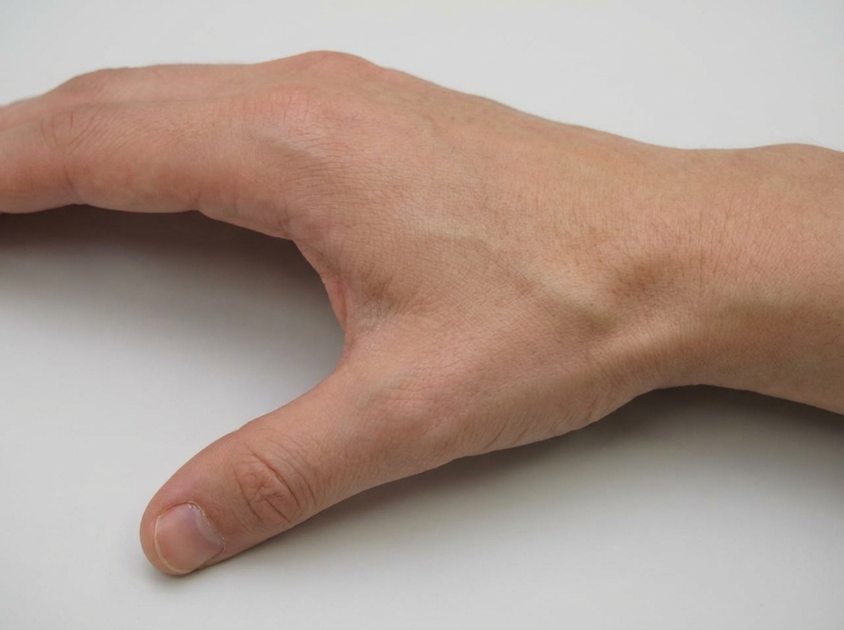
An injury to the shown area can lead to fracture of which bone?

Non-union is a complication of which of the following fractures?
Ruptured tendon is most commonly seen in which of the following conditions?
All of the following statements about dislocation of the shoulder are true, except?
Shoeing of the lower limb with abduction and internal rotation is observed in which of the following types of hip dislocation?
A 45-year-old female has a history of a slip in the bathroom, complaining of right hip pain and tenderness in Scarpa's triangle. The X-ray is normal. What is the next investigation?
Which is the most commonly injured tarsal bone?
The posterolateral lesion in the head of humerus in cases of recurrent anterior shoulder dislocation is:
Explanation: **Explanation:** Fat Embolism Syndrome (FES) typically occurs 24–72 hours after a long bone fracture (e.g., femur). The correct answer is **Hypercalcemia**, as it is not a feature of FES. In fact, **hypocalcemia** is more commonly observed because free fatty acids (released during fat degradation) bind to calcium, causing it to precipitate (saponification). **Analysis of Options:** * **Thrombocytopenia (A):** This is a classic finding. Platelets adhere to circulating fat globules, leading to sequestration and consumption, which often manifests clinically as a petechial rash. * **Fat globules in urine (B):** Lipuria occurs when fat emboli pass through the glomerular filtrate. While specific, it is seen in only about 50% of cases and usually appears within the first 3 days. * **Anemia (D):** Unexplained drop in hemoglobin is common in FES due to alveolar hemorrhage and erythrocyte aggregation/destruction triggered by free fatty acids. **Clinical Pearls for NEET-PG:** * **Gurd’s Criteria:** Used for diagnosis. Major criteria include **Respiratory insufficiency**, **Cerebral involvement** (confusion/coma), and **Petechial rash** (typically over the chest, axilla, and conjunctiva). * **Snowstorm Appearance:** The characteristic finding on a Chest X-ray (diffuse bilateral pulmonary infiltrates). * **Early Fixation:** The most effective way to prevent FES is the early stabilization/fixation of long bone fractures. * **Treatment:** Primarily supportive (Oxygenation/Ventilation). Steroids are controversial and not routinely recommended.
Explanation: **Explanation:** The **Pipkin Classification** is the standard system used to categorize **Femoral Head Fractures**, which typically occur as a result of high-energy trauma (like dashboard injuries) often associated with posterior hip dislocations. **1. Why the Correct Answer is Right:** The classification is based on the location of the fracture line relative to the **fovea capitis** and the presence of associated injuries to the acetabular rim or femoral neck. * **Type I:** Fracture inferior to the fovea capitis (small fragment). * **Type II:** Fracture superior to the fovea capitis (large fragment). * **Type III:** Type I or II fracture associated with a **femoral neck fracture** (high risk of AVN). * **Type IV:** Type I or II fracture associated with an **acetabular rim fracture**. **2. Why Incorrect Options are Wrong:** * **Acetabular Fractures:** These are most commonly classified using the **Judet-Letournel Classification** (based on columns and walls). * **Pelvis Ring Fractures:** These use the **Tile Classification** (based on stability) or the **Young-Burgess Classification** (based on mechanism of injury like APC or LC). * **Femoral Shaft Fractures:** These are typically classified using the **Winquist-Hansen Classification** (based on the degree of comminution). **Clinical Pearls for NEET-PG:** * **Mechanism:** Most femoral head fractures occur during **posterior hip dislocation**. * **Prognosis:** Pipkin Type III has the worst prognosis due to the high risk of **Avascular Necrosis (AVN)**. * **Management:** Type I and II are often treated with ORIF if displaced; Type III and IV usually require surgical intervention and carry a high risk of secondary osteoarthritis.
Explanation: ***Scaphoid*** - The **anatomical snuffbox** is the most vulnerable area for scaphoid fractures, typically occurring after a **fall on outstretched hand (FOOSH)** injury. - **Tenderness in the anatomical snuffbox** and pain with **radial deviation** of the wrist are classic clinical findings of scaphoid fracture. *Lunate* - Lunate fractures are much **rarer** and typically occur with **high-energy trauma** or **perilunate dislocations**. - The lunate is **not directly palpable** in the anatomical snuffbox area, making it less susceptible to injury from this mechanism. *Hamate* - Hamate fractures commonly occur from **direct trauma** to the **hypothenar eminence** or **hook of hamate**. - The hamate is located on the **ulnar side** of the wrist and is not anatomically related to the snuffbox area. *Trapezium* - Trapezium fractures are typically associated with **Bennett's fracture** involving the **first metacarpal base**. - Located at the **base of the thumb**, the trapezium is not directly involved in anatomical snuffbox injuries.
Explanation: ### **Explanation** The correct answer is **A. Scaphoid fracture**. **1. Why Scaphoid Fracture is the Correct Answer:** Non-union is a notorious complication of scaphoid fractures primarily due to its **precarious blood supply**. The scaphoid receives its blood supply in a **retrograde fashion** (from distal to proximal) via the dorsal carpal branch of the radial artery. A fracture across the waist or proximal pole can easily interrupt this supply, leading to ischemia of the proximal fragment. This results in a high incidence of **Avascular Necrosis (AVN)** and subsequent **non-union**. **2. Why the Other Options are Incorrect:** * **B. Colles' Fracture:** This occurs at the distal radius, which has a rich blood supply and cancellous bone. It typically heals well; the most common complication is **Mal-union** (dinner fork deformity), not non-union. * **C. Intertrochanteric Fracture:** This region is extracapsular and consists of highly vascular cancellous bone. These fractures heal readily; the primary complication is **Mal-union** (coxa vara). In contrast, *Neck of Femur* fractures (intracapsular) are prone to non-union. * **D. Supracondylar Fracture (Humerus):** This is common in children. The most frequent complications are **Mal-union** (Cubitus varus/Gunstock deformity) and vascular injury (Volkmann’s Ischemic Contracture). Non-union is extremely rare here. **3. High-Yield Clinical Pearls for NEET-PG:** * **Common sites for Non-union:** Scaphoid (waist), Neck of Femur, Talus (neck), and Lower 1/3rd of Tibia. * **Scaphoid Fact:** The most common site of fracture is the **waist (70%)**. The more proximal the fracture, the higher the risk of AVN and non-union. * **Radiology:** If a scaphoid fracture is clinically suspected (tenderness in the anatomical snuffbox) but X-rays are negative, the wrist should be immobilized and re-imaged after **10–14 days**.
Explanation: ### **Explanation** **Correct Option: C. Overuse** The most common mechanism for a tendon rupture is **chronic overuse** leading to **tendinosis** (degenerative changes). Healthy tendons are remarkably strong and rarely rupture under normal physiological loads. Rupture typically occurs when a tendon has undergone repetitive microtrauma, leading to a breakdown of collagen fibers and decreased vascularity. This weakened state makes the tendon susceptible to a complete tear during a sudden, forceful contraction or eccentric loading. Common clinical examples include: * **Achilles tendon rupture:** Often occurs in "weekend warriors" during sports. * **Supraspinatus rupture:** Usually secondary to chronic impingement and degeneration. * **Long head of Biceps rupture:** Often seen in older patients with pre-existing tendinitis. --- ### **Analysis of Incorrect Options** * **A. Stab injury:** While penetrating trauma can cause a **laceration** of a tendon, it is statistically less common than degenerative ruptures. Stab injuries more frequently involve superficial structures or neurovascular bundles rather than isolated tendon ruptures. * **B. Soft tissue tumor:** Tumors (like Synovial Sarcoma) may infiltrate or compress a tendon, but they are rare causes of spontaneous rupture compared to the high prevalence of overuse injuries. * **D. Congenital defect:** Congenital conditions (like Ehlers-Danlos syndrome) can cause generalized ligamentous laxity or collagen weakness, but they represent a very small fraction of the patient population presenting with tendon ruptures. --- ### **Clinical Pearls for NEET-PG** * **Most common site of tendon rupture:** Achilles tendon (Calcaneal tendon). * **Fluoroquinolones (e.g., Ciprofloxacin):** A high-yield pharmacological association; these drugs increase the risk of Achilles tendon rupture. * **Systemic Steroids/Rheumatoid Arthritis:** These conditions predispose patients to "spontaneous" ruptures due to weakened collagen structures. * **Simmonds/Thompson Test:** Used clinically to diagnose a ruptured Achilles tendon (absence of plantar flexion on squeezing the calf).
Explanation: **Explanation:** **1. Why Option B is the Correct Answer (The False Statement):** While the shoulder is the most commonly dislocated joint in the body, the most frequent anatomical position for the humeral head in an anterior dislocation is **Subcoracoid**, not subglenoid. In anterior dislocations (which account for >95% of cases), the head usually settles beneath the coracoid process. Subglenoid is the second most common position. **2. Analysis of Other Options:** * **Option A:** The classic mechanism of injury is a fall on an outstretched hand with the arm in **abduction, extension, and external rotation**. This forces the humeral head against the weak anterior capsule. * **Option C:** The **Axillary nerve** (C5-C6) is the most commonly injured nerve in shoulder dislocations due to its proximity to the surgical neck of the humerus. Clinically, this presents as "regimental badge" anesthesia over the lateral deltoid. * **Option D:** While many maneuvers exist (e.g., Hippocratic, Kocher’s, Stimson), the safest and easiest way to reduce a dislocation is using gentle traction/pressure under **General Anesthesia (GA)** with muscle relaxation. This overcomes muscle spasms (especially the subscapularis) and minimizes the risk of iatrogenic fractures. **3. NEET-PG High-Yield Pearls:** * **Most common type:** Anterior (Subcoracoid). * **Posterior Dislocation:** Associated with **seizures or electric shocks**. Look for the "Light bulb sign" on X-ray. * **Hill-Sachs Lesion:** Compression fracture of the posterosuperolateral humeral head. * **Bankart Lesion:** Avulsion of the anteroinferior glenoid labrum. * **Luxatio Erecta:** A rare inferior dislocation where the arm is held fixed in an overhead position.
Explanation: **Explanation:** **Central Dislocation of the Hip** occurs when the femoral head is driven medially through a fractured acetabular floor into the pelvis. This is typically a high-energy injury (e.g., a fall from height or a side-impact motor vehicle accident). 1. **Why Central Dislocation is correct:** In central dislocation, the femoral head is displaced medially. This causes the limb to appear **shortened** (due to the inward migration) and classically positioned in **abduction and internal rotation**. The term "shoeing" refers to this characteristic shortening and deformity where the limb is tucked inward toward the midline of the pelvis. 2. **Why the other options are incorrect:** * **Posterior Dislocation (Most Common):** The limb is typically **shortened, adducted, and internally rotated** (the "dashboard injury" position). * **Anterior Dislocation:** The limb is typically **abducted and externally rotated**. It may appear lengthened or shortened depending on whether it is the superior (pubic) or inferior (obturator) type. * **Lateral Dislocation:** This is not a standard anatomical classification for hip dislocations; the femoral head is blocked by the ilium/acetabulum from moving purely laterally without a fracture. **High-Yield Clinical Pearls for NEET-PG:** * **Posterior Dislocation:** Most common type (90%). Associated with Sciatic nerve injury (specifically the peroneal division). * **Anterior Dislocation:** Associated with Femoral nerve/vessel injury. * **Central Dislocation:** Always associated with an **acetabular fracture**. * **Shenton’s Line:** Broken in all types of hip dislocations. * **Avascular Necrosis (AVN):** The most serious late complication of hip dislocation; risk increases if reduction is delayed beyond 6 hours.
Explanation: ### Explanation **1. Why MRI is the Correct Choice:** The clinical presentation (slip in a bathroom, hip pain, and tenderness in Scarpa’s triangle) is highly suspicious of a **fracture of the neck of the femur**. In elderly or osteoporotic patients, these fractures can be **occult** (not visible on initial X-rays). **MRI is the gold standard** and the investigation of choice for occult hip fractures. It has a sensitivity and specificity of nearly 100%. It can detect bone marrow edema within 24 hours of injury, which indicates a fracture even when the cortical lines appear intact on a radiograph. **2. Why Other Options are Incorrect:** * **USG-guided aspiration:** This is primarily used to diagnose septic arthritis or to relieve a tense joint effusion. It has no role in diagnosing an occult fracture. * **CT Scan:** While CT is excellent for evaluating complex fracture patterns and cortical detail, it is less sensitive than MRI for detecting early trabecular microfractures or bone marrow edema. It is usually the second-line choice if MRI is contraindicated. * **Bone Scan (Technetium-99m):** Historically used for occult fractures, but it may take 48–72 hours to become positive in the elderly (delayed osteoblastic activity). It is less specific and has been largely replaced by MRI. **3. Clinical Pearls for NEET-PG:** * **Tenderness in Scarpa’s Triangle:** A classic clinical sign of a hip fracture or hip joint pathology. * **Occult Fracture Definition:** A fracture that is clinically suspected but not visible on initial plain radiographs. * **Management Rule:** If a patient has a high clinical suspicion of a hip fracture but normal X-rays, they **must not** be discharged. They require an MRI to rule out the fracture. * **MRI Contraindication:** If the patient has a pacemaker or metallic implants, a **CT scan** becomes the next best investigation.
Explanation: **Explanation:** The **calcaneus** (heel bone) is the most commonly fractured tarsal bone, accounting for approximately **60% of all tarsal fractures** and about 2% of all fractures in the body. Its vulnerability is primarily due to its weight-bearing function and its position as the first point of contact during axial loading. **Why Calcaneus is correct:** Most calcaneal fractures occur due to high-energy axial loading, such as a **fall from a height** landing on the feet (often associated with "Don Juan Syndrome"). Because it consists of a thin cortical shell surrounding cancellous bone, it is highly susceptible to compression and comminution under sudden force. **Why other options are incorrect:** * **Talus:** It is the second most common tarsal bone to be fractured. It is protected within the ankle mortise and requires significant force to fracture, often leading to avascular necrosis (AVN) due to its unique retrograde blood supply. * **Navicular:** Fractures are relatively rare and usually occur as stress fractures in athletes or due to direct trauma/twisting. * **Cuneiform:** These are rarely injured in isolation because they are tightly bound by strong interosseous ligaments within the midfoot complex. **Clinical Pearls for NEET-PG:** * **Don Juan Syndrome (Lover’s Fracture):** Calcaneal fractures are frequently associated with concurrent **compression fractures of the lumbar spine** (L1-L2) and bilateral calcaneal injuries. Always screen the spine in these patients. * **Bohler’s Angle:** A decrease in this angle (normal = 25°–40°) on a lateral X-ray suggests a calcaneal compression fracture. * **Mondor’s Sign:** Ecchymosis extending to the sole of the foot is a pathognomonic physical finding for calcaneal fractures.
Explanation: ### Explanation **Correct Option: B. Hill-Sachs lesion** **Mechanism and Pathophysiology:** In **recurrent anterior shoulder dislocation**, the humeral head is displaced anteriorly and inferiorly. As it exits the glenoid labrum, the soft **posterolateral** aspect of the humeral head strikes against the hard **anteroinferior** rim of the glenoid. This compression results in a "bookshelf" deformity or an impaction fracture on the humeral head. This specific bony defect is known as the **Hill-Sachs lesion**. It is a hallmark of recurrent instability and increases the risk of future dislocations. **Analysis of Incorrect Options:** * **A. Bankart’s lesion:** This is an injury to the **anteroinferior glenoid labrum** (and sometimes the bony rim). While it also occurs in anterior dislocations, it involves the glenoid, not the humeral head. * **C. Reverse Hill-Sachs lesion:** This is an impaction fracture on the **anterior** aspect of the humeral head, occurring during **posterior** shoulder dislocations (often seen in seizures or electric shocks). * **D. Greater tuberosity avulsion fracture:** This typically occurs in older patients during an initial traumatic dislocation due to the pull of the rotator cuff muscles, but it is not the characteristic "posterolateral lesion" associated with recurrent instability. **High-Yield Clinical Pearls for NEET-PG:** * **Bankart’s Lesion:** Most common cause of shoulder instability; involves the 3 o'clock to 6 o'clock position (right shoulder). * **Imaging:** Hill-Sachs lesions are best visualized on the **Stryker Notch view** (X-ray) or MRI. * **Engaging Hill-Sachs:** A lesion is "engaging" if it drops over the glenoid rim during functional range of motion, necessitating surgical intervention (e.g., **Remplissage procedure**). * **ALPSA Lesion:** Anterior Labral Periosteal Sleeve Avulsion (a variant of Bankart’s).
Principles of Fracture Management
Practice Questions
Upper Limb Fractures
Practice Questions
Lower Limb Fractures
Practice Questions
Spinal Trauma
Practice Questions
Pelvic and Acetabular Fractures
Practice Questions
Open Fractures
Practice Questions
Fractures in Children
Practice Questions
Fracture Complications
Practice Questions
Nonunion and Malunion
Practice Questions
Polytrauma Management
Practice Questions
Joint Dislocations
Practice Questions
Soft Tissue Injuries
Practice Questions
Get full access to all questions, explanations, and performance tracking.
Start For Free