A 32-year-old male with a history of seizures presents with pain in the left shoulder region. The left upper limb is addicted and internally rotated. What is the diagnosis?
Recurrent dislocation of the shoulder is most commonly associated with which type?
All of the following statements regarding mandibular fractures are true EXCEPT:
What is the most common type of hip dislocation?
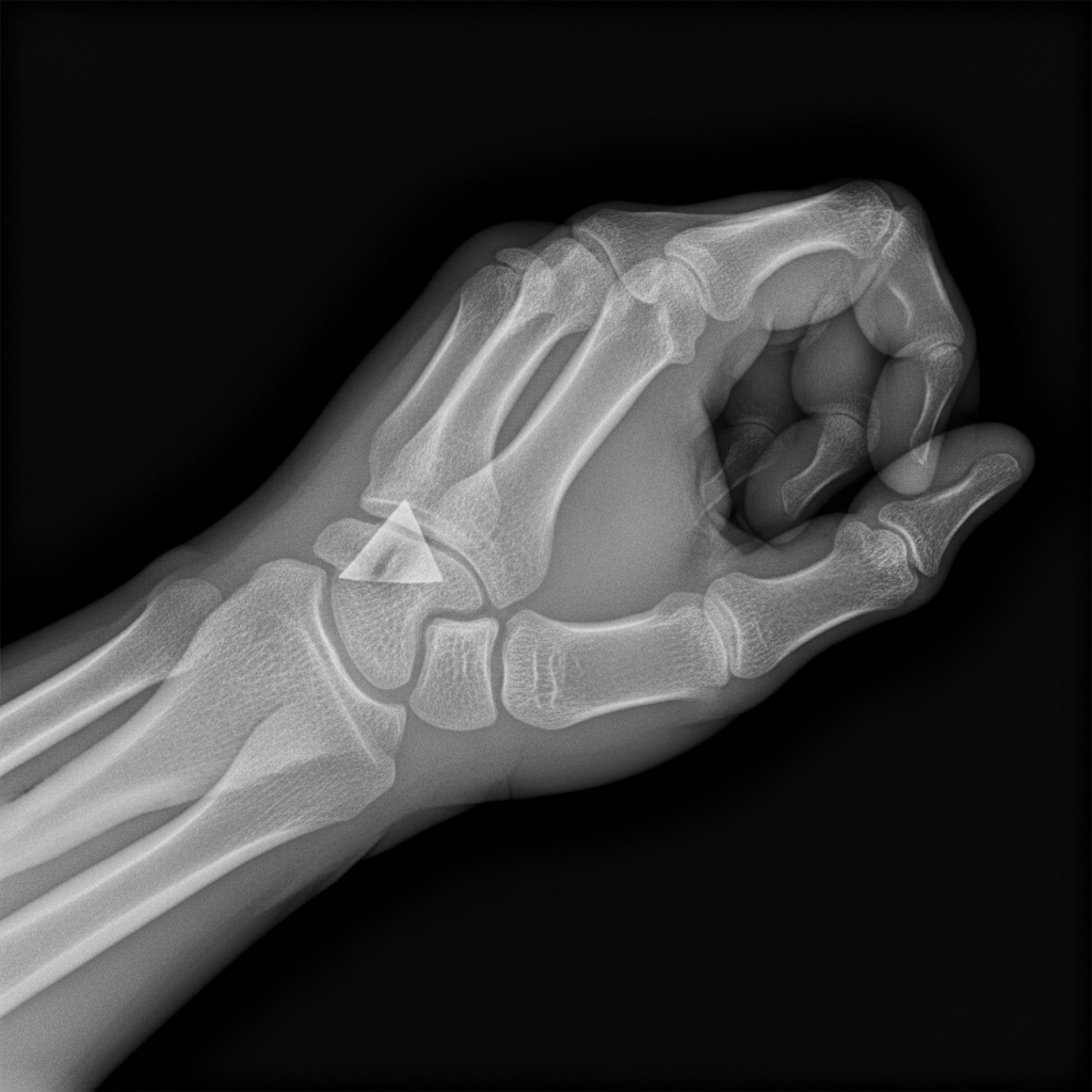
Fracture shown in this radiograph is:

Which of the following is referred to as Luxatio erecta?
Tardy ulnar nerve palsy is commonly seen in?
The vascular sign of Narath is positive in which of the following?
What is the best treatment for a trochanteric fracture of the femur?
Which complication may arise after a supracondylar fracture?
Explanation: ### Explanation **Correct Option: B. Posterior Dislocation of Shoulder** The diagnosis is based on the classic clinical presentation and history. Posterior shoulder dislocations are rare (2-5% of all dislocations) and are classically associated with **seizures** or **electric shocks**. During a seizure, the powerful internal rotators (Latissimus dorsi, Pectoralis major, and Subscapularis) overpower the weaker external rotators, forcing the humeral head posteriorly. The clinical hallmark is a limb fixed in **adduction and internal rotation**, with a pathognomonic **loss of external rotation**. On examination, there is often a palpable fullness in the posterior shoulder and a flattened anterior profile (loss of deltoid contour). **Analysis of Incorrect Options:** * **A. Anterior Dislocation:** This is the most common type. The limb is typically held in **abduction and external rotation**. It is usually caused by trauma (fall on an outstretched hand), not seizures. * **C. Inferior Dislocation (Luxatio Erecta):** A rare condition where the arm is held fixed in **full abduction** (pointing upwards) over the head. * **D. Impingement Syndrome:** A chronic overuse condition involving the rotator cuff; it does not present with an acute deformity or a fixed rotational deformity following a seizure. **High-Yield Clinical Pearls for NEET-PG:** * **Radiology:** Look for the **"Light Bulb Sign"** on AP view (the internally rotated humerus looks symmetrical like a bulb) and the **"Empty Glenoid Sign."** * **Best View:** The **Axillary view** is the gold standard to confirm posterior displacement. * **Associated Lesion:** **Reverse Hill-Sachs lesion** (impaction fracture of the anterior humeral head). * **Treatment:** Closed reduction (Modified Hippocratic method) followed by immobilization in **external rotation** (unlike anterior dislocation).
Explanation: **Explanation:** **1. Why Anterior Dislocation is Correct:** Anterior dislocation is the most common type of shoulder dislocation, accounting for approximately **95-97%** of all cases. Recurrence is a hallmark of anterior instability because the initial traumatic event often causes structural damage to the glenohumeral joint. The most significant factor is the **Bankart lesion** (avulsion of the anterior-inferior labrum), which fails to heal anatomically, leading to a permanent loss of the "chock-block" effect. Additionally, the presence of a **Hill-Sachs lesion** (compression fracture of the posterosuperior humeral head) further reduces joint stability, making recurrent episodes highly likely, especially in younger patients (age <20 years has a recurrence rate of up to 90%). **2. Why Other Options are Incorrect:** * **Posterior Dislocation:** These are rare (2-5%) and typically associated with seizures or electric shocks. While they can become chronic or "missed," they do not have the same high incidence of spontaneous recurrence as anterior dislocations. * **Inferior Dislocation (Luxatio Erecta):** This is the rarest form, characterized by the arm being locked in an overhead position. It is usually a high-energy trauma event associated with significant neurovascular injury rather than chronic recurrence. **3. Clinical Pearls for NEET-PG:** * **Most common cause of recurrence:** Patient age at the time of the first dislocation (younger = higher risk). * **Bankart Lesion:** Most common pathological finding in recurrent anterior dislocation. * **Putti-Platt/Magnuson-Stack:** Historical surgeries for recurrence (now largely replaced by Bankart repair). * **Latarjet Procedure:** Indicated when there is significant glenoid bone loss (>20-25%). * **Nerve Injury:** The **Axillary nerve** is the most commonly injured nerve in anterior dislocations (tested by sensation over the "Regimental Badge" area).
Explanation: **Explanation:** The correct answer is **D**. C.S.F. rhinorrhea is a classic sign of a **Le Fort II or III fracture** or a fracture of the anterior cranial fossa (cribriform plate). It is not a feature of mandibular fractures because the mandible is a separate bone that does not articulate with the skull base in a way that would involve the dural membranes or paranasal sinuses. **Analysis of Options:** * **Option A:** The **angle of the mandible** is one of the most common sites for fractures (along with the condyle and symphysis), especially when impacted by lateral forces. * **Option B:** The mandible is the attachment site for powerful muscles of mastication (masseter, temporalis, medial/lateral pterygoids). These muscles exert significant pull, leading to the classification of fractures as **"favorable"** (muscle pull stabilizes the fragments) or **"unfavorable"** (muscle pull displaces the fragments). * **Option C:** **Sublingual hematoma** (Coleman’s Sign) is considered a pathognomonic clinical sign of a mandibular fracture, particularly involving the body or symphysis. **High-Yield Facts for NEET-PG:** * **Most common site of Mandibular Fracture:** Condyle (overall), though the angle is highly frequent in assaults. * **Nerve Injury:** The **Inferior Alveolar Nerve** is most commonly at risk in body/angle fractures, leading to numbness of the lower lip. * **Guardsman Fracture:** A midline symphysis fracture combined with bilateral condylar fractures, usually caused by a fall on the chin. * **Management:** Most displaced fractures require **Open Reduction and Internal Fixation (ORIF)** with miniplates.
Explanation: **Explanation:** **1. Why Posterior Dislocation is Correct:** Posterior hip dislocation is the most common type, accounting for approximately **85-90%** of all traumatic hip dislocations. The underlying mechanism is typically a high-energy trauma, such as a "dashboard injury" in a motor vehicle accident. When the hip is flexed and adducted, a force applied to the knee drives the femoral head backward, rupturing the posterior capsule and dislodging it from the acetabulum. **2. Analysis of Incorrect Options:** * **Anterior Dislocation (Option B):** Much less common (approx. 10-15%). It occurs when the hip is in extreme extension, abduction, and external rotation (e.g., a fall from a height). * **Central Dislocation (Option C):** This is technically a fracture-dislocation where the femoral head is driven medially through a fractured acetabular floor into the pelvis. It is considered an acetabular fracture rather than a simple ligamentous dislocation. **3. High-Yield Clinical Pearls for NEET-PG:** * **Clinical Presentation:** * **Posterior:** Limb is **Shortened, Adducted, and Internally Rotated** (Mnemonic: "S-A-I-R"). * **Anterior:** Limb is **Abducted and Externally Rotated**. * **Associated Nerve Injury:** The **Sciatic nerve** (specifically the peroneal division) is most commonly injured in posterior dislocations. * **Vascular Complication:** **Avascular Necrosis (AVN)** of the femoral head is the most serious late complication; the risk increases if reduction is delayed beyond 6 hours. * **X-ray Sign:** In posterior dislocation, the femoral head appears smaller than the contralateral side on AP view (due to being closer to the film). * **Management:** Emergency closed reduction (e.g., Allis or Bigelow maneuver) under sedation.
Explanation: ***Bennett's fracture*** - This is a **fracture-dislocation** of the **base of the first metacarpal** involving the **carpometacarpal (CMC) joint** of the thumb. - The **abductor pollicis longus (APL)** pulls the metacarpal **proximally and radially**, causing characteristic displacement with a small **ulnar fragment** remaining attached to the trapezium. *Colles' fracture* - This involves the **distal radius** with **dorsal angulation** and typically occurs after a fall on an outstretched hand. - It does not involve the **thumb metacarpal** or **CMC joint**, making it anatomically distinct from the fracture shown. *Smith's fracture* - This is a **reverse Colles' fracture** of the **distal radius** with **volar angulation** of the distal fragment. - Like Colles', it affects the **forearm bones**, not the **thumb metacarpal** as seen in this radiograph. *Rolando's fracture* - This is a **comminuted fracture** of the **base of the first metacarpal** with **multiple fragments** involving the CMC joint. - Unlike Bennett's fracture, it shows **Y-shaped** or **T-shaped** fracture lines with **three or more fragments**, not the simple two-part fracture pattern shown.
Explanation: **Explanation:** **Luxatio Erecta** is the medical term for **Inferior Dislocation of the Shoulder**. It is the rarest form of shoulder dislocation (accounting for <1%) and occurs when a hyperabduction force is applied to the arm, levering the humeral head against the acromion and displacing it inferiorly out of the glenoid cavity. * **Why Option B is correct:** In this condition, the humeral head is displaced inferior to the glenoid. Clinically, the patient presents in a classic "salute" posture: the arm is locked in fixed abduction, with the forearm resting on or behind the head. * **Why Option A is incorrect:** A tear of the glenoid labrum (e.g., Bankart lesion) is a common consequence of shoulder dislocation but is not a type of dislocation itself. * **Why Option C is incorrect:** Anterior dislocation is the most common type of shoulder dislocation (95%). It presents with the arm held in slight abduction and external rotation, not the vertical "erect" position seen in Luxatio erecta. * **Why Option D is incorrect:** A defect in the posterosuperior humeral head is known as a **Hill-Sachs lesion**, which is a secondary bony injury resulting from anterior instability. **High-Yield Clinical Pearls for NEET-PG:** * **Mechanism:** Forceful hyperabduction. * **Clinical Sign:** Arm is locked in fixed abduction (110°–160°). * **Complications:** This type has the highest incidence of **axillary nerve** and **axillary artery** injury among all shoulder dislocations. * **Radiology:** The humeral shaft is parallel to the spine of the scapula on an X-ray.
Explanation: **Explanation:** **Tardy Ulnar Nerve Palsy** refers to a delayed-onset ulnar neuropathy occurring years after an injury. The most common cause is a **Cubitus Valgus deformity**, typically resulting from a non-union of a **lateral condyle fracture of the humerus** sustained in childhood. As the lateral condyle fails to unite, the forearm deviates laterally (valgus). This deformity increases the distance the ulnar nerve must travel around the medial epicondyle. Over time, the nerve is subjected to chronic stretching and friction within the cubital tunnel, leading to progressive ischemic changes and palsy. **Analysis of Options:** * **Cubitus Valgus (Correct):** Increases the tension and "bowstringing" of the ulnar nerve behind the medial epicondyle, leading to tardy palsy. * **Cubitus Varus (Incorrect):** Also known as "Gunstock deformity," it usually follows a malunited supracondylar fracture. While it can rarely cause ulnar nerve transposition issues, it is not the classic cause of tardy ulnar nerve palsy. * **Dinner Fork Deformity (Incorrect):** This is the characteristic clinical appearance of a **Colles’ fracture** (distal radius fracture with dorsal displacement). * **Garden Spade Deformity (Incorrect):** This is the characteristic clinical appearance of a **Smith’s fracture** (distal radius fracture with volar displacement). **High-Yield Clinical Pearls for NEET-PG:** * **Latent Period:** Tardy ulnar nerve palsy usually appears 10–20 years after the initial injury. * **First Sign:** Sensory loss/paresthesia in the little finger and the medial half of the ring finger. * **Motor Sign:** Weakness of intrinsic hand muscles (Wartenberg’s sign, Froment’s sign, and clawing). * **Treatment of Choice:** Anterior transposition of the ulnar nerve.
Explanation: **Explanation:** The **Vascular Sign of Narath** is a clinical finding used to assess the position of the femoral head in relation to the femoral artery. **1. Why Posterior Dislocation is Correct:** In a **posterior dislocation of the hip** (the most common type), the femoral head is displaced backward and upward out of the acetabulum. Normally, the femoral head lies directly behind the femoral artery in the groin, providing a solid "backing" or resistance that allows the arterial pulsations to be easily felt. When the head is dislocated posteriorly, this support is lost. Consequently, the femoral artery sinks into the empty space, and its pulsations become **diminished or impalpable**. This clinical finding—the absence or weakening of femoral pulses due to posterior displacement of the femoral head—is known as a positive Narath’s sign. **2. Why Other Options are Incorrect:** * **Anterior Dislocation:** The femoral head moves forward, often becoming more prominent near the inguinal ligament. This would typically make the pulse more palpable or shift its position, but it does not produce the "hollow" sign of Narath. * **Central Dislocation:** This involves the femoral head being driven through the acetabular floor into the pelvis. While the head moves away from the artery, this is a fracture-dislocation pattern and is not classically associated with Narath’s sign. * **Lateral Dislocation:** This is not a standard anatomical classification for hip dislocations. **3. Clinical Pearls for NEET-PG:** * **Position of Limb:** Posterior dislocation presents with **Internal Rotation, Adduction, and Shortening** (The "F-IR-AD" mnemonic: Flexion, Internal Rotation, Adduction). * **Most Common Nerve Injury:** Sciatic nerve (specifically the peroneal division). * **Associated Fracture:** Often associated with a fracture of the posterior lip of the acetabulum. * **Emergency:** Hip dislocation is an orthopedic emergency due to the high risk of **Avascular Necrosis (AVN)** of the femoral head.
Explanation: **Explanation:** Trochanteric (intertrochanteric) fractures are extracapsular fractures that occur in a highly vascular area. The primary goal of treatment is early mobilization to prevent complications of prolonged recumbency (like DVT or pneumonia). **1. Why Dynamic Hip Screw (DHS) is the Correct Answer:** The **Dynamic Hip Screw (DHS)** is the gold standard for stable intertrochanteric fractures. The underlying medical concept is the **"sliding principle."** The DHS allows the femoral head fragment to collapse and slide along the barrel of the plate during weight-bearing. This controlled compression at the fracture site promotes secondary bone healing and increases stability. **2. Why the Other Options are Incorrect:** * **Inlay plates:** These are not used for trochanteric fractures as they do not provide the necessary dynamic compression or angular stability required to withstand the biomechanical stresses of the proximal femur. * **Plaster in abduction / Internal rotation:** Conservative management with hip spica or traction was used historically but is now obsolete. It leads to high rates of malunion (coxa vara) and life-threatening complications due to prolonged immobilization in elderly patients. **Clinical Pearls for NEET-PG:** * **Classification:** Boyd and Griffin or Evans classification is commonly used for these fractures. * **Stable vs. Unstable:** For **stable** fractures, DHS is preferred. For **unstable** fractures (e.g., reverse oblique type or loss of posteromedial cortex), a **Proximal Femoral Nail (PFN)** is the treatment of choice because it is an intramedullary device with a shorter lever arm. * **Complication:** The most common deformity following a poorly treated trochanteric fracture is **Coxa Vara** (decreased neck-shaft angle) leading to a short-limbed gait.
Explanation: **Explanation:** Supracondylar fractures of the humerus (specifically the extension type, which accounts for 95% of cases) are among the most common pediatric fractures and are notorious for both immediate neurovascular and late structural complications. 1. **Nerve Injury (Option A):** The displaced proximal fragment can stretch or contuse adjacent nerves. The **Median nerve** (specifically the Anterior Interosseous Nerve branch) is the most commonly injured in extension-type fractures. The Radial nerve is the second most common, while the Ulnar nerve is typically injured in flexion-type fractures or during surgical pinning. 2. **Vascular Injury (Option B):** The **Brachial artery** lies in close proximity to the sharp edge of the proximal bone fragment. Injury or spasm can lead to pulselessness and, if untreated, **Volkmann’s Ischemic Contracture (VIC)**. 3. **Deformity (Option C):** **Cubitus varus** (Gunstock deformity) is the most common late complication. It usually results from malunion (inadequate reduction of medial tilt or rotation) rather than physeal arrest. **High-Yield Clinical Pearls for NEET-PG:** * **Most common nerve injured:** Anterior Interosseous Nerve (AIN) – test by asking the patient to make an "OK" sign. * **Most common deformity:** Cubitus varus (primarily a cosmetic issue, rarely affects function). * **Gartland Classification:** Used to grade displacement (Type I: Undisplaced; Type II: Angulated but posterior cortex intact; Type III: Completely displaced). * **Urgency:** A "pink pulseless hand" is a surgical emergency requiring immediate reduction.
Principles of Fracture Management
Practice Questions
Upper Limb Fractures
Practice Questions
Lower Limb Fractures
Practice Questions
Spinal Trauma
Practice Questions
Pelvic and Acetabular Fractures
Practice Questions
Open Fractures
Practice Questions
Fractures in Children
Practice Questions
Fracture Complications
Practice Questions
Nonunion and Malunion
Practice Questions
Polytrauma Management
Practice Questions
Joint Dislocations
Practice Questions
Soft Tissue Injuries
Practice Questions
Get full access to all questions, explanations, and performance tracking.
Start For Free