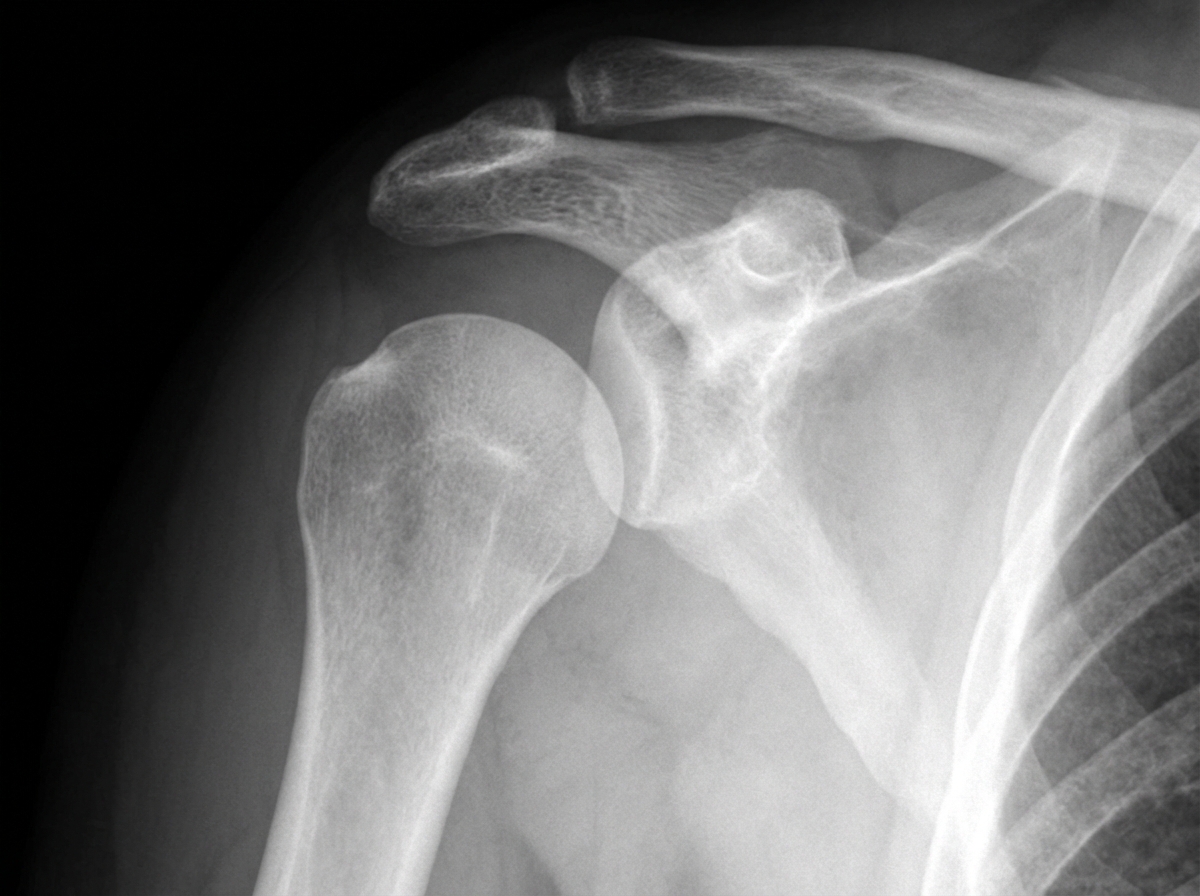
A 54-year-old male patient sustained a right upper limb injury in a road traffic accident and presented to the emergency room unable to move his right arm. Radiographic findings are shown. Which of the following nerves is most likely injured?

Which of the following is NOT a major criterion for Gurd?
What is the commonest site for osteochondritis dissecans in the elbow?
High-stepping gait is characteristic of which condition?
For which of the following conditions is a TT splint NOT indicated?
A person presents 3 days post Colles' fracture with inability to extend the thumb. What is the most likely cause?
Long compression is used for which fracture?
What is the most common fracture in the elderly?
Cubital tunnel syndrome involves which nerve?
In a posteriorly dislocated elbow, which nerve is most commonly involved?
Explanation: ***Axillary nerve*** - **Anterior shoulder dislocation** or **surgical neck of humerus fracture** commonly injures the axillary nerve due to its anatomical course around the surgical neck. - Injury results in **deltoid muscle paralysis** (inability to abduct arm) and **loss of sensation** over the lateral shoulder (regimental badge area). *Radial nerve* - Most commonly injured with **midshaft humerus fractures** due to its spiral course in the radial groove. - Causes **wrist drop** (inability to extend wrist) and **weakness of finger extension**, not complete arm immobility. *Ulnar nerve* - Typically injured with **medial epicondyle fractures** or **elbow trauma** affecting the cubital tunnel. - Results in **claw hand deformity** and **weakness of intrinsic hand muscles**, preserving shoulder and arm movement. *Median nerve* - Commonly injured in **supracondylar fractures** of the humerus or **carpal tunnel syndrome**. - Causes **ape thumb deformity** and **thenar muscle weakness**, but does not affect shoulder or arm movement.
Explanation: To diagnose **Fat Embolism Syndrome (FES)**, Gurd and Wilson’s criteria are widely used. The diagnosis requires at least **one Major criterion** and **four Minor criteria**. ### **Why Thrombocytopenia is the Correct Answer** **Thrombocytopenia** is classified as a **Minor criterion**, not a major one. While the classic triad of FES includes respiratory distress, cerebral signs, and petechiae, the laboratory findings like a drop in platelet count (<150,000/mm³) or a drop in hemoglobin are considered supportive (minor) rather than primary diagnostic pillars. ### **Analysis of Incorrect Options (Major Criteria)** * **Subconjunctival petechiae (Option A):** This is a hallmark **Major criterion**. Petechiae typically appear 24–36 hours after injury in a "vest distribution" (neck, axilla, and conjunctiva) due to capillary occlusion by fat globules. * **PaO2 < 60 mmHg (Option B):** Respiratory insufficiency is the most common early sign. A partial pressure of oxygen below 60 mmHg on room air is a **Major criterion**. * **Pulmonary Edema (Option D):** Acute pulmonary changes, often manifesting as "snowstorm" appearance on X-ray or diffuse bilateral edema, constitute a **Major criterion** (often grouped under respiratory insufficiency). * *Note: The third Major criterion not listed here is **Cerebral involvement** (non-traumatic confusion, agitation, or coma).* ### **Clinical Pearls for NEET-PG** * **The Triad:** Hypoxemia, Neurological abnormalities, and Petechial rash. * **Gurd’s Minor Criteria:** Tachycardia (>110 bpm), Pyrexia (>38.5°C), Retinal changes (fat globules/hemorrhages), Jaundice, Renal changes (oliguria/anuria), and laboratory findings (Thrombocytopenia, Anemia, Elevated ESR, Fat globules in sputum/urine). * **Treatment:** Primarily supportive (Oxygenation/Ventilation). Early stabilization of long bone fractures (especially femur) is the best preventive measure. * **Free Fatty Acids:** The chemical theory suggests FES is caused by the toxic effect of free fatty acids on the lung parenchyma.
Explanation: **Explanation:** **Osteochondritis Dissecans (OCD)** of the elbow is an idiopathic condition characterized by focal subchondral bone necrosis and subsequent fragmentation of the overlying articular cartilage. **Why Capitulum is Correct:** The **capitulum** is the most common site for OCD in the elbow (specifically the anterolateral aspect). This is primarily due to its unique vascular anatomy—it is supplied by only one or two end-arteries that enter posteriorly, making it susceptible to ischemic necrosis. It is frequently seen in adolescent athletes (12–15 years), particularly "overhead throwers" (e.g., baseball pitchers) or gymnasts. The repetitive valgus stress causes compressive forces between the radial head and the capitulum, leading to microtrauma and focal ischemia. **Why Other Options are Incorrect:** * **Trochlea:** While OCD can occur here, it is extremely rare. The trochlea has a more robust blood supply compared to the capitulum. * **Radial Head:** The radial head is usually the site of secondary changes (like hypertrophy) or fractures (Mason classification) rather than primary OCD. * **Olecranon:** This site is more commonly associated with stress fractures or traction apophysitis (in the pediatric population) rather than OCD. **High-Yield Clinical Pearls for NEET-PG:** * **Panner’s Disease:** Often confused with OCD, this is osteochondrosis of the *entire* capitulum in younger children (5–10 years). It carries a better prognosis and usually heals with rest. * **Clinical Presentation:** Vague lateral elbow pain, clicking, or "locking" if a loose body is present. * **Radiology:** The "Fragment" or "Crater" sign on X-ray. MRI is the gold standard for assessing stability. * **Management:** Conservative (rest) if the fragment is stable; surgical (debridement/fixation) if unstable or loose bodies are present.
Explanation: ### Explanation **Correct Answer: B. Common Peroneal Nerve Palsy** The **Common Peroneal Nerve (CPN)**, specifically its deep branch, innervates the muscles of the anterior compartment of the leg (Tibialis anterior, Extensor digitorum longus, and Extensor hallucis longus). These muscles are responsible for **dorsiflexion** of the foot and extension of the toes. In CPN palsy, the loss of dorsiflexion leads to **Foot Drop**. To prevent the toes from dragging on the ground during the swing phase of walking, the patient compensates by excessively flexing the hip and knee, lifting the foot high off the ground. This results in the characteristic **High-stepping gait**. **Analysis of Incorrect Options:** * **A. CTEV:** Characterized by a "Cave" (Cavus, Adductus, Varus, Equinus) deformity. The gait is typically a "stumbling" gait or walking on the outer border of the foot, not high-stepping. * **C. Poliomyelitis:** While it can cause various paralytic deformities (including foot drop), it typically presents with a **Hand-knee gait** (due to quadriceps weakness) or a **Trendelenburg gait** (due to gluteal weakness). * **D. Cerebral Palsy:** Most commonly presents with a **Scissor gait** (due to adductor spasticity) or a **Crouch gait**. **Clinical Pearls for NEET-PG:** * **Most common site of injury:** The CPN is the most frequently injured nerve in the lower limb because it winds superficially around the **neck of the fibula**. * **Sensory Loss:** Occurs over the lateral aspect of the leg and the dorsum of the foot (sparing the web space between the 1st and 2nd toes if only the superficial branch is involved). * **Trendelenburg Gait:** Seen in Superior Gluteal Nerve injury (weakness of Gluteus Medius/Minimus). * **Waddling Gait:** Seen in bilateral developmental dysplasia of the hip (DDH) or muscular dystrophy.
Explanation: **Explanation:** The **Thomas Splint (TT Splint)** is a traction splint primarily designed to provide immobilization and maintain longitudinal traction for injuries involving the femur and the knee joint. **Why Infective Arthritis is the Correct Answer:** In **Infective (Septic) Arthritis** of the knee, the primary goal of splintage is absolute rest and immobilization to prevent joint destruction, but **traction is not indicated**. Instead, these patients are typically managed with a **Bohler-Braun splint** or a simple posterior slab/plaster. A Thomas splint is cumbersome for the frequent clinical monitoring and joint aspirations required in septic cases. Furthermore, the pressure from the ring of the Thomas splint can cause discomfort and skin breakdown in patients who are already systemically ill. **Analysis of Incorrect Options:** * **Injuries around the knee joint & Knee dislocation:** The Thomas splint is excellent for stabilizing the knee joint, preventing further neurovascular damage, and maintaining alignment during transport or definitive healing. * **Fracture of the femur:** This is the **classic indication** for a Thomas splint. It provides the necessary traction to overcome the powerful pull of the thigh muscles, preventing shortening and reducing pain by stabilizing the fracture fragments. **NEET-PG High-Yield Pearls:** * **Measurement:** The Thomas splint ring size is "1.5 inches (or 4 cm) more than the circumference of the thigh at the groin." The length is "6 inches (15 cm) beyond the heel." * **Modifications:** The **Keller-Blake splint** is a modification of the Thomas splint with a half-ring, making it easier to apply in emergencies. * **Fixed vs. Sliding Traction:** Thomas splint can be used for both fixed traction (tied to the end of the splint) and sliding traction (using weights and pulleys).
Explanation: ### Explanation **Correct Option: A. Rupture of extensor pollicis longus (EPL) tendon** The inability to extend the thumb following a distal radius fracture (Colles' fracture) is a classic presentation of **delayed rupture of the EPL tendon**. * **Mechanism:** The EPL tendon passes through a narrow fibro-osseous tunnel at **Lister’s tubercle** (3rd dorsal compartment). Following a fracture, the tendon is compromised either by direct mechanical friction against bony irregularities or, more commonly, by **avascular necrosis**. The swelling within the tight compartment compromises the blood supply (via the vincula), leading to delayed attrition and rupture. * **Timing:** This typically occurs **weeks to months** after the injury, but can manifest as early as a few days post-trauma. **Why other options are incorrect:** * **B. Volkmann's Ischemic Contracture (VIC):** This is a late sequela of compartment syndrome (usually after supracondylar fractures). It involves ischemic necrosis of the forearm flexors, leading to a characteristic flexion deformity of the wrist and fingers, not isolated thumb extension loss. * **C. Radial Nerve Injury:** While the radial nerve supplies the thumb extensors, a Colles' fracture is a distal injury. Radial nerve palsy (e.g., Saturday Night Palsy) would result in a complete **wrist drop** and loss of extension at the MCP joints of all fingers. * **D. Finger Stiffness:** This is a common complication of immobilization but would cause a global decrease in the range of motion rather than a sudden, specific loss of the ability to extend the thumb. **NEET-PG High-Yield Pearls:** * **Treatment of Choice:** Direct end-to-end repair is usually not possible due to tendon fraying. The gold standard is **Extensor Indicis Proprius (EIP) tendon transfer**. * **Lister’s Tubercle:** Acts as a pulley for the EPL; it is the anatomical landmark most associated with this complication. * **Risk:** Interestingly, EPL rupture is more common in **undisplaced** or minimally displaced distal radius fractures because the intact periosteum keeps the hematoma (and subsequent pressure) confined.
Explanation: **Explanation:** **Long Compression** (also known as the **Essex-Lopresti method**) is a specific technique used for the reduction and fixation of **Calcaneum fractures**, particularly those involving the joint surface (intra-articular). The calcaneum is composed primarily of cancellous bone with a thin cortical shell. When it fractures, it often undergoes significant compression and loss of height (measured by **Bohler’s angle**). The "Long Compression" technique involves using a heavy-duty pin (Schanz screw or Gissane spike) inserted through the posterior aspect of the tuberosity into the displaced fragment. This allows the surgeon to apply longitudinal traction and leverage to restore the height and alignment of the bone before securing it. **Analysis of Incorrect Options:** * **Talus:** Fractures of the talus (e.g., Aviator’s fracture) are typically treated with ORIF using lag screws to achieve interfragmentary compression, but the specific "Long Compression" maneuver is not a standard term for this bone. * **Fibula:** Most fibular fractures are treated with plate fixation (e.g., lateral malleolus) or are managed conservatively if they are non-weight-bearing. * **Femur:** Femoral fractures usually require intramedullary nailing or heavy plating. While compression is used in plating, the specific clinical term "Long Compression" is not associated with femoral trauma. **High-Yield Clinical Pearls for NEET-PG:** * **Bohler’s Angle:** Normal is 25°–40°. A decrease indicates a calcaneal burst fracture. * **Angle of Gissane:** Normal is 120°–145°. * **Mondor’s Sign:** Ecchymosis extending to the sole of the foot; pathognomonic for calcaneal fracture. * **Associated Injuries:** Always check the **Lumbar Spine (L1)** in calcaneal fractures (Don Juan Syndrome/Lover’s Fracture) due to axial loading.
Explanation: **Explanation:** The most common fracture in the elderly population is **Colles' fracture** (distal radius fracture). This is primarily due to the high prevalence of postmenopausal osteoporosis and the mechanism of injury: a low-energy fall on an outstretched hand (FOOSH). While hip fractures are associated with higher morbidity and mortality, epidemiological studies consistently show that distal radius fractures occur with the highest frequency in the geriatric age group. **Analysis of Options:** * **Colles' fracture (Correct):** Defined as a distal radius fracture within 2.5 cm of the wrist joint with dorsal displacement and angulation. It is often the first "sentinel" fracture that signals underlying osteoporosis in the elderly. * **Trochanteric/Intertrochanteric fracture (Incorrect):** These are very common hip fractures in the elderly, often occurring a decade later than Colles' fractures. While they carry a higher clinical significance due to surgical requirements, their absolute incidence is lower than distal radius fractures. * **Supracondylar fracture (Incorrect):** This is the most common fracture in **children** (typically aged 5–8 years) following a fall, but it is relatively rare in the elderly. **NEET-PG High-Yield Pearls:** * **Colles' Deformity:** Classically described as a **"Dinner Fork Deformity."** * **Most common fracture in children:** Supracondylar fracture of the humerus. * **Most common site of osteoporosis-related fracture:** Vertebral body (often asymptomatic/compression fractures), followed by the distal radius and hip. * **Management:** Most Colles' fractures are managed via closed reduction and a Colles' cast (below-elbow cast in slight palmar flexion and ulnar deviation).
Explanation: **Explanation:** **Cubital Tunnel Syndrome** is the second most common compression neuropathy of the upper limb (after Carpal Tunnel Syndrome). It occurs due to the compression or entrapment of the **Ulnar nerve** as it passes through the cubital tunnel at the elbow. The tunnel is bordered medially by the medial epicondyle and laterally by the olecranon, with the roof formed by **Osborne’s ligament** (arcuate ligament). * **Why Ulnar Nerve is Correct:** The ulnar nerve is most vulnerable at the elbow because it lies superficially in the retrocondylar groove. Chronic pressure, repetitive flexion, or valgus deformity (Tardy Ulnar Palsy) leads to paresthesia in the small finger and the ulnar half of the ring finger, along with weakness of the intrinsic muscles of the hand. **Analysis of Incorrect Options:** * **Radial Nerve:** Compression typically occurs in the spiral groove (Saturday Night Palsy) or at the arcade of Frohse (Posterior Interosseous Nerve syndrome), leading to wrist drop or finger extension weakness. * **Median Nerve:** Most commonly compressed at the wrist (Carpal Tunnel Syndrome) or between the two heads of the pronator teres (Pronator Syndrome). * **Axillary Nerve:** Usually injured during anterior shoulder dislocations or fractures of the surgical neck of the humerus, affecting the deltoid muscle. **Clinical Pearls for NEET-PG:** * **Tardy Ulnar Palsy:** Delayed ulnar nerve palsy occurring years after a lateral condyle fracture of the humerus due to resultant **cubitus valgus** deformity. * **Froment’s Sign:** Positive in ulnar nerve palsy due to weakness of the Adductor Pollicis (compensated by Flexor Pollicis Longus). * **Wartenberg’s Sign:** Persistent abduction of the small finger due to weak palmar interossei.
Explanation: **Explanation:** In a **posterior dislocation of the elbow** (the most common type), the radius and ulna are displaced posteriorly and often laterally relative to the distal humerus. The **ulnar nerve** is the most commonly injured nerve because of its vulnerable anatomical position in the fibro-osseous tunnel (cubital tunnel) behind the medial epicondyle. During posterior displacement, the nerve is subjected to significant traction or direct compression as the joint capsule and medial collateral ligaments are disrupted. **Analysis of Options:** * **Ulnar Nerve (Correct):** Its proximity to the medial joint complex makes it highly susceptible to traction injuries during the deformity. * **Median Nerve:** This is the second most commonly injured nerve. It is typically affected in **posterolateral** dislocations or when there is an associated "entrapment" within the joint or a concomitant brachial artery injury. * **Radial Nerve:** Injury is rare in simple elbow dislocations. It is more commonly associated with fractures of the humeral shaft (Holstein-Lewis fracture) or radial head. * **Musculocutaneous Nerve:** This nerve is well-protected by the overlying musculature and is rarely involved in elbow trauma. **NEET-PG High-Yield Pearls:** * **Most common direction:** Posterolateral. * **Most common complication:** Stiffness (Loss of terminal extension). * **Terrible Triad of the Elbow:** Elbow dislocation + Coronoid fracture + Radial head fracture. * **Clinical Sign:** The "Three-point relationship" (between the olecranon and the two epicondyles) is lost in dislocation but maintained in supracondylar fractures of the humerus. * **Vascular Injury:** The **Brachial artery** is the most common vessel injured, especially in open dislocations or severe anterior displacement of the humerus.
Principles of Fracture Management
Practice Questions
Upper Limb Fractures
Practice Questions
Lower Limb Fractures
Practice Questions
Spinal Trauma
Practice Questions
Pelvic and Acetabular Fractures
Practice Questions
Open Fractures
Practice Questions
Fractures in Children
Practice Questions
Fracture Complications
Practice Questions
Nonunion and Malunion
Practice Questions
Polytrauma Management
Practice Questions
Joint Dislocations
Practice Questions
Soft Tissue Injuries
Practice Questions
Get full access to all questions, explanations, and performance tracking.
Start For Free