
Trauma — MCQs

On this page
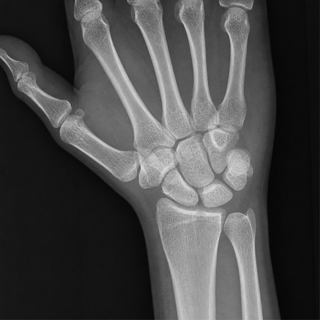
Which bone is commonly fractured when a person falls on outstretched hands?
What is the first sign of Volkmann's ischemia?
Elephant foot deformity is indicative of:
What is the most appropriate management option for an intra-articular fracture?
In the case of an inferior shoulder dislocation, which nerve is most commonly injured?
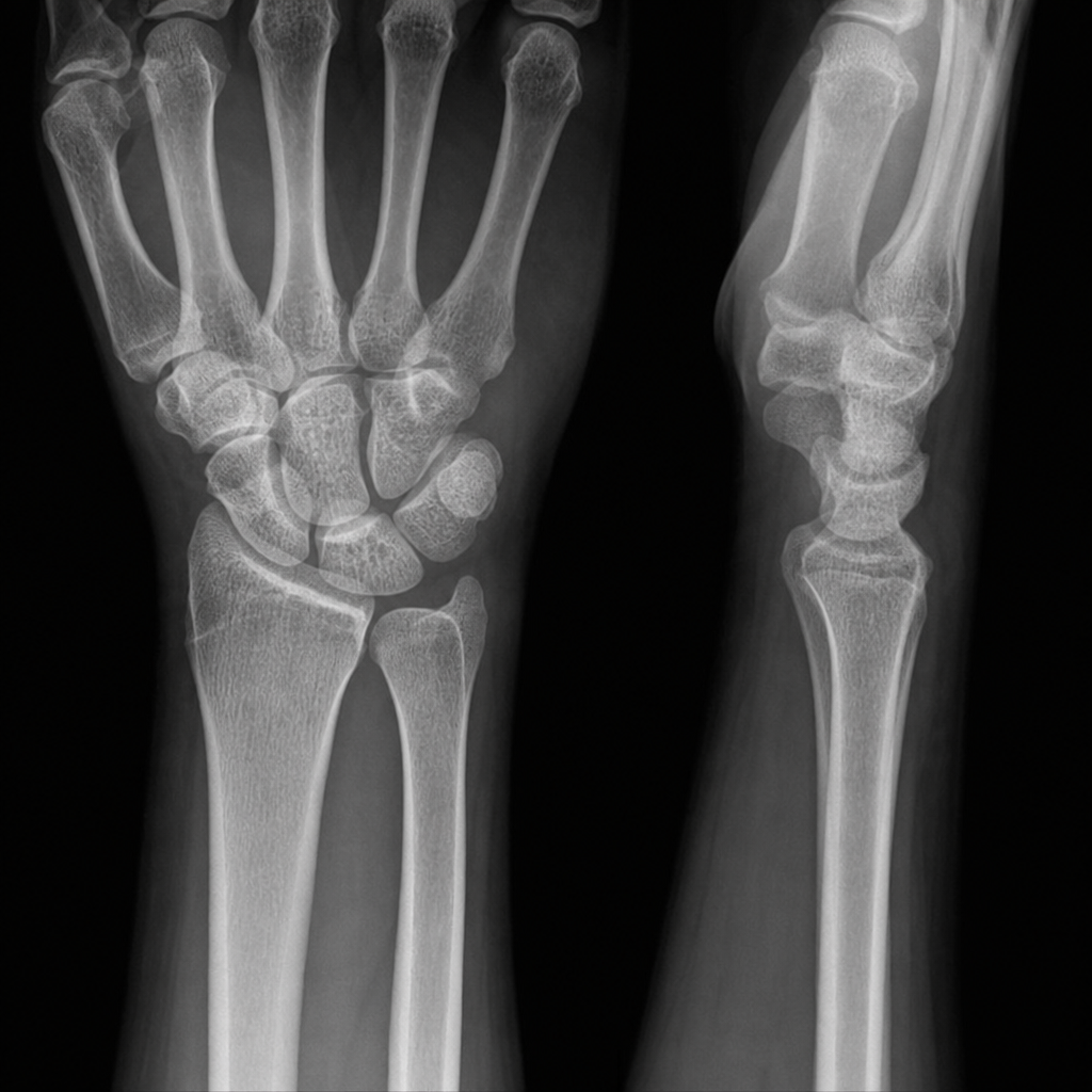
A 25-year-old man presents to the emergency department after falling onto an outstretched hand while playing cricket. He has tenderness in the anatomical snuffbox and pain with axial loading of the thumb. What is the most likely diagnosis based on the X-ray findings?
Holstein-Lewis fracture is associated with injury to which nerve?
A 36-year-old man presents with an ankle injury sustained during a soccer match. What urgent intervention is required?
In fracture of the olecranon, excision of the proximal fragment is indicated in all of the following situations, EXCEPT:
Displaced transverse patella; what is the treatment?
Practice by Chapter
Principles of Fracture Management
Practice Questions
Upper Limb Fractures
Practice Questions
Lower Limb Fractures
Practice Questions
Spinal Trauma
Practice Questions
Pelvic and Acetabular Fractures
Practice Questions
Open Fractures
Practice Questions
Fractures in Children
Practice Questions
Fracture Complications
Practice Questions
Nonunion and Malunion
Practice Questions
Polytrauma Management
Practice Questions
Joint Dislocations
Practice Questions
Soft Tissue Injuries
Practice Questions
Want unlimited practice?
Get full access to all questions, explanations, and performance tracking.
Scan to download app