
Trauma — MCQs

On this page
Which of the following is NOT a clinical feature of fat embolism?
True about proximal fragment in subtrochanteric fracture is?
The treatment of choice for a 65-year-old man who presented with a fracture of the neck of femur three days after injury is:
Which of the following injuries can be classified as Gustilo-Anderson Grade III injuries?
Which clinical sign is consistently present in all bone fractures?
Which of the following attitudes will be seen in a patient with posterior dislocation of the hip?
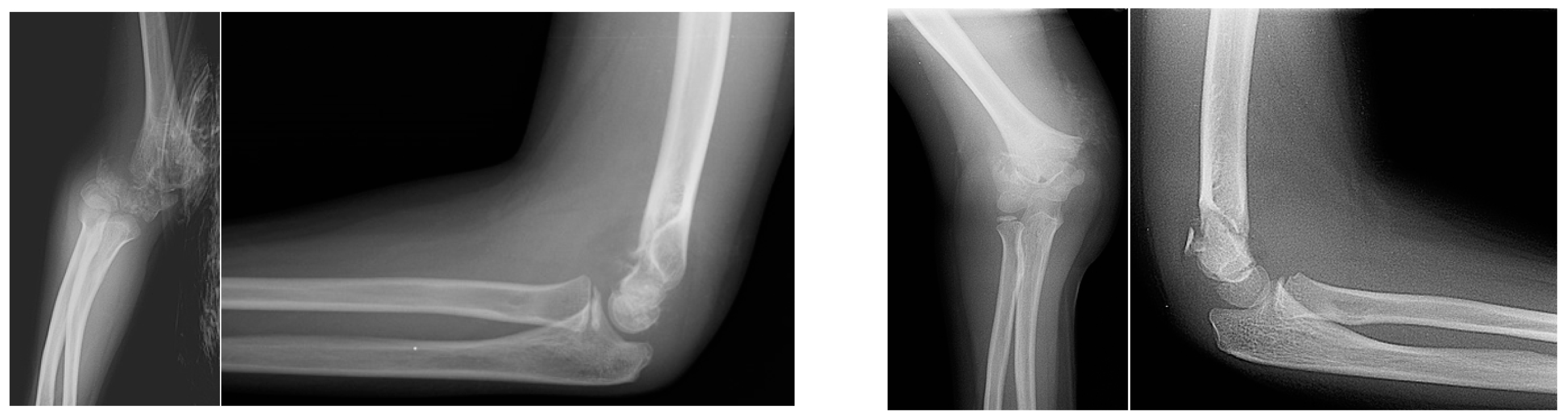
Which of the following classifications is used to assess the fracture shown in the provided image?
Which of the following statements is false regarding Colles' fracture?
A 42-year-old man is brought to the trauma center after a fall from a ladder. Physical examination reveals a slightly deformed left lower extremity with a 0.5 cm soft tissue defect over the anterolateral aspect of his leg. The wound appears relatively clean with no gross contaminants present. Radiographs depict a short oblique proximal one-third diaphyseal tibia fracture. What is his Gustilo open fracture classification grade?
Velpeau bandage and Sling and Swathe splint are used in?
Practice by Chapter
Principles of Fracture Management
Practice Questions
Upper Limb Fractures
Practice Questions
Lower Limb Fractures
Practice Questions
Spinal Trauma
Practice Questions
Pelvic and Acetabular Fractures
Practice Questions
Open Fractures
Practice Questions
Fractures in Children
Practice Questions
Fracture Complications
Practice Questions
Nonunion and Malunion
Practice Questions
Polytrauma Management
Practice Questions
Joint Dislocations
Practice Questions
Soft Tissue Injuries
Practice Questions
Want unlimited practice?
Get full access to all questions, explanations, and performance tracking.
Scan to download app