
Trauma — MCQs

On this page
Most common complication of mid shaft humerus fracture is ?
Thomas splint is used for immobilizing fractures of ?
Which of the following factors does NOT indicate an unstable pelvis?
Patient comes with crush injury to upper limb, doctor is concerned about gangrene and sepsis. What scoring system can help decide between amputation and limb salvage?
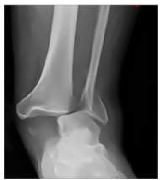
An RTA patient presented to the emergency department with severe pain in the ankle. An X-ray was performed, given below. What is the best next step in management?
12 yr old Child admitted to ICU with blunt trauma and femur fracture- Pao2 60% despite 100%o2 and rebreather mask, CXR shows lung fields clear but the patient remains confused. What is most likely the diagnosis?
As an intern in the emergency department, you encounter four patients with different types of fractures. Which patient should you prioritize for orthopedic consultation based on the severity of their condition?
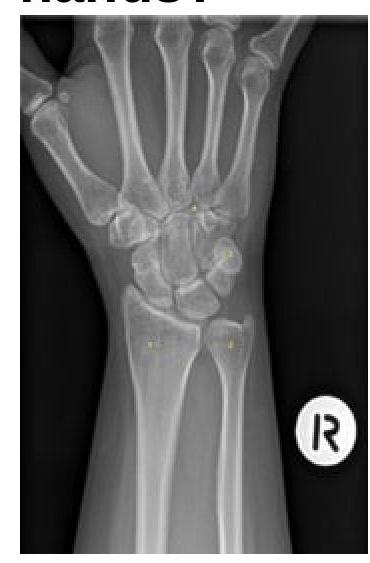
Identify the bone numbered in the X-ray that most commonly fractures when a person falls on outstretched hands.
What is meant by perilunate dislocations?
The K nail can be used for all of the following types of fractures except -
Practice by Chapter
Principles of Fracture Management
Practice Questions
Upper Limb Fractures
Practice Questions
Lower Limb Fractures
Practice Questions
Spinal Trauma
Practice Questions
Pelvic and Acetabular Fractures
Practice Questions
Open Fractures
Practice Questions
Fractures in Children
Practice Questions
Fracture Complications
Practice Questions
Nonunion and Malunion
Practice Questions
Polytrauma Management
Practice Questions
Joint Dislocations
Practice Questions
Soft Tissue Injuries
Practice Questions
Want unlimited practice?
Get full access to all questions, explanations, and performance tracking.
Scan to download app