
Trauma — MCQs

On this page
Tibial Fracture with > 1 cm wound, slight comminution and moderate crushing is
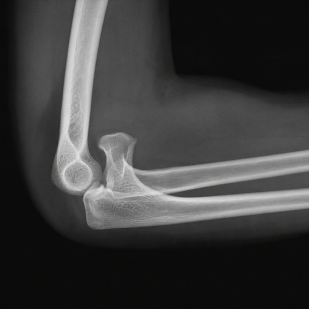
What is seen on x-ray with posterior elbow dislocation
Russell and Taylor classification is used for:
Clinical feature of fat embolism includes all except:
Avascular necrosis of head of femur occurs commonly at :
A 27-year-old right hand dominant man sustains a right distal radius fracture after a fall. He is treated with closed reduction. Which radiographic parameter has the greatest bearing on functional outcome
Which of the following fractures is associated with high mortality and morbidity?
What is luxatio erecta ?
Immediate treatment of compound fracture of tibia includes:
All are true regarding brachial plexus injury, except:
Practice by Chapter
Principles of Fracture Management
Practice Questions
Upper Limb Fractures
Practice Questions
Lower Limb Fractures
Practice Questions
Spinal Trauma
Practice Questions
Pelvic and Acetabular Fractures
Practice Questions
Open Fractures
Practice Questions
Fractures in Children
Practice Questions
Fracture Complications
Practice Questions
Nonunion and Malunion
Practice Questions
Polytrauma Management
Practice Questions
Joint Dislocations
Practice Questions
Soft Tissue Injuries
Practice Questions
Want unlimited practice?
Get full access to all questions, explanations, and performance tracking.
Scan to download app