
Trauma — MCQs

On this page
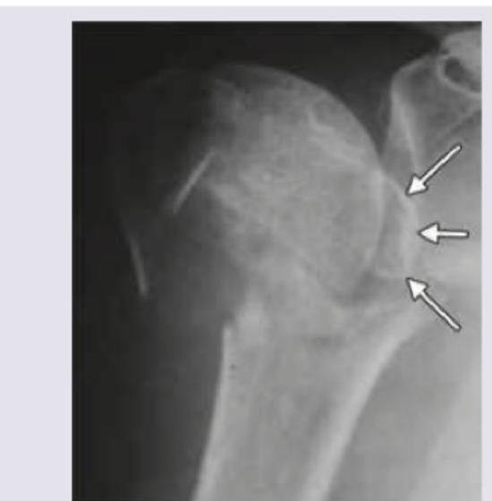
What type of fracture is shown in X-ray of left shoulder?
Consider the following : 1. Pain relief 2. Prevention of infection 3. Anaesthesia 4. Restoration of anatomy Which of the features given above are priorities for fracture treatment?
The commonest complication of fracture of clavicle is :
The nerve most likely to get injured in patients with fracture of upper end of radius is :
Which one of the following fractures is most often complicated by fat embolism ?
Avascular necrosis may develop in the following fractures except
What is the most common injury sustained due to fall on outstretched hand by a person aged 65 years?
An 18 year old boy had a closed lower limb injury while riding his motorbike. He was brought to hospital where on examination he had severe pain which increased on passive movement of affected limb with distal sensory disturbances. What is the probable diagnosis?
An 8 year old girl sustained a fall on the outstretched right hand 6 hours ago and was treated with egg albumen bandages by a village bone setter. She presented with gross swelling of the right elbow and forearm. The first essential intervention in this case would be to
Which one of the following statements about Compartment Syndrome is NOT correct?
Practice by Chapter
Principles of Fracture Management
Practice Questions
Upper Limb Fractures
Practice Questions
Lower Limb Fractures
Practice Questions
Spinal Trauma
Practice Questions
Pelvic and Acetabular Fractures
Practice Questions
Open Fractures
Practice Questions
Fractures in Children
Practice Questions
Fracture Complications
Practice Questions
Nonunion and Malunion
Practice Questions
Polytrauma Management
Practice Questions
Joint Dislocations
Practice Questions
Soft Tissue Injuries
Practice Questions
Want unlimited practice?
Get full access to all questions, explanations, and performance tracking.
Scan to download app