
Trauma — MCQs

On this page
A 34-year-old male presents with a femur shaft fracture. Four days after the injury, he develops petechiae over his chest. What is the most probable diagnosis?
Bryant's triangle is useful in the diagnosis of which of the following conditions except?
Which of the following is NOT true regarding fracture neck of femur?
Which is the most common site of pelvic apophyseal avulsion fractures?
What is true about compartment syndrome?
A patient presents with a fracture of the femur. On the 3rd day of admission, he develops breathlessness. What is the most probable diagnosis?
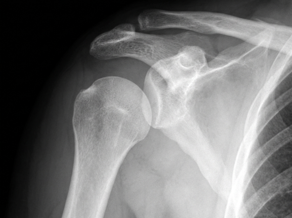
A 54-year-old male patient sustained a right upper limb injury in a road traffic accident and presented to the emergency room unable to move his right arm. Radiographic findings are shown. Which of the following nerves is most likely injured?
Which of the following is NOT a major criterion for Gurd?
High-stepping gait is characteristic of which condition?
For which of the following conditions is a TT splint NOT indicated?
Practice by Chapter
Principles of Fracture Management
Practice Questions
Upper Limb Fractures
Practice Questions
Lower Limb Fractures
Practice Questions
Spinal Trauma
Practice Questions
Pelvic and Acetabular Fractures
Practice Questions
Open Fractures
Practice Questions
Fractures in Children
Practice Questions
Fracture Complications
Practice Questions
Nonunion and Malunion
Practice Questions
Polytrauma Management
Practice Questions
Joint Dislocations
Practice Questions
Soft Tissue Injuries
Practice Questions
Want unlimited practice?
Get full access to all questions, explanations, and performance tracking.
Scan to download app