
Trauma — MCQs

On this page
What is a straddle fracture?
Which of the following statements is NOT true regarding anterior dislocation of the hip?
What is the most common direction of elbow dislocation?
Which of the following fractures can cause cubitus varus deformity as a complication?
All of the following are types of avascular nonunion of fracture except?
A 32-year-old biker sustained an injury over his left hip joint. X-ray revealed a posterior dislocation of the right hip joint. What is the clinical attitude of the affected lower limb?
What is the treatment of choice in a fracture neck of femur in a 40-year-old male presenting after 2 days?
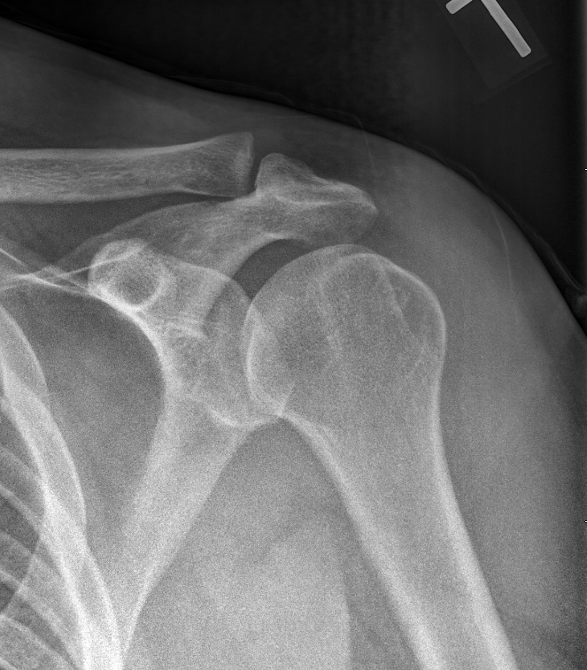
A 21-year-old man presents with shoulder pain after falling off his bike. What is your diagnosis?
What is the pathognomic sign of a traumatic fracture?
What is the absolute pressure threshold for indication of surgical compartment release in compartment syndrome?
Practice by Chapter
Principles of Fracture Management
Practice Questions
Upper Limb Fractures
Practice Questions
Lower Limb Fractures
Practice Questions
Spinal Trauma
Practice Questions
Pelvic and Acetabular Fractures
Practice Questions
Open Fractures
Practice Questions
Fractures in Children
Practice Questions
Fracture Complications
Practice Questions
Nonunion and Malunion
Practice Questions
Polytrauma Management
Practice Questions
Joint Dislocations
Practice Questions
Soft Tissue Injuries
Practice Questions
Want unlimited practice?
Get full access to all questions, explanations, and performance tracking.
Scan to download app