
Trauma — MCQs

On this page
Which of the following conditions shows the vascular sign of Narath?
What is Pauvel's angle?
The Maudsley test is performed to assess for which condition?
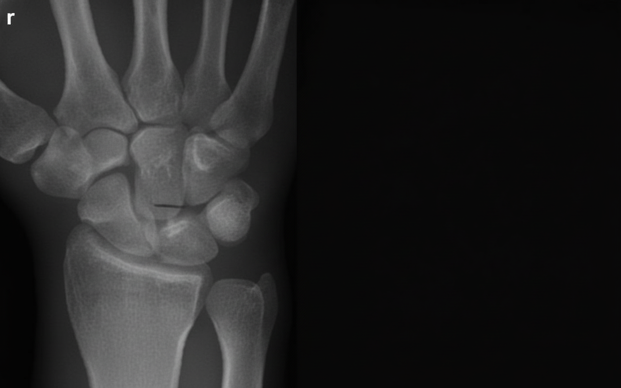
This diagnosis is?
A 40-year-old construction worker is pulled from the rubble after a building collapse, sustaining a comminuted fracture of the right tibia and fibula in his right lower leg. The dorsal pedis and posterior tibial pulses are palpable. The patient reports severe pain, which is worsened with dorsiflexion of the foot, and the calf feels tense. What is the appropriate next step?
Intramedullary fixation is ideal in a case of fracture of the shaft of the femur when there is which of the following conditions?
A segmental compound fracture of the tibia with a 12 cm skin wound, crushed tissue, contamination, and adequate soft tissue coverage, occurring in a farm environment, is classified as which type?
Which of the following is not a complication of Colles fracture?
What is the most common type of anterior shoulder dislocation?
Tardy ulnar nerve palsy is typically seen after which type of injury?
Practice by Chapter
Principles of Fracture Management
Practice Questions
Upper Limb Fractures
Practice Questions
Lower Limb Fractures
Practice Questions
Spinal Trauma
Practice Questions
Pelvic and Acetabular Fractures
Practice Questions
Open Fractures
Practice Questions
Fractures in Children
Practice Questions
Fracture Complications
Practice Questions
Nonunion and Malunion
Practice Questions
Polytrauma Management
Practice Questions
Joint Dislocations
Practice Questions
Soft Tissue Injuries
Practice Questions
Want unlimited practice?
Get full access to all questions, explanations, and performance tracking.
Scan to download app