
Trauma — MCQs

On this page
Regarding the displacement of the distal fragment in a Colles fracture, which statement is true?
Myositis ossificans is due to:
Sudeck's atrophy is more common in which of the following conditions?
Which of the following is NOT a complication of Colles fracture?
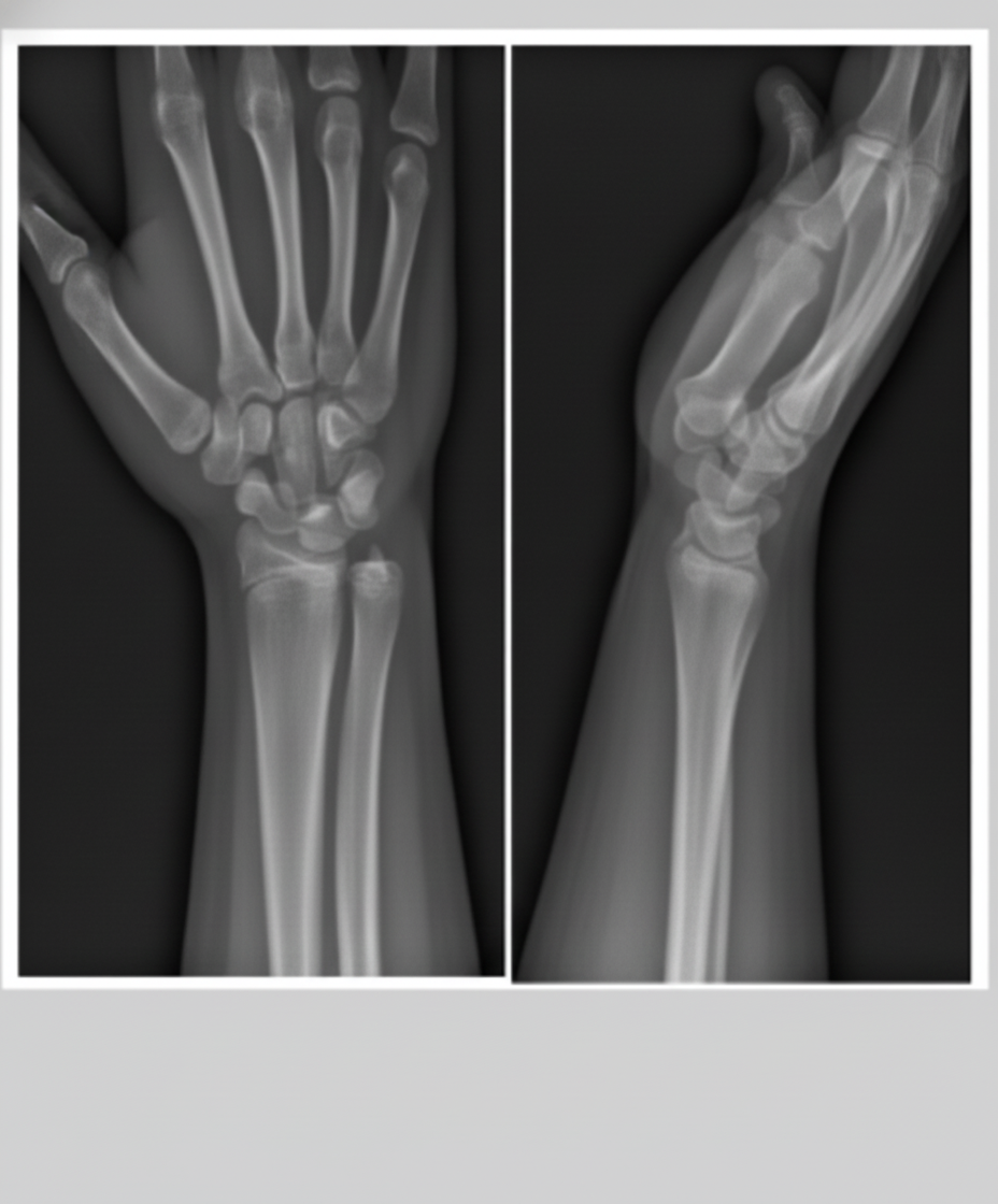
A boy fell from a height onto his outstretched hand. The X-ray of his forearm is shown below. What is the most likely diagnosis?
A 20-year-old male patient presented with acute pain in the left hip region and inability to bear weight on the left hip. On examination, deformity of the left lower limb was noticed. The lower limb was in slight flexion, adduction, and internal rotation. The patient gave a history of a road accident with sudden braking. Neurovascular examination was normal. Which of the following x-rays will most likely correspond to the above condition?
What is the most common complication of a talar neck fracture?
What is the commonest hip injury in elderly patients?
A man operated for fracture femur developed dyspnea, severe chest pain, streaky hemoptysis, and hypotension on the 4th day. What is the most likely cause?
All are true about Barton's fracture except?
Practice by Chapter
Principles of Fracture Management
Practice Questions
Upper Limb Fractures
Practice Questions
Lower Limb Fractures
Practice Questions
Spinal Trauma
Practice Questions
Pelvic and Acetabular Fractures
Practice Questions
Open Fractures
Practice Questions
Fractures in Children
Practice Questions
Fracture Complications
Practice Questions
Nonunion and Malunion
Practice Questions
Polytrauma Management
Practice Questions
Joint Dislocations
Practice Questions
Soft Tissue Injuries
Practice Questions
Want unlimited practice?
Get full access to all questions, explanations, and performance tracking.
Scan to download app