What is a Colles' fracture a fracture of?
Arrange the following nerves according to the incidence of their involvement in a supracondylar fracture of the humerus:
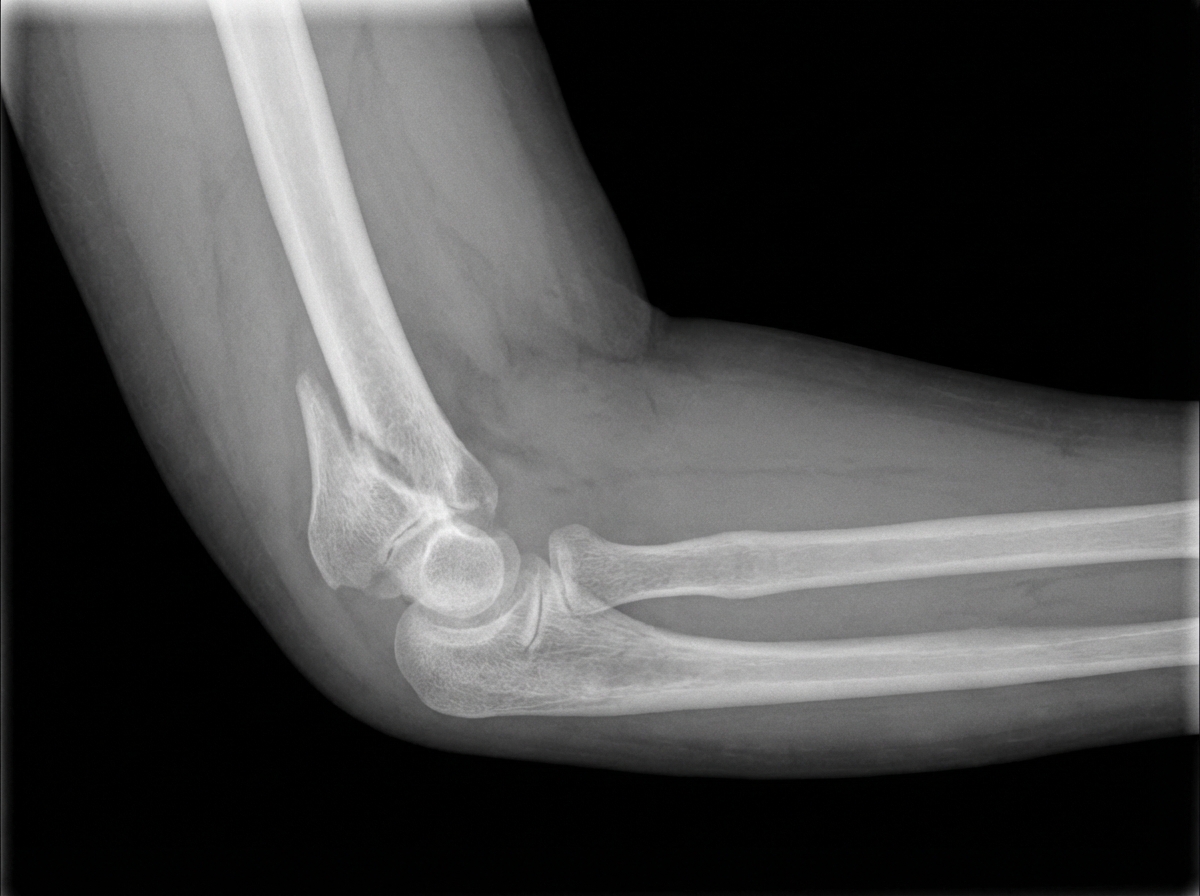
What is the most important factor in fracture healing?
A patient operated for a forearm fracture under general anesthesia with a tourniquet was unable to move his fingers and had sensory loss over the entire hand postoperatively. What is the most common type of nerve injury in this scenario?
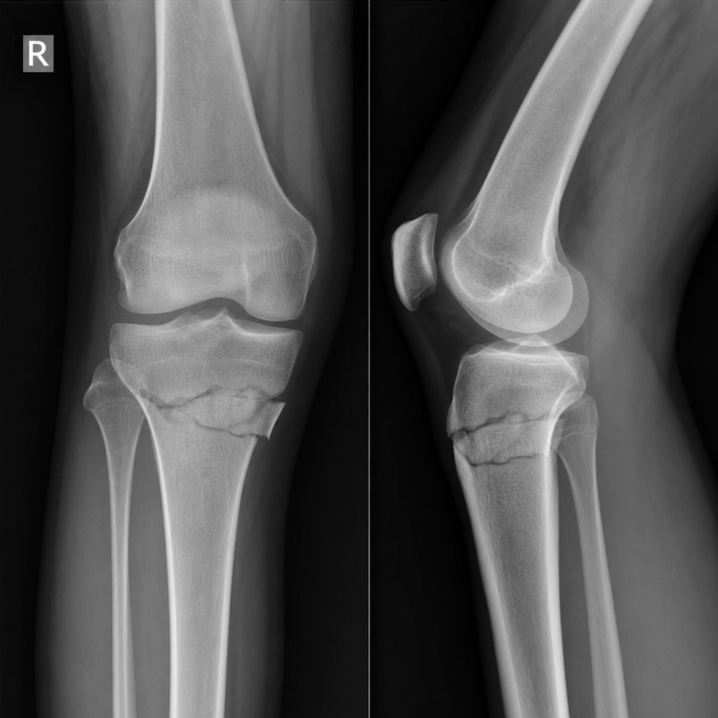
What is the X-ray diagnosis?

A 25-year-old man presents with a blue right arm with absent radial pulse and painful passive finger extension following a supracondylar fracture of the humerus. What condition is he most likely suffering from?
A Tillaux fracture involves which part of the bone?
What is a crescent fracture?
Tinel's sign indicates which of the following?
What is Neurapraxia?
Explanation: **Explanation:** A **Colles' fracture** is classically defined as an **extra-articular fracture of the distal radius** (approximately 2 cm proximal to the radiocarpal joint) with dorsal displacement and angulation. It typically occurs due to a fall on an outstretched hand (FOOSH). **Analysis of Options:** * **Correct Answer (B/D Note):** There appears to be a discrepancy in the provided key. Traditionally, a Colles' fracture is **Extra-articular (Option B)**. However, if the question follows specific recent exam patterns where "Barton’s fracture" (intra-articular) is being contrasted, or if the option marked "Correct" in your prompt is D, it is important to note that **Option D describes a Barton’s fracture**, not a classic Colles'. In standard textbooks (Maheshwari/Apley), Colles' is strictly extra-articular. * **Option A:** Incorrect. This refers to supracondylar or distal humerus fractures, common in children but unrelated to the wrist. * **Option C:** Incorrect. Intra-articular fractures of the distal radius are classified as Barton’s, Chauffeur’s, or Die-punch fractures. **Clinical Pearls for NEET-PG:** 1. **Deformity:** Characterized by the **"Dinner Fork Deformity"** due to dorsal displacement. 2. **Displacements:** There are six classic displacements: Dorsal displacement, Dorsal tilt, Lateral displacement, Lateral tilt, Impaction, and Supination. 3. **Complications:** The most common late complication is **Malunion**. The most common nerve involvement is the **Median nerve** (Carpal Tunnel Syndrome). A specific late complication is the rupture of the **Extensor Pollicis Longus (EPL)** tendon. 4. **Reverse Colles':** Known as **Smith’s fracture**, where the displacement is volar (Garden Spade deformity).
Explanation: **Explanation:** Supracondylar fractures of the humerus are the most common pediatric elbow fractures. Nerve injuries occur in approximately 10–15% of cases, primarily due to the displacement of bone fragments. **1. Why the Correct Answer (B) is Right:** The incidence of nerve involvement follows a specific pattern based on the direction of displacement: * **Anterior Interosseous Nerve (AIN):** This is the **most common** nerve injured overall, particularly in **extension-type** fractures with posterolateral displacement. The AIN is a branch of the median nerve and is susceptible to traction or entrapment. * **Median Nerve:** The main trunk of the median nerve is the second most common, often injured in posterolateral displacement. * **Radial Nerve:** This is typically involved in fractures with **posteromedial** displacement. * **Ulnar Nerve:** This is the **least common** in extension-type fractures but is the most common nerve injured in **flexion-type** fractures (which account for only 5% of cases) or via iatrogenic injury during medial pinning. **2. Why Other Options are Wrong:** * **Option A & C:** These incorrectly place the main trunk of the Median nerve or Radial nerve ahead of the AIN. Clinical studies consistently show AIN palsy (tested by the "OK sign") as the most frequent neurological deficit. * **Option D:** This suggests the Ulnar nerve is the most common, which is only true for the rare flexion-type injury or post-operative pinning complications, not the general incidence. **3. NEET-PG High-Yield Pearls:** * **AIN Testing:** Ask the patient to make an **"OK sign."** Inability to flex the distal phalanx of the thumb and index finger indicates AIN palsy. * **Most common type:** Extension-type (95%). * **Gartland Classification:** Used to grade displacement (Type I: Undisplaced; Type II: Displaced with intact posterior cortex; Type III: Completely displaced). * **Vascular Complication:** Brachial artery injury is the most common vascular concern, potentially leading to **Volkmann’s Ischemic Contracture**.
Explanation: **Explanation:** The primary requirement for fracture healing is **Immobilization**. For a fracture to heal, the "callus" (the bridge of new bone) must form across the gap. Excessive movement at the fracture site creates shear forces that disrupt the delicate capillary ingrowth and the formation of the fibrocartilaginous bridge. If the fracture site is not sufficiently immobilized, the body may form a "non-union" or a "pseudoarthrosis" (false joint) instead of solid bone. **Analysis of Options:** * **A. Good alignment:** While alignment is important for functional recovery and preventing deformity (malunion), a fracture can still heal (unite) even if it is poorly aligned. * **B. Organization of blood clot:** While the hematoma is the first stage of fracture healing and provides a fibrin scaffold, it is not the *most* important factor. Healing can still occur in the absence of a significant clot (e.g., in primary bone healing via internal fixation). * **C. Accurate reduction and 100% apposition:** This is a common misconception. While 100% apposition is ideal, many fractures (like the clavicle or ribs) heal perfectly well with minimal contact or significant displacement, provided they are kept still. **High-Yield Clinical Pearls for NEET-PG:** * **Primary Bone Healing:** Occurs with absolute stability (e.g., compression plating). There is **no callus formation**. * **Secondary Bone Healing:** Occurs with relative stability (e.g., POP cast, intramedullary nailing). This involves the classic stages: Hematoma → Inflammation → Soft Callus → Hard Callus → Remodeling. * **The "Rule of Two":** For adequate immobilization, a cast should generally include the joint above and the joint below the fracture site. * **Most common cause of Non-union:** Inadequate immobilization or poor blood supply.
Explanation: ### Explanation The correct answer is **Neuropraxia**. **1. Why Neuropraxia is correct:** The clinical scenario describes a **Tourniquet Palsy**. This occurs due to mechanical compression and local ischemia caused by the tourniquet cuff. In such cases, the nerve injury is almost always **Neuropraxia** (Seddon’s Classification). * **Mechanism:** The pressure causes a focal conduction block due to segmental demyelination, but the **axon remains intact**. * **Clinical Presentation:** There is motor paralysis and loss of proprioception/vibration, while pain and temperature sensation are often spared (though the question mentions "entire hand" sensory loss, the transient nature post-op points to neuropraxia). * **Prognosis:** Since there is no Wallerian degeneration, recovery is spontaneous and complete, usually within weeks. **2. Why other options are incorrect:** * **Axonotmesis:** This involves the disruption of the axon but preservation of the connective tissue sheath (endoneurium). It leads to Wallerian degeneration and requires regeneration (1mm/day). It is less common in simple tourniquet compression and usually results from more severe crush injuries or fractures. * **Neurotmesis:** This is the most severe form, where both the axon and the connective tissue sheath are completely severed. It requires surgical intervention for recovery and is not caused by standard tourniquet use. **3. Clinical Pearls for NEET-PG:** * **Seddon’s Classification:** Neuropraxia (Mildest) → Axonotmesis → Neurotmesis (Most severe). * **Sunderland’s Classification:** Expands this into 5 degrees (1st degree = Neuropraxia; 5th degree = Neurotmesis). * **Tourniquet Safety:** To prevent palsy, the recommended pressure is **100 mmHg above systolic BP** for the upper limb and **2x systolic BP** (or 100-150 mmHg above) for the lower limb. * **Time Limit:** The safe tourniquet time is generally **up to 2 hours**. If longer is needed, the cuff should be deflated for 10–15 minutes (breathing period) before re-inflation.
Explanation: ***Supracondylar humerus fracture*** - The **anterior humeral line** does not pass through the middle third of the **capitellum**, a key radiographic sign for supracondylar fractures. - This is the **most common pediatric elbow fracture**, typically occurring after a fall on an outstretched hand with hyperextension injury. *Olecranon fracture* - Would show a clear **fracture line** through the **olecranon process** of the ulna, which is not present here. - Typically causes inability to actively extend the elbow due to **triceps muscle disruption**. *Humerus shaft fracture* - The fracture line would be located in the **mid-shaft** of the humerus, not near the elbow joint. - Associated with **radial nerve palsy** causing wrist drop and loss of thumb extension. *Elbow dislocation* - Shows **displacement** of the radius and ulna relative to the humerus with loss of normal joint alignment. - The **radiocapitellar line** would not pass through the center of the capitellum in true dislocations.
Explanation: ### Explanation The patient is presenting with the classic signs of **Volkmann’s Ischemia**, a precursor to Volkmann’s Ischemic Contracture (VIC), which is a manifestation of **Compartment Syndrome**. **Why Compartment Syndrome is correct:** Supracondylar fractures of the humerus (especially the displaced extension type) are the most common cause of compartment syndrome in the forearm. The "blue arm" and "absent radial pulse" indicate vascular compromise (brachial artery involvement), while **painful passive finger extension** is the **most sensitive and earliest clinical sign** of muscle ischemia within the deep flexor compartment. This occurs because stretching the ischemic muscles causes intense pain. **Why the other options are incorrect:** * **Sudeck’s Atrophy (Complex Regional Pain Syndrome):** This is a chronic condition characterized by post-traumatic autonomic dysfunction, leading to burning pain, swelling, and trophic skin changes. It does not present acutely with pulse loss. * **Median Nerve Injury:** While the median nerve is commonly injured in supracondylar fractures, it would present with sensory loss (lateral 3.5 fingers) and motor weakness (ape thumb deformity), not a cold, pulseless, blue limb. * **Myositis Ossificans:** This is a late complication involving heterotopic ossification within muscles (usually the brachialis). It presents as progressive stiffness and a palpable mass weeks after the injury, often aggravated by forceful massage. **High-Yield Clinical Pearls for NEET-PG:** * **The 5 P’s of Compartment Syndrome:** Pain (out of proportion), Pallor, Paresthesia, Pulselessness, and Paralysis. * **Earliest Sign:** Pain on passive stretching of muscles. * **Most Common Site:** Deep posterior compartment of the leg and the volar compartment of the forearm. * **Pressure Threshold:** Diagnosis is confirmed if the intracompartmental pressure is **>30 mmHg** or if the Delta pressure (Diastolic BP - Compartment Pressure) is **<30 mmHg**. * **Management:** Immediate removal of tight bandages/casts; if no improvement, urgent **fasciotomy**.
Explanation: **Explanation:** A **Tillaux fracture** is a specific type of avulsion fracture involving the **anterolateral tubercle of the distal (lower end) tibia**. **1. Why Option A is correct:** The fracture occurs due to an external rotation force of the foot, which puts extreme tension on the **Anterior Inferior Tibiofibular Ligament (AITFL)**. Instead of the ligament tearing, it pulls off a bony fragment from its insertion site on the lateral aspect of the distal tibia. In pediatric populations, this is known as a **Juvenile Tillaux fracture** (a Salter-Harris Type III injury), occurring because the medial part of the distal tibial growth plate closes before the lateral part, leaving the lateral side vulnerable to avulsion. **2. Why the other options are incorrect:** * **Upper end tibia (B):** Fractures here are typically Tibial Plateau fractures or Segond fractures (avulsion of the lateral capsule). * **Lower end femur (C):** Common injuries include Supracondylar or Intercondylar fractures, often due to high-energy trauma. * **Upper end femur (D):** This involves Neck of Femur or Intertrochanteric fractures, common in geriatric falls or high-impact trauma. **Clinical Pearls for NEET-PG:** * **Mechanism:** Forced external rotation of the ankle. * **Radiology:** Best seen on an AP view of the ankle; however, **CT scans** are the gold standard to assess the degree of displacement and articular involvement. * **Management:** Displaced fractures (>2mm) require Open Reduction and Internal Fixation (ORIF) to prevent secondary osteoarthritis. * **Differentiate:** Do not confuse this with a **Wagstaffe-Le Fort fracture**, which is an avulsion of the AITFL from the **fibula** rather than the tibia.
Explanation: **Explanation:** A **Crescent fracture** is a specific type of unstable pelvic ring injury. It is a fracture-dislocation of the sacroiliac (SI) joint where a vertical fracture line runs through the posterior portion of the **iliac crest** (the posterior superior iliac spine), leaving a "crescent-shaped" fragment attached to the sacrum via the posterior SI ligaments. **Analysis of Options:** * **A. Fracture of the iliac crest (Correct):** This is the defining feature of the crescent fracture. It typically occurs due to a Lateral Compression (LC) injury (Young-Burgess Type II). The force causes the iliac bone to fracture posteriorly while the anterior SI ligaments are disrupted. * **B. Fracture of the coccyx:** These are usually isolated injuries resulting from a direct fall on the buttocks and are not associated with the "crescent" eponym. * **C. Fracture of the calcaneum:** Common eponyms for calcaneal fractures include "Lover’s fracture" or "Don Juan fracture." * **D. Fracture of the Atlas:** A fracture of the C1 vertebra is known as a **Jefferson fracture**. **High-Yield Clinical Pearls for NEET-PG:** * **Classification:** Crescent fractures are categorized under the **Young-Burgess classification** as **Lateral Compression Type II (LC-II)**. * **Stability:** These are considered rotationally unstable but vertically stable injuries. * **Management:** Surgical stabilization (ORIF) is often required to restore the pelvic ring integrity and prevent long-term gait abnormalities or chronic pain. * **Radiology:** On an AP view of the pelvis, look for a vertical fracture line through the ilium that spares the sacroiliac joint surface itself.
Explanation: **Explanation:** **Tinel’s sign** is a clinical indicator of **nerve regeneration**. It is elicited by percussing along the course of a damaged nerve. A positive sign is characterized by a "pins and needles" sensation or tingling in the distal distribution of the nerve. 1. **Why Regeneration is Correct:** When a peripheral nerve undergoes repair, the regenerating axonal sprouts (growth cones) are thin and lack a mature myelin sheath. These immature axons are **hyperexcitable** and highly sensitive to mechanical pressure. Percussing these regenerating fibers triggers an electrical impulse, signaling that the nerve is successfully growing distally (typically at a rate of 1 mm/day). 2. **Why Other Options are Incorrect:** * **Atrophy of nerves:** Atrophy refers to the wasting of muscles or degeneration of nerve tissue, which does not produce an evocative sensory response to percussion. * **Neuroma:** While tapping a neuroma (a disorganized bulb of nerve fibers) can cause pain, Tinel’s sign specifically refers to the *progression* of the tingling sensation down the limb, which tracks recovery rather than just the presence of a lesion. * **Injury to nerve:** Immediately after an injury (like neurotmesis), Tinel’s sign is absent. It only becomes positive once regeneration begins. **NEET-PG High-Yield Pearls:** * **Hoffmann-Tinel Sign:** The full name of the sign. * **Advancing Tinel’s Sign:** If the point of maximum tingling moves distally over time, it is a **good prognostic sign** indicating recovery. * **Static Tinel’s Sign:** If the tingling remains at the site of injury and does not move distally, it suggests a **neuroma** and poor regenerative progress. * **Carpal Tunnel Syndrome:** Tinel’s sign is also used diagnostically here to indicate nerve compression/irritation at the wrist.
Explanation: **Explanation:** **Neurapraxia** is the mildest form of nerve injury according to the **Seddon Classification**. It is characterized by a **reversible physiological conduction block** without any anatomical disruption of the axon or its connective tissue coverings (endoneurium, perineurium, or epineurium). 1. **Why Option D is Correct:** The underlying mechanism is typically focal demyelination or local ischemia caused by mild compression or traction. Since the axon remains intact, there is no Wallerian degeneration. Once the pressure is relieved or the myelin is repaired, nerve conduction is fully restored. 2. **Why Other Options are Incorrect:** * **Option A (Complete division):** This describes **Neurotmesis**, the most severe form of injury where the entire nerve trunk is severed. Recovery is impossible without surgical intervention. * **Option B (Axonal interruption):** This describes **Axonotmesis**. Here, the axon is damaged, leading to Wallerian degeneration, but the supporting connective tissue framework remains intact, allowing for guided regeneration. * **Option C (Irreversible injury):** Neurapraxia is inherently **reversible**. Recovery is usually spontaneous and rapid, occurring within days to a few weeks (typically 3–6 weeks). **High-Yield Clinical Pearls for NEET-PG:** * **Wallerian Degeneration:** Absent in Neurapraxia; present in Axonotmesis and Neurotmesis. * **Tinel’s Sign:** Negative in Neurapraxia (as there is no regenerating axonal bud); Positive in Axonotmesis. * **Common Example:** "Saturday Night Palsy" (radial nerve compression) is a classic clinical presentation of neurapraxia. * **EMG/NCV:** In neurapraxia, there is a conduction block at the site of injury, but distal stimulation remains normal.
Principles of Fracture Management
Practice Questions
Upper Limb Fractures
Practice Questions
Lower Limb Fractures
Practice Questions
Spinal Trauma
Practice Questions
Pelvic and Acetabular Fractures
Practice Questions
Open Fractures
Practice Questions
Fractures in Children
Practice Questions
Fracture Complications
Practice Questions
Nonunion and Malunion
Practice Questions
Polytrauma Management
Practice Questions
Joint Dislocations
Practice Questions
Soft Tissue Injuries
Practice Questions
Get full access to all questions, explanations, and performance tracking.
Start For Free