Thomas splint is used for immobilizing fractures of ?
Callus formation is seen between what duration of fracture healing?
Most common complication of mid shaft humerus fracture is ?
Garden's classification used for which fracture?
Which of the following factors does NOT indicate an unstable pelvis?
Which part of scaphoid fracture is most susceptible to avascular necrosis?
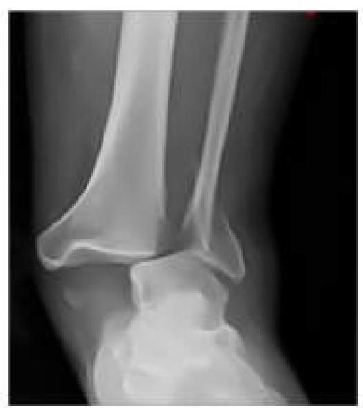
An RTA patient presented to the emergency department with severe pain in the ankle. An X-ray was performed, given below. What is the best next step in management?

As an intern in the emergency department, you encounter four patients with different types of fractures. Which patient should you prioritize for orthopedic consultation based on the severity of their condition?
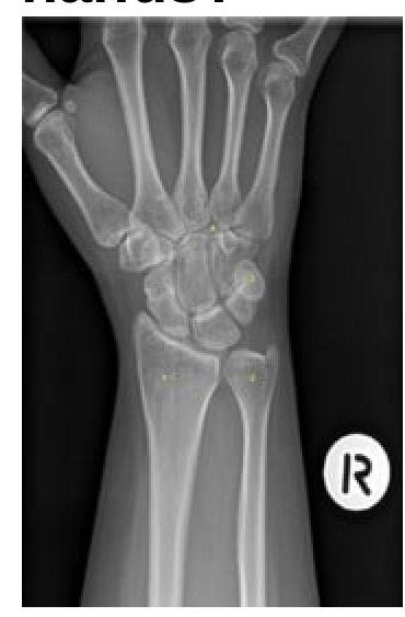
Identify the bone numbered in the X-ray that most commonly fractures when a person falls on outstretched hands.

What is meant by perilunate dislocations?
Explanation: ***Femur*** - The **Thomas splint** is primarily designed for the temporary immobilization and traction of **femoral shaft fractures**. - Its design allows for effective stabilization and helps reduce muscle spasms and pain associated with these types of fractures. *Tibia* - While it can be used in some lower limb injuries, the Thomas splint is not the primary or most effective device for isolated **tibial fractures**. - Other splints, such as a **long leg posterior splint** or an air splint, are typically preferred for tibial immobilization. *Radius* - The **radius** is a bone in the forearm, and its fractures are typically managed with a variety of **short arm casts** or splints. - The Thomas splint is specifically designed for the much longer and larger **femur** and would be completely unsuitable for a forearm injury. *Ulna* - Similar to the radius, the **ulna** is a forearm bone, and its fractures require immobilization techniques appropriate for the upper limb. - The **Thomas splint's bulk** and mechanism are inappropriate for the immobilization of forearm bones.
Explanation: ***4 - 12 weeks*** - This period aligns with the **reparative phase** of fracture healing, where both **soft callus** (fibrocartilaginous) and then **hard callus** (woven bone) are formed. - Callus formation during this time significantly contributes to the **mechanical stability** of the fracture site. *0 - 2 weeks* - This initial phase primarily involves the **inflammatory stage**, characterized by **hematoma formation** and the recruitment of inflammatory cells. - While essential for initiating healing, significant structural callus formation does not typically occur within this timeframe. *2 - 4 weeks* - This period marks the transition from the inflammatory to the reparative phase, with the initial development of a **soft callus**. - However, the more robust and radiographically evident **hard callus formation** extends beyond this period. *12 - 16 weeks* - By this stage, the fracture is generally in the **remodeling phase**, where the woven bone of the hard callus is gradually replaced by **lamellar bone**. - While callus is present, its formation is more accurately described in the earlier 4-12 week period; this stage focuses on refinement and strengthening.
Explanation: ***Radial nerve palsy*** - The **radial nerve** runs in close proximity to the **spiral groove** of the humerus, making it highly vulnerable to injury in mid-shaft fractures. - This results in the characteristic **wrist drop** and loss of sensation over the dorsal aspect of the hand. *Median nerve palsy* - The **median nerve** is not typically affected by mid-shaft humeral fractures as it is located more medially and anteriorly. - Injury to the median nerve is more common with supracondylar fractures of the humerus or carpal tunnel syndrome. *Nonunion* - While **nonunion** (failure of bone healing) occasionally occurs after mid-shaft humerus fractures, **radial nerve palsy** is a more immediate and frequent complication observed at the time of injury. - Risks for nonunion include severe trauma, soft tissue interposition, and inadequate immobilization. *Malunion* - **Malunion** (healing in an unacceptable alignment) can occur, especially with conservative management, but like nonunion, it is a complication of the healing process rather than an acute injury presentation. - Functional outcomes are generally good even with some degree of angulation in humeral shaft fractures.
Explanation: ***Neck of femur*** - **Garden's classification** is a widely used system to categorize **femoral neck fractures** based on displacement. - This classification helps guide treatment decisions, as different grades of displacement have varying prognoses for **avascular necrosis** and **non-union**. *Fracture of the surgical neck of the humerus* - Fractures of the surgical neck of the humerus are typically classified using the **Neer classification system**. - The Neer classification is based on the number of displaced parts, such as the **humeral head**, **greater tuberosity**, **lesser tuberosity**, and **humeral shaft**. *Fracture of the shaft of the humerus* - Fractures of the humeral shaft are generally classified by their **location** (e.g., proximal, middle, distal third), **morphology** (e.g., spiral, transverse, oblique, comminuted), and the presence of **open vs. closed injuries**. - There is no specific, widely recognized classification system comparable to Garden's used exclusively for these fractures. *Fracture of the shaft of the femur* - Fractures of the femoral shaft are commonly classified based on their **location** (e.g., proximal, middle, distal third), **morphology** (e.g., transverse, oblique, spiral, comminuted), and the presence of **segmental or open fractures**. - The **Winquist and Hansen classification** is sometimes used for comminuted femoral shaft fractures, but Garden's classification is not applicable here.
Explanation: ***Avulsion fractures of the L5 transverse process*** - While significant, avulsion fractures of the **L5 transverse process** are generally considered indicative of a **high-energy trauma** and potential for associated injuries, but they do not directly represent **pelvic instability** in the same way as disruptions to the pelvic ring itself. - These fractures point to powerful shearing forces but don't disrupt the **structural integrity** of the pelvic ring that dictates its stability. *Posterior sacroiliac complex displacement by > 1 cm* - This degree of displacement signifies a substantial injury to the **strong posterior ligamentous structures** of the sacroiliac joint. - Such displacement indicates a highly unstable pelvic ring, often associated with significant hemorrhage and requiring surgical stabilization. *Avulsion fracture of sacral or ischial end of the sacrospinous ligament* - An avulsion fracture at either the sacral or ischial attachment of the **sacrospinous ligament** suggests a severe disruption of the posterior pelvic ring. - The **sacrospinous ligament** is a key stabilizer of the pelvis, and injury to it indicates pelvic instability due to the loss of its anchoring function between the sacrum and ischium. *Isolated disruption of pubic symphysis with pubic diastasis of 2 cm.* - A **pubic diastasis of 2 cm** signifies a significant opening of the pubic symphysis, which is a critical anterior component of the pelvic ring. - This indicates **anterior pelvic instability**, even in the absence of posterior injuries, as the normal interlocking mechanism of the symphysis is compromised.
Explanation: ***Proximal 1/3rd*** - The **scaphoid bone** has a **retrograde blood supply**, meaning blood vessels enter distally and flow towards the proximal pole. - A fracture in the **proximal 1/3rd** can disrupt the blood supply to the **proximal fragment**, making it highly susceptible to **avascular necrosis**. *Distal 1/3rd* - Fractures in the **distal 1/3rd** of the scaphoid generally have a robust blood supply due to the entry of vessels from the distal pole. - While still requiring proper management, the risk of **avascular necrosis** is significantly lower compared to proximal fractures. *Middle 1/3rd* - Fractures in the **middle 1/3rd** (waist) of the scaphoid are common and can still compromise blood flow to the proximal segment, but the risk of **avascular necrosis** is intermediate. - The more proximal the fracture within the middle third, the higher the risk of **avascular necrosis**. *Scaphoid Tubercle* - The **scaphoid tubercle** is a distal projection of the scaphoid bone. - Fractures of the **scaphoid tubercle** are extra-articular and typically have an excellent blood supply; thus, they are at very low risk for **avascular necrosis**.
Explanation: ***Neurovascular Assessment and Closed reduction with slab application*** - The X-ray shows an **ankle dislocation without an obvious fracture**, making **closed reduction** the appropriate initial treatment. - A **slab (splint)** is preferred over a full cast initially for acute injuries to accommodate for swelling, reducing the risk of compartment syndrome, and allowing for serial neurovascular checks. *Neurovascular Assessment and Closed reduction with cast application* - While closed reduction is correct, applying a **full cast** immediately after an acute injury carries a risk of **compartment syndrome** due to potential swelling that cannot be accommodated by a rigid cast. - A cast would typically be applied after the initial swelling has subsided, usually a few days to a week after initial reduction and splinting. *Neurovascular Assessment and Immediate surgery* - **Immediate surgery** is generally reserved for **open fractures/dislocations**, dislocations that cannot be reduced closed (irreducible dislocations), or those with significant associated fractures that require surgical fixation to stabilize the joint. - In this case, the dislocation appears to be isolated and amenable to closed reduction, making surgery not the immediate next step. *Neurovascular Assessment and Immediate open reduction* - **Open reduction** is performed when closed reduction fails or is contraindicated, for example, due to soft tissue interposition or highly unstable fracture patterns. - Since closed reduction has not yet been attempted, immediate open reduction is premature and unnecessary for an apparently simple dislocation.
Explanation: ***Patient's finger shows signs of severe vascular compromise*** - **Vascular compromise** is an orthopedic emergency as it can lead to **tissue ischemia**, necrosis, and permanent loss of function if not addressed immediately. - Urgent consultation is needed to restore **blood flow** and prevent irreversible damage to the limb. *Patient can't extend his arm* - This symptom suggests a potential **nerve injury** (e.g., radial nerve) or **tendon rupture**, which requires prompt evaluation but is generally less immediately life-threatening than vascular compromise. - While important, **nerve injuries** typically do not require the same emergent intervention as severe vascular compromise unless there's a rapidly deteriorating neurological deficit. *Intra articular fracture of Elbow Joint* - **Intra-articular fractures** involve the joint space and can lead to long-term complications like **arthrosis** and stiffness if not managed properly, often requiring surgical fixation. - Although significant, it is not as urgent as vascular compromise, which can lead to rapid **tissue death**. *Simple closed fracture of the radius with good distal circulation* - A **simple closed fracture** with **good distal circulation** is a stable injury that is typically managed non-surgically or with elective surgical intervention. - This type of fracture does not present an immediate threat to limb viability.
Explanation: ***Distal radius*** - The **distal radius** is the bone most frequently fractured when a person falls on an **outstretched hand**. This is commonly known as a **Colles fracture** if the fragment is displaced dorsally. - The force of impact is transmitted directly to the distal end of the radius, making it susceptible to fracture, especially in cases of **osteoporosis**. *Scaphoid* - The **scaphoid** bone is also commonly fractured with a fall on an outstretched hand, but it is less frequent than the distal radius. - A scaphoid fracture is concerning due to its **precarious blood supply**, which can lead to **avascular necrosis** and non-union. *Lunate* - The **lunate** bone is centrally located in the wrist and is less commonly fractured but can be dislocated or injured in high-impact trauma. - Injuries to the lunate are often associated with **perilunate dislocations**, which are severe wrist injuries. *Capitate* - The **capitate** is the largest carpal bone, located in the center of the wrist, and is rarely fractured in isolation from a fall on an outstretched hand. - Fractures of the capitate are often associated with more extensive carpal injuries due to its robust nature and central, protected position.
Explanation: ***Lower radius, scaphoid, and lunate in alignment; capitate alone is out of plane.*** - In a **perilunate dislocation**, the lunate maintains its normal relationship with the **distal radius**, and the scaphoid often remains aligned with the lunate. - The other **carpal bones**, particularly the **capitate**, are dislocated dorsally or radially relative to the lunate. *Lower radius, scaphoid, and capitate in alignment; lunate alone out of plane.* - This description corresponds to a **lunate dislocation**, where the lunate is displacedvolarly while the other carpal bones remain aligned with the radius. - In a lunate dislocation, the **capitate** remains aligned with the distal radius and scaphoid, but the **lunate** is displaced. *Both lunate and capitate are out of plane.* - While possible in severe wrist trauma, this specific combined displacement doesn't precisely define a typical perilunate dislocation, where the **lunate** is usually the stable point. - This scenario would represent a more complex or severely disrupted carpal injury. *Lower radius, scaphoid, lunate, and capitate all in the same plane.* - This describes a **normal alignment** of the carpal bones and distal radius. - None of the bones are dislocated or out of their usual anatomical plane relative to each other.
Principles of Fracture Management
Practice Questions
Upper Limb Fractures
Practice Questions
Lower Limb Fractures
Practice Questions
Spinal Trauma
Practice Questions
Pelvic and Acetabular Fractures
Practice Questions
Open Fractures
Practice Questions
Fractures in Children
Practice Questions
Fracture Complications
Practice Questions
Nonunion and Malunion
Practice Questions
Polytrauma Management
Practice Questions
Joint Dislocations
Practice Questions
Soft Tissue Injuries
Practice Questions
Get full access to all questions, explanations, and performance tracking.
Start For Free