A 25-year-old male presents with a three-month history of lower back pain, following a fall. He reports mild weakness in both lower limbs but can walk without support. He has approximately 30% sensory loss and exhibits bladder symptoms. Physical examination reveals tenderness over D12-L1 vertebrae. X-ray shows paradiscal destruction of vertebrae, and MRI reveals destruction with indentation of the thecal sac. What is the most appropriate management?
Which of the following spine deformities is being evaluated?

Which of the following statements about spinal injuries is false?
Spondylolysis is more common in which anatomical location?
In a case of tuberculosis of the thoracic spine, what is the earliest sign of cord compression?
The Denis classification is used to assess which of the following?
In which of the following conditions does pseudoclaudication occur?
What is the most common cause of acute sciatica?
Adam's test is performed for which of the following?
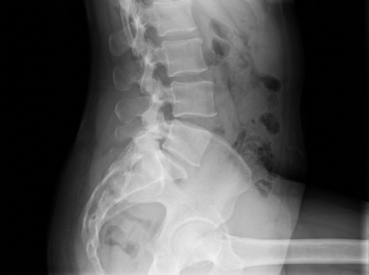
A patient with traumatic paraplegia due to injury of the T3 thoracic cord level presents with a blood pressure of 210/120. What should be the initial management?
Explanation: ### Explanation The clinical presentation of chronic back pain, **paradiscal destruction** on X-ray, and MRI evidence of thecal sac indentation in a young patient is highly suggestive of **Pott’s Spine (Tuberculous Spondylitis)**. **Why Option D is Correct:** The management of Pott’s spine is primarily medical (ATT); however, surgery is indicated in specific scenarios. This patient exhibits **neurological deficits** (lower limb weakness and sensory loss) and, most critically, **bladder symptoms**. Bladder involvement in spinal tuberculosis indicates significant cord or cauda equina compression and is an absolute indication for **urgent surgical decompression** along with ATT to prevent permanent neurological damage. **Why Other Options are Wrong:** * **Option A (Observation):** Pott’s spine is a progressive infectious disease; observation would lead to worsening deformity and paralysis. * **Option B & C (ATT alone):** While ATT is the mainstay of treatment, "middle-path" conservative management is only suitable for patients without significant neurological deficits. Once bladder involvement or progressive motor weakness occurs, medical therapy alone is insufficient. **Clinical Pearls for NEET-PG:** * **Most common site of Pott’s Spine:** Lower Thoracic and Upper Lumbar (D12-L1 is a classic "high-yield" location). * **Earliest X-ray sign:** Narrowing of the disc space (paradiscal type is most common). * **Indications for Surgery (The "4 Ps"):** **P**aralysis (onset/worsening), **P**us (large cold abscess), **P**ain (intractable), and **P**osture (progressive kyphosis/deformity). * **Bladder involvement** is a late sign in spinal TB but signifies an emergency requiring decompression.
Explanation: ***Scoliosis*** - **Lateral curvature** of the spine visible on posterior view, often with **rotational component** causing rib prominence - Diagnosed using **Cobb angle measurement** on X-ray and **Adam's forward bend test** to assess rib hump *Kyphosis* - **Anterior flexion** or **forward curvature** of the spine, typically in the thoracic region, creating a **hunched back** appearance - Best visualized on **lateral view** rather than posterior view, unlike the lateral deviation seen here *Gibbus* - **Sharp angular kyphosis** caused by vertebral collapse from **tuberculosis** or **fracture**, creating a distinct angular prominence - Presents as a **localized hump** rather than the smooth lateral curvature pattern observed *Lordosis* - **Exaggerated inward curvature** of the lumbar spine creating increased **lumbar concavity** - Appears as **swayback posture** on lateral view, not as lateral spinal deviation seen in this evaluation
Explanation: ### Explanation **1. Why Option B is False (The Correct Answer):** In the cervical spine, **fractures are more common than dislocations.** While the cervical spine is the most mobile segment and has relatively horizontal facet joints (making it more susceptible to subluxation compared to the lumbar spine), the mechanical forces required to cause injury typically result in bony fractures (e.g., vertebral body or arch fractures) more frequently than pure ligamentous disruptions or facet dislocations. **2. Analysis of Other Options:** * **Option A (True):** Epidemiological data indicates that spinal injuries account for approximately **6% of all trauma admissions**. * **Option C (True):** The **cervical spine** is the most commonly injured region of the spinal column due to its high mobility and the weight of the head (the "bowling ball" effect). Within the cervical spine, C2 is the most common site of injury, followed by C5-C6. * **Option D (True):** Approximately **40-50%** of patients with spinal column injuries present with some degree of neurological deficit (complete or incomplete spinal cord injury). **3. High-Yield Clinical Pearls for NEET-PG:** * **Most common site of spinal fracture:** The **Thoracolumbar junction (T12-L1)** is the most common site for fractures due to the transition from the rigid thoracic spine to the mobile lumbar spine. * **Most common cervical fracture:** The **Atlas (C1)** and **Axis (C2)** are frequently involved, with Odontoid fractures being highly significant. * **Jefferson Fracture:** A burst fracture of C1 (Atlas) caused by axial loading. * **Hangman’s Fracture:** Traumatic spondylolisthesis of C2 (Axis) involving the pars interarticularis. * **Chance Fracture:** A "seatbelt injury" resulting in a horizontal distraction fracture of the thoracolumbar spine.
Explanation: **Explanation:** **Spondylolysis** refers to a bony defect or stress fracture in the **pars interarticularis** of the vertebral arch. The pars interarticularis is the specific segment of bone between the superior and inferior articular processes of a vertebra. This area is anatomically vulnerable to repetitive mechanical stress, particularly hyperextension, which leads to micro-fractures. It most commonly occurs at the **L5 level (90%)**, followed by L4. **Analysis of Options:** * **Pars interarticularis (Correct):** This is the classic site of the defect. On an oblique X-ray of the lumbar spine, this defect appears as a "break in the neck" of the **Scotty Dog sign**. * **Intervertebral disc & Annulus fibrosus (Incorrect):** These are soft tissue components of the spine. While they are involved in disc herniations (prolapse), spondylolysis is strictly a bony pathology. * **Anterior pars (Incorrect):** This is not a standard anatomical term used to describe the site of this fracture. The defect is specifically localized to the posterior elements of the vertebra. **Clinical Pearls for NEET-PG:** 1. **Spondylolisthesis:** If the bilateral pars defect leads to the forward slipping of one vertebra over another, the condition is termed Spondylolisthesis. 2. **Most Common Site:** L5 is the most common site for spondylolysis; however, for **degenerative** spondylolisthesis, **L4 over L5** is more common. 3. **Imaging:** The **Oblique view** X-ray is best for visualizing the "Scotty Dog," but **CT scan** is the gold standard for diagnosing the bony defect. 4. **Population:** It is frequently seen in young athletes involved in gymnastics, diving, or fast bowling (cricket) due to repeated lumbar hyperextension.
Explanation: In Tuberculosis of the spine (Pott’s disease), cord compression typically occurs due to an abscess, granulation tissue, or bony sequestration. Because the thoracic spine is the most common site for Pott's paraplegia, the compression affects the **Upper Motor Neuron (UMN)** pathways. ### Why "Extensor Plantar Response" is Correct The earliest clinical manifestation of spinal cord compression is the loss of inhibitory control from the corticospinal tracts. This leads to **Babinski’s sign (Extensor Plantar Response)**. It precedes subjective motor weakness or sensory changes because the long tracts are highly sensitive to early mechanical pressure or ischemia. At this stage, the patient may be asymptomatic or have only "clumsy" gaits, but the physical exam will reveal the extensor response. ### Why Other Options are Incorrect * **Motor Weakness:** This is usually the first *symptom* the patient notices (heaviness or dragging of feet), but it occurs after the reflex changes are already present. * **Sensory Loss:** Sensory fibers are generally more resistant to pressure than motor fibers. Sensory loss (numbness/paresthesia) typically appears after motor deficits. * **Bladder Dysfunction:** This is a **late sign** of cord compression. Its presence indicates significant, advanced compression and carries a poorer prognosis for recovery. ### High-Yield Clinical Pearls for NEET-PG * **Sequence of involvement:** Reflex changes (Extensor plantar) → Motor weakness → Sensory loss → Bladder/Bowel involvement. * **Most common site of Pott's Spine:** Lower Thoracic / Dorso-lumbar junction. * **Earliest Radiological Sign:** Diminution/narrowing of the disc space (due to destruction of the subchondral bone). * **Cold Abscess in Thoracic Spine:** Appears as a "Bird's Nest" or fusiform shadow on X-ray.
Explanation: **Explanation:** The **Denis Classification** is the most widely used system for assessing **spinal stability** and injury patterns in the thoracolumbar spine. It is based on the **Three-Column Concept**, which divides the spinal segment into: 1. **Anterior Column:** Anterior longitudinal ligament (ALL) and the anterior two-thirds of the vertebral body/disc. 2. **Middle Column:** Posterior one-third of the vertebral body/disc and the posterior longitudinal ligament (PLL). 3. **Posterior Column:** Posterior elements (pedicles, laminae, facets) and the posterior ligamentous complex. **Why Option A is correct:** According to Denis, the **middle column** is the key to spinal stability. If two or more columns are disrupted, the spine is considered unstable. This classification helps surgeons decide between conservative management and surgical stabilization. **Why other options are incorrect:** * **Option B:** Calcium content is typically assessed via DEXA scans (T-scores) or Singh’s Index (for osteoporosis). * **Option C:** Tumor invasion is often graded using the **Enneking classification** (for staging) or the **SINS score** (Spinal Instability Neoplastic Score). * **Option D:** Femur head subluxation in dysplastic hips (DDH) is assessed using the **Crowe** or **Hartofilakidis** classifications. **High-Yield Clinical Pearls for NEET-PG:** * **The "Middle Column" Rule:** If the middle column is intact, the injury is usually stable (e.g., simple Wedge Compression fracture). If the middle column is disrupted, it is unstable (e.g., Burst fracture). * **Four types of injuries defined by Denis:** Compression, Burst, Seat-belt (Chance), and Fracture-dislocation. * **Holdsworth's Classification:** An earlier two-column model that Denis improved upon.
Explanation: **Explanation:** **Lumbar Canal Stenosis (LCS)** is the correct answer because it is the classic cause of **pseudoclaudication** (also known as neurogenic claudication). This condition occurs due to the narrowing of the spinal canal, which compresses the nerve roots of the cauda equina. During walking or standing, the lumbar spine undergoes extension, further narrowing the canal and increasing venous congestion around the nerve roots. This leads to ischemia of the nerves, manifesting as pain, heaviness, or paresthesia in the lower limbs. **Why the other options are incorrect:** * **Options A and B (Femoral and Popliteal artery stenosis):** These cause **true vascular claudication**. While the symptoms (leg pain on walking) are similar, the underlying mechanism is arterial insufficiency (decreased blood supply to muscles) rather than nerve compression. * **Option D (Radial artery stenosis):** This affects the upper limb and would not present with claudication symptoms in the legs. **Clinical Pearls for NEET-PG:** * **The "Shopping Cart Sign":** Patients with LCS feel relief when leaning forward (flexion), as this increases the functional diameter of the spinal canal. This distinguishes it from vascular claudication. * **Bicycle Test of van Gelderen:** Patients with neurogenic claudication can cycle longer than they can walk because the spine is in a flexed position while cycling. * **Pulses:** In pseudoclaudication, peripheral pulses (Dorsalis pedis/Posterior tibial) are **normal**, whereas they are absent or feeble in vascular claudication. * **Investigation of Choice:** MRI Spine is the gold standard for diagnosing Lumbar Canal Stenosis.
Explanation: **Explanation:** **Correct Answer: C. Acute prolapsed intervertebral disc (PIVD)** Sciatica refers to radiating pain along the distribution of the sciatic nerve (L4-S3), typically caused by compression or irritation of the nerve roots. The **most common cause** of acute sciatica is a **Prolapsed Intervertebral Disc (PIVD)**, specifically at the **L4-L5 or L5-S1** levels. In an acute setting, the nucleus pulposus herniates through the annulus fibrosus, leading to mechanical compression and chemical irritation (via inflammatory mediators like prostaglandin E2) of the adjacent nerve root. **Analysis of Incorrect Options:** * **A. Trauma:** While trauma can cause fractures or dislocations leading to nerve compression, it is a less frequent cause of isolated sciatica compared to degenerative disc disease. * **B. Spinal Metastases:** These typically present with "red flag" symptoms such as nocturnal pain, weight loss, and constitutional symptoms. While they can cause radiculopathy, they are statistically far less common than PIVD. * **C. Tuberculosis of the Spine (Pott’s Disease):** This is a common cause of chronic back pain and paraplegia in developing countries, but it usually presents with cold abscesses or kyphotic deformity (gibbus) rather than acute sciatica. **High-Yield Clinical Pearls for NEET-PG:** * **Most common levels for PIVD:** L4-L5 (most common) followed by L5-S1. * **Lasegue’s Sign (Straight Leg Raising Test):** The most sensitive physical exam finding for L4-S1 nerve root irritation. * **Gold Standard Investigation:** MRI of the Spine. * **Management:** 90% of cases resolve with conservative management (NSAIDs, activity modification). Surgery (Discectomy) is indicated for "Cauda Equina Syndrome" (Surgical Emergency) or progressive neurological deficits.
Explanation: **Explanation:** **Adam’s Forward Bend Test** is the standard clinical screening tool used to differentiate between **structural scoliosis** and **non-structural (postural/functional) scoliosis**. * **The Mechanism:** The patient is asked to bend forward at the waist with feet together and arms hanging down. * In **structural scoliosis**, the vertebral bodies are rotated. This rotation causes the ribs to push posteriorly on the convex side, creating a visible **rib hump**. This deformity persists or becomes more prominent during the forward bend. * In **postural scoliosis**, the curvature is flexible and lacks vertebral rotation; therefore, the spine straightens out and the rib hump disappears upon bending. **Analysis of Incorrect Options:** * **Option A (Thoracic Outlet Syndrome):** This is evaluated using tests like **Adson’s test**, Roo’s test, or Wright’s test, which assess neurovascular compression at the superior thoracic aperture. * **Option C (Perfusion of the hand):** This is assessed using the **Allen’s test**, which checks the patency of the radial and ulnar arteries. * **Option D (Nerve regeneration):** This is monitored using the **Tinel’s sign**, where distal tingling is elicited by percussing over a regenerating nerve trunk (Hoffmann-Tinel sign). **NEET-PG High-Yield Pearls:** * **Scoliometer:** Used during Adam’s test to measure the Angle of Trunk Rotation (ATR). An ATR **>7°** usually warrants radiological evaluation. * **Cobb’s Angle:** The gold standard for quantifying scoliosis on an X-ray. * **Risser’s Sign:** Used to grade skeletal maturity on a pelvic X-ray to predict the risk of scoliosis progression. * **Most common type:** Adolescent Idiopathic Scoliosis (AIS) is the most frequent form, typically presenting with a right-sided thoracic curve.
Explanation: ### **Explanation** The patient is presenting with **Autonomic Dysreflexia (AD)**, a life-threatening medical emergency seen in spinal cord injuries at or above the **T6 level**. **1. Why Nifedipine is Correct:** Autonomic dysreflexia occurs when a noxious stimulus (like a full bladder or impacted bowel) below the level of the injury triggers an uninhibited sympathetic response. This leads to severe vasoconstriction and a dangerous spike in blood pressure (hypertension). * **Initial Management:** The first step is to sit the patient upright (to induce orthostatic pressure drop) and remove the inciting stimulus (e.g., catheterization). * **Pharmacotherapy:** If the systolic BP remains >150 mmHg, rapid-acting antihypertensives are indicated. **Nifedipine (immediate-release)** or Nitroglycerin paste are the preferred agents to prevent intracranial hemorrhage or seizures. **2. Why Other Options are Incorrect:** * **A. LMWH:** Used for DVT prophylaxis in spinal cord injuries, but it does not address the acute hypertensive crisis. * **B. Steroids:** Methylprednisolone was historically used for acute spinal cord injury (NASICS trials), but it is no longer the standard of care due to complications and has no role in managing hypertension. * **D. Normal Saline/Dextrose:** Fluid resuscitation is indicated for *Neurogenic Shock* (which presents with hypotension and bradycardia). Giving fluids in AD would worsen the hypertension. **3. High-Yield Clinical Pearls for NEET-PG:** * **Level of Injury:** AD typically occurs in injuries at **T6 or above**. * **The "Rule of 3 B’s":** Most common triggers are **B**ladder (distension/UTI), **B**owel (impaction), and **B**reak in skin (pressure sores/ingrown toenails). * **Clinical Presentation:** Hypertension and headache (above the lesion) + Bradycardia (compensatory vagal response) + Flushing/Sweating (above the lesion) + Dry/Pale skin (below the lesion). * **Neurogenic Shock vs. Autonomic Dysreflexia:** * *Neurogenic Shock:* Occurs in the acute phase; presents with **Hypotension**. * *Autonomic Dysreflexia:* Occurs in the chronic phase (after spinal shock resolves); presents with **Hypertension**.
Cervical Spine Disorders
Practice Questions
Thoracic Spine Disorders
Practice Questions
Lumbar Spine Disorders
Practice Questions
Intervertebral Disc Disease
Practice Questions
Spinal Stenosis
Practice Questions
Spondylolisthesis
Practice Questions
Spinal Deformities
Practice Questions
Spinal Infections
Practice Questions
Spinal Tumors
Practice Questions
Spinal Cord Injuries
Practice Questions
Minimally Invasive Spine Surgery
Practice Questions
Rehabilitation of Spine Conditions
Practice Questions
Get full access to all questions, explanations, and performance tracking.
Start For Free