
Spine Disorders — MCQs

On this page
A 46-year-old male, a known alcoholic, presents with pain in the dorsal spine. Examination reveals tenderness at the dorso-lumbar junction. Radiographs show destruction of the 12th dorsal vertebra with loss of disc space between D12 and L1 vertebrae. What is the most probable diagnosis?
Prolapsed intervertebral disc is most common at which spinal level?
A 44-year-old woman suffers a fall while rock climbing, landing on her buttocks and falling forward. Despite prolonged airlift to the ED, she is hemodynamically stable. She complains of bilateral pain in her legs, distal to her knees. She has profound weakness in her bilaterally extensor hallucis longi and gastrocsoleus complexes and has marked saddle anesthesia. MRI shows a large, midline herniated disc, compressing each of the traversing nerve roots and entire cauda equina below its level, but sparing the exiting nerve roots. Which disc is most likely involved in this injury?
Schmorl's nodule is a radiographic finding seen in which of the following conditions?
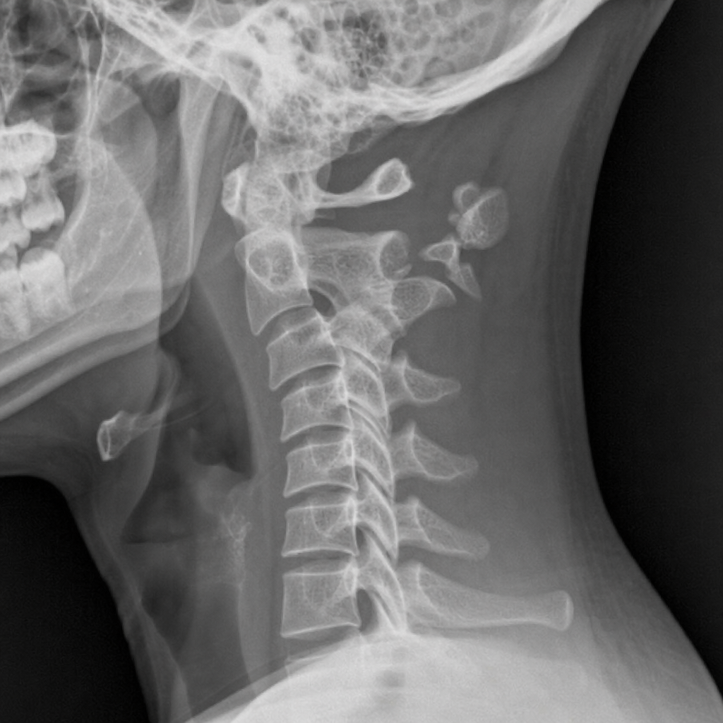
What is the type of fracture shown?
A 10-year-old boy presents with scoliosis, a tuft of hair over the skin of the lumbar spine, and weakness with features of lower motor neuron paralysis in both lower limbs. An X-ray of the spine reveals fusion of the two lumbar vertebrae at that level. What is the most likely diagnosis?
Which of the following are indicators of poor prognosis in Pott's spine?
Arrange the following conditions according to the worsening stages in Tuli's clinical staging: paraplegia in extension, ankle clonus present with extensor plantar response and no sensory deficit, no sensory deficit with motor deficit and ambulatory with support, paraplegia in flexion?
Minerva jacket is used for which of the following conditions?
Which of the following is NOT an indication for surgery in Pott's spine?
Practice by Chapter
Cervical Spine Disorders
Practice Questions
Thoracic Spine Disorders
Practice Questions
Lumbar Spine Disorders
Practice Questions
Intervertebral Disc Disease
Practice Questions
Spinal Stenosis
Practice Questions
Spondylolisthesis
Practice Questions
Spinal Deformities
Practice Questions
Spinal Infections
Practice Questions
Spinal Tumors
Practice Questions
Spinal Cord Injuries
Practice Questions
Minimally Invasive Spine Surgery
Practice Questions
Rehabilitation of Spine Conditions
Practice Questions
Want unlimited practice?
Get full access to all questions, explanations, and performance tracking.
Scan to download app