
Spine Disorders — MCQs

On this page
A 50-year-old male with a history of chronic low back pain presents with new-onset bowel and bladder incontinence. What is the next best step in management?
Which radiological feature is a hallmark of adolescent idiopathic scoliosis?
A 35-year-old male presents with sharp pain in his lower back radiating to the posterior thigh and leg. Physical examination reveals a positive straight leg raise test. Which nerve root is most likely affected?
A 70-year-old woman with a history of osteoporosis presents with sudden severe back pain after lifting a heavy object. What is the most likely diagnosis?
What is the characteristic postural deformity observed in advanced ankylosing spondylitis?
Which radiographic sign is characteristic of scoliosis?
A 14-year-old girl presents with a curvature of the spine that progresses with growth. Which condition is she likely to have?
During an examination of the back, a palpable step-off in the lower lumbar region suggests a disruption of which structure?
A 40-year-old athlete experiences sudden back pain while lifting heavy weights. An MRI reveals a herniated lumbar disc. What is the best initial management?
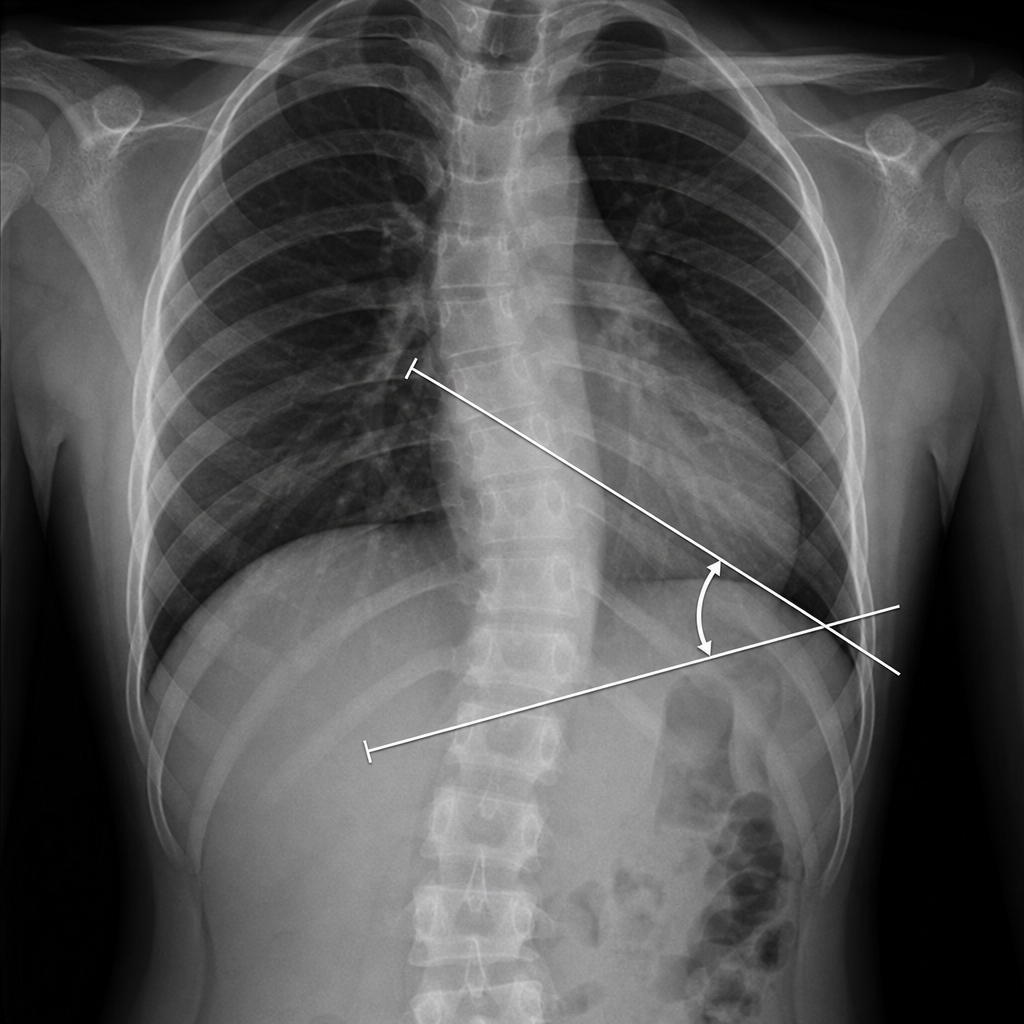
The angle shown in the image is known as:
Practice by Chapter
Cervical Spine Disorders
Practice Questions
Thoracic Spine Disorders
Practice Questions
Lumbar Spine Disorders
Practice Questions
Intervertebral Disc Disease
Practice Questions
Spinal Stenosis
Practice Questions
Spondylolisthesis
Practice Questions
Spinal Deformities
Practice Questions
Spinal Infections
Practice Questions
Spinal Tumors
Practice Questions
Spinal Cord Injuries
Practice Questions
Minimally Invasive Spine Surgery
Practice Questions
Rehabilitation of Spine Conditions
Practice Questions
Want unlimited practice?
Get full access to all questions, explanations, and performance tracking.
Scan to download app