
Spine Disorders — MCQs

On this page
Jefferson fracture is:
A patient presents with a burning sensation in the middle finger and weak triceps reflex. Which cervical disc level is most likely involved?
Prolapsed intervertebral Disc (PID) is most common at -
A patient while lifting a heavy weight presents with sudden onset pain in the lower back radiating along the postero-lateral thigh and lateral leg to the big toe with numbness. The most likely diagnosis is:
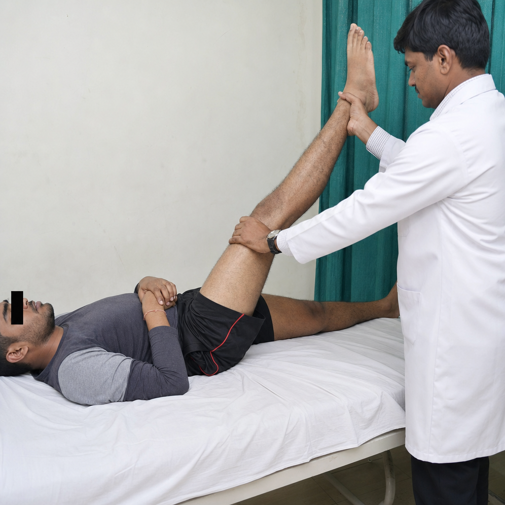
The given test is used for-
What is the most common cause of a single vertebral collapse?
Tuberculosis of the spine commonly affects all of the following parts of the vertebra except:
False about fracture of vertebrae
Block vertebrae are seen in
All of the following are the classical presentation of Craniovertrebral junction anomalies except
Practice by Chapter
Cervical Spine Disorders
Practice Questions
Thoracic Spine Disorders
Practice Questions
Lumbar Spine Disorders
Practice Questions
Intervertebral Disc Disease
Practice Questions
Spinal Stenosis
Practice Questions
Spondylolisthesis
Practice Questions
Spinal Deformities
Practice Questions
Spinal Infections
Practice Questions
Spinal Tumors
Practice Questions
Spinal Cord Injuries
Practice Questions
Minimally Invasive Spine Surgery
Practice Questions
Rehabilitation of Spine Conditions
Practice Questions
Want unlimited practice?
Get full access to all questions, explanations, and performance tracking.
Scan to download app