Which of the following is NOT a clinical feature of Tuberculosis of the Spine?
What is the commonest extradural spinal tumor?
Boutonniere deformity involves:
Flowing wax appearance on anterior and posterior borders of vertebrae with normal intervertebral disc space, occurring due to ligament calcification, is seen in which condition?
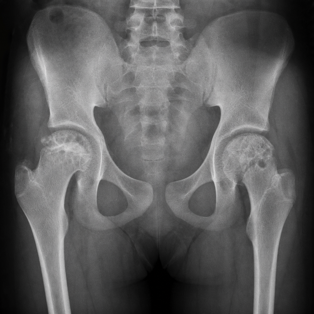
What is the earliest radiological change in rheumatoid arthritis?
Which of the following is NOT true regarding Scheuermann's osteochondritis?
During intervertebral disc prolapse, which part of the disc prolapses into the spinal canal?
What is the most common cause of vertebra plana?
A 38-year-old male presents with pain and tenderness in the dorsolumbar junction. Radiographs show destruction of the 12th dorsal vertebra with loss of disc space between D12 and L1. What is the most probable diagnosis?
In Spondylolisthesis, which of the following is NOT a useful investigation?
Explanation: **Explanation:** Tuberculosis (TB) of the spine, also known as **Pott’s Disease**, is a chronic granulomatous infection caused by *Mycobacterium tuberculosis*. Understanding its systemic and local manifestations is crucial for NEET-PG. **Why "Weight Gain" is the correct answer:** Tuberculosis is a **chronic catabolic state**. The body’s immune response to the infection increases the basal metabolic rate, leading to significant **weight loss** and anorexia (loss of appetite). Therefore, weight gain is clinically inconsistent with a diagnosis of active spinal TB. **Analysis of other options:** * **Loss of lordosis (Option A):** This is a classic early sign. The infection typically affects the anterior part of the vertebral body, leading to bone destruction and collapse. This causes the normal inward curve (lordosis) of the cervical or lumbar spine to straighten or reverse, eventually leading to a **kyphotic deformity (Gibbus)**. * **Night sweats (Option B) & Evening rise of temperature (Option D):** These are hallmark **constitutional symptoms** of TB. They are mediated by cytokines like TNF-alpha and Interleukins released during the chronic inflammatory process. **High-Yield Clinical Pearls for NEET-PG:** 1. **Most common site:** Lower thoracic and upper lumbar vertebrae (Thoracolumbar junction). 2. **Earliest sign:** Loss of joint space (disc space narrowing) on X-ray, though the disc is initially spared as it is avascular. 3. **Paradiscal type:** The most common pattern of involvement where the infection starts in the metaphysis and spreads under the anterior longitudinal ligament. 4. **Cold Abscess:** A collection of pus without the typical signs of inflammation (heat/redness), often presenting as a psoas abscess in lumbar TB.
Explanation: **Explanation:** Spinal tumors are anatomically classified into three compartments based on their relationship to the dura mater and the spinal cord. **Metastasis** is the correct answer because it is the most common tumor involving the spinal column overall and specifically the **extradural** space. **1. Why Metastasis is Correct:** Extradural tumors (located outside the dura) account for approximately 80% of all spinal neoplasms. The vast majority of these are secondary deposits (metastases) from primary cancers, most commonly originating from the **lung, breast, prostate, and kidney**. These tumors typically reach the vertebral bodies via the Batson venous plexus. **2. Analysis of Incorrect Options:** * **Neurofibroma (Option A):** This is the most common **Intradural-Extramedullary** tumor. While it can occasionally have an extradural component (forming a "dumb-bell" shape), its primary classification is intradural. * **Glioma (Option B):** These (specifically Ependymomas and Astrocytomas) are **Intramedullary** tumors, meaning they arise within the substance of the spinal cord itself. Ependymomas are the most common intramedullary tumors in adults. * **Meningioma (Option C):** This is the second most common **Intradural-Extramedullary** tumor, frequently seen in middle-aged women in the thoracic spine. **Clinical Pearls for NEET-PG:** * **Most common spinal tumor overall:** Metastasis (Extradural). * **Most common primary Intradural-Extramedullary tumor:** Neurofibroma (followed by Meningioma). * **Most common Intramedullary tumor (Adults):** Ependymoma. * **Most common Intramedullary tumor (Children):** Astrocytoma. * **Red Flag:** New-onset back pain in an elderly patient with a history of weight loss should always be investigated for spinal metastasis.
Explanation: **Explanation:** The **Boutonniere deformity** is a characteristic finger deformity most commonly seen in Rheumatoid Arthritis or following trauma. The primary pathology is the **rupture or attenuation of the central slip** of the extensor tendon at its insertion on the base of the middle phalanx. 1. **Why Option C is correct:** When the central slip is damaged, the lateral bands of the extensor mechanism slide volarly (towards the palm) past the axis of the PIP joint. These displaced lateral bands now act as flexors of the PIP joint. Simultaneously, the increased tension on these bands at their distal insertion causes **hyperextension of the DIP joint**. Thus, the classic presentation is **PIP flexion and DIP extension**. 2. **Why other options are incorrect:** * **Option A & B:** These describe uniform flexion or extension across both joints, which does not occur in isolated extensor mechanism injuries. * **Option D:** This describes **Swan-neck deformity** (PIP extension and DIP flexion), which is the functional opposite of Boutonniere deformity and is caused by volar plate laxity or terminal extensor tendon rupture (Mallet finger). **High-Yield Clinical Pearls for NEET-PG:** * **Elson’s Test:** The clinical test used to diagnose an acute central slip injury before the deformity becomes fixed. * **Etiology:** Often caused by a "jammed finger" (forced flexion of an actively extended PIP joint) or inflammatory erosion in Rheumatoid Arthritis. * **Management:** Initial treatment usually involves splinting the PIP joint in full extension for 6 weeks while allowing active DIP motion. * **Mnemonic:** **B**outonniere = **B**ent (Flexed) PIP.
Explanation: **Explanation:** The correct answer is **Diffuse Idiopathic Skeletal Hypertrophy (DISH)**, also known as Forestier’s disease. **1. Why DISH is correct:** DISH is a non-inflammatory systemic condition characterized by the ossification of ligaments and entheses. The hallmark radiological feature is the **"flowing wax"** or **"melted candle wax"** appearance, caused by the ossification of the **Anterior Longitudinal Ligament (ALL)**. * **Key Diagnostic Criteria:** * Involvement of at least four contiguous vertebral bodies. * **Preservation of intervertebral disc height** (unlike degenerative disc disease). * **Absence of sacroiliac (SI) joint involvement** (distinguishes it from Ankylosing Spondylitis). **2. Why other options are incorrect:** * **Ankylosing Spondylitis (AS):** Characterized by thin, vertical **syndesmophytes** (Bamboo spine) and mandatory **SI joint involvement** (sacroiliitis). It involves the destruction of disc spaces and is an inflammatory condition (HLA-B27 positive). * **Psoriatic Spondyloarthropathy:** Features asymmetric, coarse, and bulky non-marginal syndesmophytes. It usually presents with skin lesions and "pencil-in-cup" deformities in peripheral joints. * **Rheumatoid Arthritis:** Primarily affects the cervical spine (atlantoaxial subluxation). It does not cause flowing calcification of the thoracolumbar ligaments and typically spares the rest of the spine. **3. High-Yield Clinical Pearls for NEET-PG:** * **Demographics:** Most common in elderly males (>50 years). * **Metabolic Association:** Strongly associated with **Diabetes Mellitus** and obesity. * **Clinical Presentation:** Often asymptomatic or presents with stiffness; large cervical osteophytes may rarely cause **dysphagia**. * **Radiology Tip:** If you see "Bamboo spine," think AS; if you see "Flowing wax" with normal discs and normal SI joints, think DISH.
Explanation: **Explanation:** In Rheumatoid Arthritis (RA), the primary pathology is **synovitis**. The inflamed synovium (pannus) releases pro-inflammatory cytokines (like IL-1, IL-6, and TNF-α) and increases local blood flow (hyperemia). This process stimulates osteoclast activity and leads to the resorption of bone minerals in the immediate vicinity of the joint. Therefore, **periarticular osteopenia** (also known as juxta-articular osteoporosis) is the earliest radiological sign, appearing even before structural damage to the cartilage or bone occurs. **Analysis of Incorrect Options:** * **Decreased joint space:** This occurs later in the disease progression as the pannus destroys the articular cartilage. * **Articular erosion:** These are "marginal erosions" that occur at the "bare areas" (where the bone is not covered by cartilage). While characteristic of RA, they appear after the initial osteopenic phase. * **Subchondral cyst:** These are more characteristic of Osteoarthritis (Geodes) due to hydraulic pressure of synovial fluid through cracked cartilage; they are not a primary or early feature of RA. **NEET-PG High-Yield Pearls:** * **Earliest Sign overall:** Periarticular soft tissue swelling. * **Earliest Bone change:** Periarticular osteopenia. * **Most characteristic sign:** Marginal erosions. * **Spine involvement:** RA typically affects the **Cervical Spine** (atlanto-axial subluxation) but characteristically **spares** the Lumbar spine. * **Radiological hallmark:** Symmetrical joint involvement and absence of osteophytes (unlike Osteoarthritis).
Explanation: **Scheuermann’s Disease** (Juvenile Kyphosis) is a developmental disorder of the spine characterized by a structural increase in thoracic or thoracolumbar kyphosis. ### **Explanation of Options** * **Option B (Correct Answer):** The inheritance pattern of Scheuermann's disease is **Autosomal Dominant** with variable expressivity, not autosomal recessive. This makes Option B the false statement. * **Option A:** **Dorsal kyphosis** is the hallmark clinical feature. Unlike postural kyphosis, this is a "fixed" deformity (rigid) that becomes more apparent on the forward bending test (Adam’s test). * **Option C:** The condition typically presents in **adolescents** (13–16 years). While some studies suggest a near-equal gender distribution, classic teaching and many clinical series indicate a slight **predominance in boys**. * **Option D:** Pathologically, there is **irregular ossification of the vertebral endplates** and disc space narrowing. This leads to the classic radiographic finding of **Schmorl’s nodes** (herniation of the nucleus pulposus into the vertebral body). ### **NEET-PG High-Yield Pearls** * **Sorensen’s Radiographic Criteria:** Diagnosis requires at least **3 adjacent vertebrae** wedged by **5° or more** each. * **Clinical Sign:** Compensatory cervical and lumbar hyperlordosis is often present. * **Radiological Signs:** 1. **Schmorl’s nodes** (protrusion of disc into the vertebral body). 2. **Limbus vertebrae** (bone fragment at the anterior-superior edge of the vertebral body). 3. **Wedging** of vertebral bodies. * **Management:** Conservative (exercises/bracing) for curves <60°; surgery (posterior fusion) is considered for curves >70–75° or progressive neurological deficits.
Explanation: ### Explanation **Concept Overview:** The intervertebral disc consists of two primary components: the **Nucleus Pulposus** (a central, gelatinous core derived from the embryonic notochord) and the **Annulus Fibrosus** (a peripheral ring of dense collagenous fibers). In Intervertebral Disc Prolapse (IVDP), the Annulus Fibrosus undergoes degeneration or develops a tear (fissure). This allows the pressurized, semi-fluid **Nucleus Pulposus** to herniate or "prolapse" through the fibers into the spinal canal or intervertebral foramen, leading to nerve root compression. **Analysis of Options:** * **D. Nucleus Pulposus (Correct):** This is the actual material that herniates. It is rich in proteoglycans and water, acting as a shock absorber. When it prolapses, it causes both mechanical compression and chemical irritation of the nerve roots. * **A. Annulus Fibrosis:** This is the outer containment structure. While it may bulge (disc bulge), it is the *failure* of this structure that allows the nucleus to prolapse. * **B & C (Nucleus Fibrosis / Annular Pulposus):** These are anatomically incorrect terms created by mixing the names of the two disc components. They do not exist in human anatomy. **Clinical Pearls for NEET-PG:** * **Most Common Site:** L4-L5 or L5-S1 (Lumbar spine is most affected due to weight-bearing). * **Direction of Prolapse:** Usually **Postero-lateral**, as the Posterior Longitudinal Ligament (PLL) is thinner at the sides, making it the path of least resistance. * **Level of Compression:** A posterolateral prolapse usually compresses the **traversing nerve root** (e.g., an L4-L5 disc prolapse typically affects the L5 nerve root). * **Schmorl’s Nodes:** Prolapse of the nucleus pulposus vertically into the vertebral body.
Explanation: **Explanation:** **Vertebra plana** (also known as Calvé disease) refers to the complete symmetrical collapse of a vertebral body, resulting in a thin, wafer-like or "pancake" appearance, while the adjacent intervertebral discs remain preserved. **1. Why Eosinophilic Granuloma (EG) is correct:** Eosinophilic granuloma, a localized form of **Langerhans Cell Histiocytosis (LCH)**, is the most common cause of vertebra plana, particularly in children (ages 2–10). The pathological process involves the infiltration of the vertebral body by histiocytes, leading to osteolysis and rapid collapse. A classic radiographic feature is that despite the severe collapse, the bone density often remains normal, and the height can partially recover over time. **2. Why other options are incorrect:** * **Leukemia:** While it can cause generalized osteopenia and vertebral compression fractures in children, it rarely leads to the classic, isolated "wafer-thin" vertebra plana seen in EG. * **Tuberculosis (TB):** TB of the spine (Pott’s disease) typically involves the **intervertebral disc** (discitis) and adjacent vertebrae. It leads to wedge-shaped collapse and angulation (Gibbus deformity) rather than the uniform, flat collapse of a single body with disc preservation. * **Metastasis:** In adults, metastases (e.g., from lung or breast) are a common cause of pathological collapse, but they usually present as asymmetric destruction or "winking owl" sign (pedicle involvement) rather than classic vertebra plana. **Clinical Pearls for NEET-PG:** * **Mnemonic:** "MELT" for causes of Vertebra Plana (**M**etastasis/Myeloma, **E**osinophilic Granuloma, **L**ymphoma/Leukemia, **T**uberculosis). * **Key Radiographic Sign:** Preservation of disc space is the hallmark of EG, distinguishing it from infection. * **Age Factor:** If a question mentions a child with a "pancake vertebra," always suspect Eosinophilic Granuloma first.
Explanation: ### Explanation **Pott’s Spine (Tuberculous Spondylitis)** is the most probable diagnosis based on the classic triad of clinical and radiological findings: the location, the destruction of the vertebral body, and the involvement of the intervertebral disc space. 1. **Why Pott’s Spine is correct:** * **Location:** The dorsolumbar junction (D12-L1) is the most common site for spinal tuberculosis. * **Disc Space Involvement:** Tuberculosis typically starts in the paradiscal region. It destroys the subchondral bone and crosses the disc space to involve the adjacent vertebra. The loss of disc space is a hallmark of infectious etiology (TB or pyogenic) because the infection spreads via the paradoxical route, depriving the disc of nutrition. * **Demographics:** It commonly affects young to middle-aged adults in endemic regions. 2. **Why other options are incorrect:** * **Trauma:** While trauma causes vertebral collapse, it typically presents with an acute history of injury and does not cause "loss of disc space" unless there is a chronic associated infection. * **Vertebral Osteomyelitis (Pyogenic):** While it also involves the disc space, it usually presents with a more acute, febrile onset and rapid progression compared to the more indolent course of TB. * **Metastasis:** This is the most important differential. However, **metastases characteristically spare the intervertebral disc**. Malignant cells do not produce the proteolytic enzymes required to cross the fibrocartilage of the disc; hence, the disc space remains preserved on radiographs. ### High-Yield Pearls for NEET-PG: * **Earliest sign of Pott's spine:** Rarefaction of the paradiscal margin. * **Cold Abscess:** A hallmark of TB; it is "cold" because it lacks traditional signs of inflammation (heat/redness). * **Pott’s Paraplegia:** Most commonly occurs in the thoracic spine due to the narrow spinal canal and the physiological kyphosis. * **Radiological Sign:** "Girdle Pain" or "Bird’s Nest appearance" (due to paravertebral abscess in the thoracic region).
Explanation: **Explanation:** **Spondylolisthesis** is defined as the forward slippage of one vertebra over the one below it (most commonly L5 over S1). To diagnose and grade this condition, the clinician must visualize the sagittal alignment of the vertebral column. 1. **Why Anteroposterior (AP) X-ray is the correct answer (NOT useful):** An AP view provides a frontal look at the spine. While it may show secondary signs in severe cases (like the "inverted Napoleon hat sign"), it cannot demonstrate the **forward displacement** of the vertebral body. Since the slippage occurs in the sagittal plane, an AP view is insufficient for diagnosis, grading, or assessing the stability of the slip. 2. **Why the other options are wrong (Useful investigations):** * **Lateral View X-ray:** This is the **investigation of choice** and the most useful initial tool. it allows for the measurement of the degree of slip (Meyerding Grading) and visualization of the defect in the pars interarticularis. * **CT Scan:** Excellent for detailed bony anatomy. It is the best modality to visualize the **pars defect** (spondylolysis) and associated bony changes. * **MRI Scan:** The gold standard for evaluating **neural structures**. It is essential to assess the degree of canal stenosis, foraminal narrowing, and nerve root compression. **High-Yield Clinical Pearls for NEET-PG:** * **Meyerding Grading:** Based on the percentage of slip (Grade I: <25%, Grade II: 25-50%, Grade III: 50-75%, Grade IV: 75-100%, Grade V: Spondyloptosis). * **Oblique X-rays:** Used to see the **"Scotty Dog"** sign; a break in the dog's neck represents a pars interarticularis defect. * **Inverted Napoleon Hat Sign:** Seen on **AP X-rays** only in severe cases (Grade IV or V) where the L5 vertebra tilts downward and overlaps the sacrum. * **Most common site:** L5-S1 (Isthmic type); L4-L5 (Degenerative type).
Cervical Spine Disorders
Practice Questions
Thoracic Spine Disorders
Practice Questions
Lumbar Spine Disorders
Practice Questions
Intervertebral Disc Disease
Practice Questions
Spinal Stenosis
Practice Questions
Spondylolisthesis
Practice Questions
Spinal Deformities
Practice Questions
Spinal Infections
Practice Questions
Spinal Tumors
Practice Questions
Spinal Cord Injuries
Practice Questions
Minimally Invasive Spine Surgery
Practice Questions
Rehabilitation of Spine Conditions
Practice Questions
Get full access to all questions, explanations, and performance tracking.
Start For Free