In walking, gravity tends to tilt pelvis and trunk to the unsupported side, the major factor in preventing this unwanted movement is?
All of the following factors affect osseointegration EXCEPT:
High stepping gait is due to
Patellar tendon-bearing P.O.P. cast is indicated in the following fracture:
Displaced transverse patella; what is the treatment?
Which prosthesis is shown below in the X-ray?

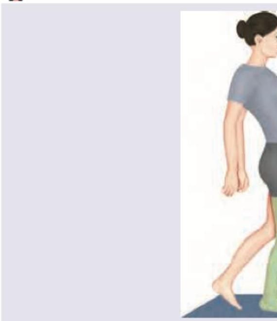
The following gait is seen due to weakness of:

The Milwaukee brace is used in the treatment of which of the following conditions?
Taylor's Brace is used for which of the following?
Which of the following is NOT true regarding the SACH Foot?
Explanation: ***Gluteus medius and minimus*** - The **gluteus medius** and **gluteus minimus** are essential **abductors** of the hip, primarily responsible for stabilizing the pelvis during the **single-limb support phase of gait**. - When one leg is lifted during walking, these muscles on the **stance leg side** contract to prevent the pelvis from tilting downwards on the unsupported swing leg side. *Adductor muscles* - **Adductor muscles** (adductor longus, brevis, magnus, pectineus, gracilis) primarily function to bring the thigh toward the midline of the body. - While they play a role in gait stability, their main action is not to prevent the lateral pelvic tilt described. *Quadriceps* - The quadriceps femoris group (rectus femoris, vastus lateralis, medialis, intermedius) are powerful **extensors of the knee**. - They are crucial for weight acceptance and propulsion during walking but do not directly prevent lateral pelvic tilt [1]. *Gluteus maximus* - The **gluteus maximus** is the largest and most powerful muscle of the hip, primarily responsible for **hip extension** and **external rotation**. - It is crucial for activities like climbing stairs or running, but its main role in normal walking is not to prevent lateral pelvic tilt; that function is more specific to the gluteus medius and minimus.
Explanation: ***Patient's blood type*** - A patient's **blood type** (e.g., A, B, AB, O) is determined by antigens present on red blood cells and plays no direct role in the biological processes of bone healing or the integration of a dental implant with bone. - While systemic factors can influence osseointegration, blood type itself does not affect the cellular and molecular mechanisms required for direct bone-to-implant contact. *Biocompatibility of implant material* - The **biocompatibility** of the implant material (e.g., **titanium**) is crucial for osseointegration, as it must not elicit adverse reactions and must permit host bone growth on its surface. - Materials that are cytotoxic or inflammatory will prevent bone apposition and lead to fibrous encapsulation rather than direct bone contact. *Implant design* - **Implant design**, including features like **surface roughness**, thread pitch, and macro-geometry, significantly influences the initial stability and long-term success of osseointegration. - A greater surface area and appropriate surface treatments can enhance bone cell attachment and differentiation, promoting faster and stronger bone integration. *Status of the host bed* - The **status of the host bone bed** refers to its quality and quantity (e.g., bone density, vascularity), which are critical for the biological processes of osseointegration. - Adequate bone volume and good bone quality provide a stable foundation and sufficient blood supply for bone regeneration around the implant.
Explanation: ***Foot drop*** - **Foot drop** causes the patient to lift the leg higher during walking to prevent the toes from dragging on the ground, resulting in a **high stepping gait**. - This condition is often due to weakness or paralysis of the **dorsiflexor muscles** of the foot, typically from **peroneal nerve injury** or **L4/L5 radiculopathy**. *Gluteus maximum paralysis* - **Gluteus maximus paralysis** causes difficulty with hip extension and is often compensated by a **backward lurch** of the trunk during gait. - It results in a **Trendelenburg gait** (if the gluteus medius is also affected) or instability during standing, but not typically a high stepping gait. *CDH* - **Congenital hip dysplasia (CDH)** involves abnormal development of the hip joint. - It usually leads to a **waddling gait** due to instability and pain, or limb length discrepancy, not a high stepping gait. *Quadriceps paralysis* - **Quadriceps paralysis** results in weakness or inability to extend the knee. - Patients typically compensate by hyperextending the knee or leaning forward over the affected leg during gait, which is not a high stepping gait.
Explanation: ***Fracture of the tibia*** - A **patellar tendon-bearing (PTB) cast** is specifically designed to bypass the knee joint and transfer weight from the patellar tendon to the cast, offloading the tibia. - This design is particularly useful for **stable, distal tibia fractures** where partial weight-bearing is desired to promote healing. *Fracture of the patella* - A PTB cast would place direct pressure on the **patella**, which is contraindicated in a patellar fracture. - Patellar fractures often require a **cylinder cast** or surgical fixation to immobilize the knee. *Fracture of the femur* - Femoral fractures are typically **more proximal** and require **traction**, **internal fixation**, or a **spica cast** for stabilization. - A PTB cast would not provide adequate immobilization or weight-bearing relief for a femoral fracture due to its design. *Fracture of the medial malleolus* - Medial malleolus fractures involve the **ankle joint**, which is distal to the area covered by a PTB cast. - These fractures typically require a **short leg cast** or surgical repair, focusing on ankle stabilization.
Explanation: ***Tension band wiring*** - This is the standard treatment for **displaced patellar fractures**, especially transverse ones, as it converts distractive forces into compressive ones, promoting healing. - It involves using K-wires and a cerclage wire to achieve **stable fixation** and allow for early range of motion. *POP cast* - A **Plaster of Paris (POP) cast** is typically used for non-displaced or minimally displaced fractures that do not require surgical stabilization. - It would not provide adequate stability for a **displaced transverse patella fracture**, which is prone to further displacement due to quadriceps pull. *Non-operative* - **Non-operative treatment** is reserved for **non-displaced** or minimally displaced patellar fractures where the extensor mechanism remains intact. - A **displaced transverse patella fracture** disrupts the extensor mechanism, making non-operative treatment unsuitable as it would lead to poor functional outcomes and a high risk of nonunion. *Patellectomy* - **Patellectomy** (partial or total removal of the patella) is considered for severely comminuted fractures where reconstruction is not possible or for chronic symptomatic nonunion. - It is generally avoided as a primary treatment for displaced transverse fractures due to the importance of the patella in **knee extension mechanics** and the risk of quadriceps weakness.
Explanation: ***Austin Moore's prosthesis*** - The image clearly shows a **femoral stem with a long intramedullary component** and an **integrated prosthetic head** that articulates directly with the native acetabulum. This is characteristic of a hemiarthroplasty design, specifically resembling an Austin Moore prosthesis. - This type of prosthesis is commonly used for **femoral neck fractures** in older patients, replacing only the femoral head and neck rather than the entire hip joint. *Articular resurfacing* - **Articular resurfacing** involves capping the femoral head and lining the acetabulum with metallic implants, preserving more bone than a traditional total hip replacement. - The X-ray image does not show a cap on the femoral head or a separate acetabular component, which are features of resurfacing. *Thompson prosthesis* - The **Thompson prosthesis** is another type of hemiarthroplasty, but it typically has a **shorter, bulkier femoral stem** and a **relatively smaller head** compared to the Austin Moore prosthesis shown. - While both Thompson and Austin Moore prostheses are hemiarthroplasties, the specific shape and length of the stem in the X-ray are more consistent with an Austin Moore design. *Birmingham hip replacement* - The **Birmingham hip replacement** is a type of **hip resurfacing arthroplasty**, which, as explained earlier, involves capping the femoral head and is not depicted in this image. - It maintains more of the patient's original bone structure compared to conventional total hip replacement but still requires both femoral and acetabular components.
Explanation: ***Gluteus medius*** - Weakness of the **gluteus medius** leads to a **Trendelenburg gait**, where the pelvis drops on the unsupported side during the swing phase of gait. - The image suggests pelvic tilting, which is characteristic of the body attempting to compensate for the inability of the gluteus medius to stabilize the pelvis. *Gluteus maximus* - Weakness of the gluteus maximus causes difficulty in **hip extension**, resulting in a **lurching gait** where the trunk is thrown backward at heel strike. - This is commonly known as a **gluteus maximus lurch**, which is not depicted in an obvious manner here. *Psoas major* - Weakness of the psoas major would primarily affect **hip flexion**, making it difficult to lift the leg off the ground (e.g., during the swing phase). - This would result in compensatory movements such as circumduction or hiking the hip, rather than the characteristic pelvic drop. *Tibialis anterior* - Weakness of the tibialis anterior causes **foot drop**, leading to a **steppage gait** where the knee is lifted high to avoid dragging the foot. - The image does not show a foot drop or high stepping, thus ruling out tibialis anterior weakness.
Explanation: **Explanation:** The **Milwaukee brace** (also known as a Cervico-Thoraco-Lumbo-Sacral Orthosis or CTLSO) is a classic active corrective orthosis designed specifically for the non-operative management of **Scoliosis**. **1. Why Scoliosis is Correct:** The brace is used for curves with an apex above T7. It works on the principle of **longitudinal traction** and **lateral pressure**. It consists of a pelvic mold, three upright metal stays (one anterior, two posterior), and a neck ring with a throat mold and occipital pads. This design encourages the patient to pull away from the pads, thereby actively correcting the spinal curvature. It is typically indicated for progressive curves between 25° and 40° (Cobb’s angle) in a skeletally immature child (Risser sign 0-II). **2. Why Other Options are Incorrect:** * **Kyphosis:** While a modified Milwaukee brace can be used for Scheuermann’s kyphosis, it is primarily and classically associated with Scoliosis in medical examinations. For lower thoracic kyphosis, a Boston brace or Taylor’s brace is more common. * **Cubitus varus:** This is a coronal plane deformity of the elbow (Gunstock deformity), usually a late complication of supracondylar fractures. It is treated surgically (e.g., French osteotomy), not with a spinal brace. * **Genu varum:** This refers to "bow legs." Treatment involves observation, Vitamin D (if rachitic), or corrective braces like the **HKAFO** or medial upright orthotics, but never a spinal brace. **High-Yield Clinical Pearls for NEET-PG:** * **Boston Brace:** A TLSO (Thoraco-Lumbo-Sacral Orthosis) used for curves with an apex below T7; it is "low-profile" and lacks the neck ring. * **Charleston Bending Brace:** A nocturnal (night-time) brace used for scoliosis. * **Somerset/SOMI Brace:** Used for cervical spine stabilization. * **Indication Rule:** Bracing is generally indicated when the Cobb’s angle is **25°–40°**. If the angle exceeds **40°–45°**, surgical intervention (e.g., spinal fusion with pedicle screws) is usually required.
Explanation: **Explanation:** **Taylor’s Brace** is a high-yield spinal orthosis in orthopaedics. It is a **thoraco-lumbo-sacral orthosis (TLSO)** designed specifically for **dorsolumbar immobilization**. The brace consists of two vertical posterior bars (paraspinal bars) and a pelvic band. It works on the principle of **three-point pressure**, providing hyperextension to the spine. By limiting flexion and rotation of the thoracolumbar junction, it stabilizes the spine, making it the gold standard for conditions like **Pott’s disease (Spinal TB)** and stable compression fractures of the lower thoracic or upper lumbar vertebrae. **Analysis of Options:** * **A. Cervical immobilization:** This requires a cervical collar (e.g., Philadelphia collar) or a Halo-vest. Taylor’s brace does not extend high enough to stabilize the neck. * **C. Scoliosis:** While some TLSOs are used for scoliosis, the specific brace of choice is usually the **Milwaukee brace** (for high curves) or the **Boston brace** (for lower curves). Taylor’s brace is for immobilization, not for corrective lateral forces. * **D. Fracture femur:** Femur fractures are managed with traction, intramedullary nails, or plates; orthotic management involves a Thomas splint or a Knee-Ankle-Foot Orthosis (KAFO), not a spinal brace. **Clinical Pearls for NEET-PG:** * **Ashman’s/Knight’s Brace:** Used for Lumbo-sacral immobilization (LSO). * **Milwaukee Brace:** A CTLSO used for Scoliosis with an apex above T8. * **Somersault/SOMI Brace:** Used for cervical spine injuries (C4-C5 level). * **Key Component:** Taylor’s brace specifically restricts **flexion and extension** of the dorsolumbar spine.
Explanation: **Explanation:** The **SACH (Solid Ankle Cushion Heel) Foot** is the most commonly used non-articulated prosthetic foot. The correct answer is **Option C** because the SACH foot is rigid and lacks a functional ankle joint, which makes **squatting very difficult** for the user. In the Indian context, where squatting is culturally significant, specialized modifications or different prostheses (like the Jaipur Foot) are preferred. **Analysis of Options:** * **A. It has a solid ankle cushion heel:** This is **true**. The SACH foot consists of a rigid internal wooden or plastic keel surrounded by a molded foam rubber shell. The "cushion heel" compresses at heel strike to simulate plantarflexion. * **B. It is a type of prosthesis:** This is **true**. It is a distal component of a lower limb prosthesis used for transtibial (below-knee) and transfemoral (above-knee) amputees. * **D. It does not look like a normal foot:** This is **true**. While it is shaped like a foot, it is a static, cosmetic block. It lacks the realistic texture, toe movements, and flexibility of a natural foot or more advanced lifelike prosthetics. **High-Yield Clinical Pearls for NEET-PG:** * **Mechanism:** It simulates "pseudo-plantarflexion" through heel compression during the early stance phase. * **Indication:** Best for elderly patients or those with low activity levels due to its high stability and low maintenance. * **Contraindication:** Not ideal for uneven terrain or activities requiring squatting/pivoting. * **Comparison:** Unlike the SACH foot, the **Jaipur Foot** allows for squatting and cross-legged sitting, making it more suitable for the Indian lifestyle.
Get full access to all questions, explanations, and performance tracking.
Start For Free