
Arthrology and Infections — MCQs

On this page
What is the most common cause of fracture in hemophilic arthropathy?
What is true about frozen shoulder?
A patient with nephrotic syndrome on long-term steroid therapy for 6 years presents with a limp gait and limitation of hip abduction and internal rotation. What is the most probable diagnosis?
Melon seed bodies are found in which of the following conditions?
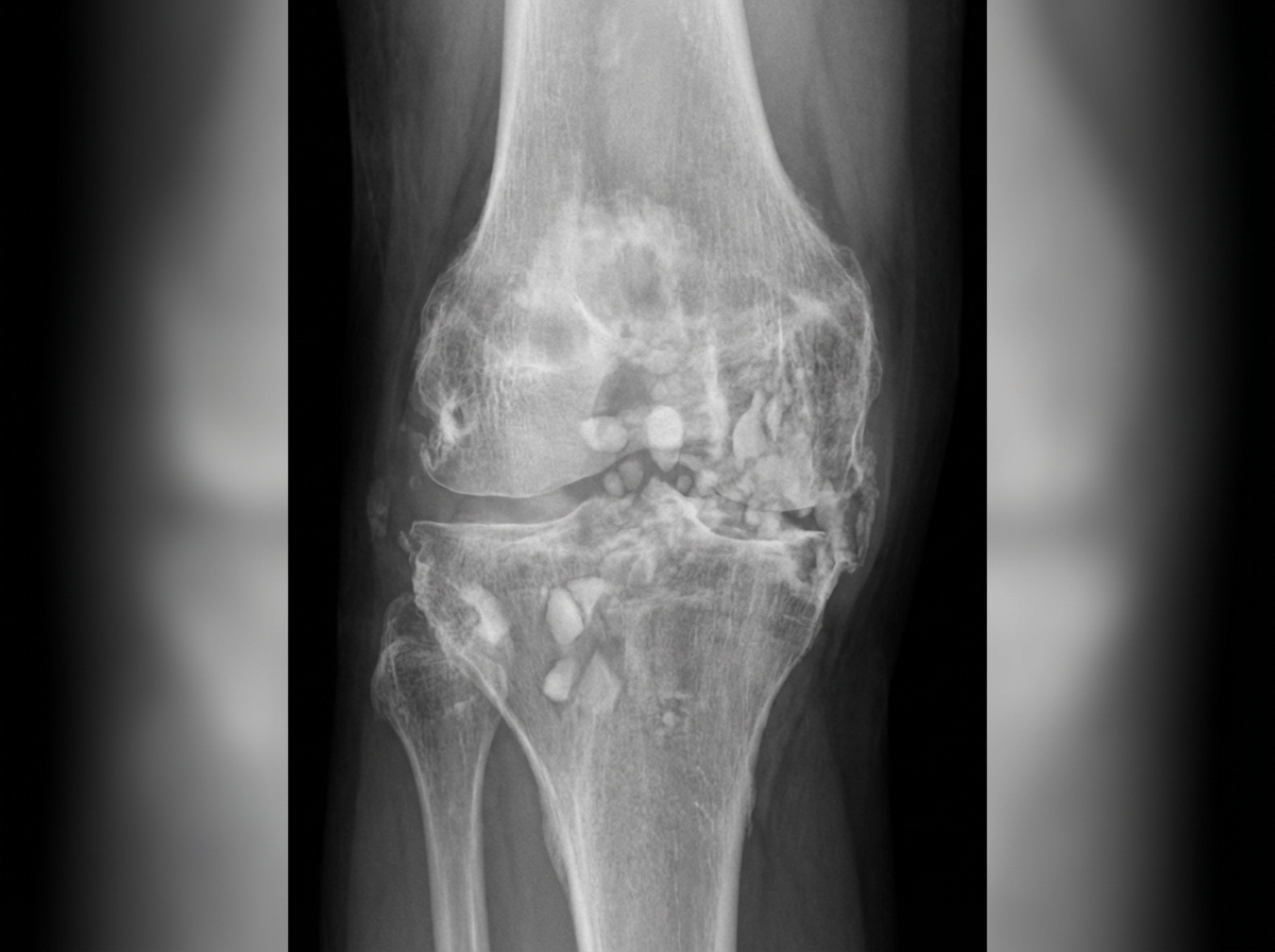
Examine the provided knee X-ray carefully. What is the most likely diagnosis?
What is the recommended total duration of antibiotic therapy for acute osteomyelitis?
A pregnant woman presents with a 6-week history of right hip pain. On examination, she has guarding on passive hip movements. Plain X-ray showed osteoporosis of the proximal femur. What is the most likely diagnosis?
What is the radiographic appearance of dead bone on an X-ray?
A 7-year-old boy presents with abrupt onset of hip pain, with the hip held in abduction. A hernogram is normal, and the ESR is elevated. What is the next line of management?
Which of the following drugs is NOT known to produce osteonecrosis?
Practice by Chapter
Septic Arthritis
Practice Questions
Osteomyelitis
Practice Questions
Tuberculosis of Bones and Joints
Practice Questions
Fungal and Parasitic Infections
Practice Questions
Diabetic Foot Infections
Practice Questions
Prosthetic Joint Infections
Practice Questions
Reactive Arthritis
Practice Questions
Management of Joint Infections
Practice Questions
Prevention of Orthopaedic Infections
Practice Questions
Biofilms in Orthopaedic Infections
Practice Questions
Antibiotic Prophylaxis
Practice Questions
Implant-Related Infections
Practice Questions
Want unlimited practice?
Get full access to all questions, explanations, and performance tracking.
Scan to download app