
Arthrology and Infections — MCQs

On this page
In chronic osteomyelitis, which complication may result in a non-healing sinus?
Which sign is commonly associated with spinal tuberculosis?
A 20-year-old patient presents with a fever and a swollen right knee. Joint aspiration reveals purulent fluid. What is the first line of treatment?
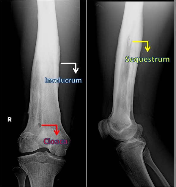
A 28-year-old male with a history of trauma presents with a non-healing sinus on the tibia. An X-ray shows a sequestrum. What is the appropriate next step in management?
A 10-year-old boy presents with a painful limp. X-rays reveal a mixed lytic and sclerotic lesion in the metaphysis of the femur. What is the most likely diagnosis?
What is the next best step in managing a child presenting with fever and metaphyseal bone pain, suspected of having acute osteomyelitis?
A 3-year-old child presents with a history of high fever, irritability, and refusal to move the right hip. What is the most likely diagnosis?
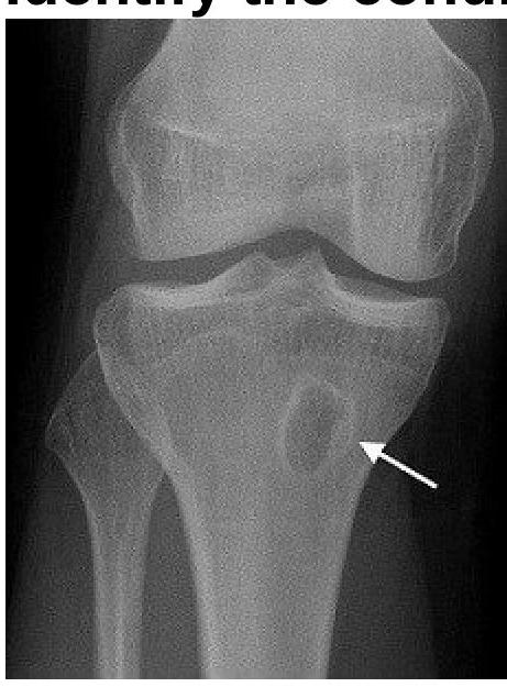
What is the most common presenting complaint of a patient with subacute osteomyelitis (Brodie's abscess)?
Which classification is most appropriate for assessing chronic osteomyelitis?
A 25-year-old male presents with localized pain in the tibia and swelling. Imaging reveals a bone abscess. Identify the condition.
Practice by Chapter
Septic Arthritis
Practice Questions
Osteomyelitis
Practice Questions
Tuberculosis of Bones and Joints
Practice Questions
Fungal and Parasitic Infections
Practice Questions
Diabetic Foot Infections
Practice Questions
Prosthetic Joint Infections
Practice Questions
Reactive Arthritis
Practice Questions
Management of Joint Infections
Practice Questions
Prevention of Orthopaedic Infections
Practice Questions
Biofilms in Orthopaedic Infections
Practice Questions
Antibiotic Prophylaxis
Practice Questions
Implant-Related Infections
Practice Questions
Want unlimited practice?
Get full access to all questions, explanations, and performance tracking.
Scan to download app