
Arthrology and Infections — MCQs

On this page
Which part of the bone is most commonly affected in children with acute osteomyelitis?
A 62-year-old diabetic patient presents with burning pain of the right foot, which began 3 weeks after an inversion injury of the ankle. Examination reveals flat arches and decreased proprioception bilaterally. What is the most likely diagnosis?
Onion skin appearance is seen in which of the following conditions?
Which of the following conditions can be responsible for heterotopic calcification?
Which of the following is NOT true about septic arthritis?
Heterotopic calcification occurs in which of the following conditions?
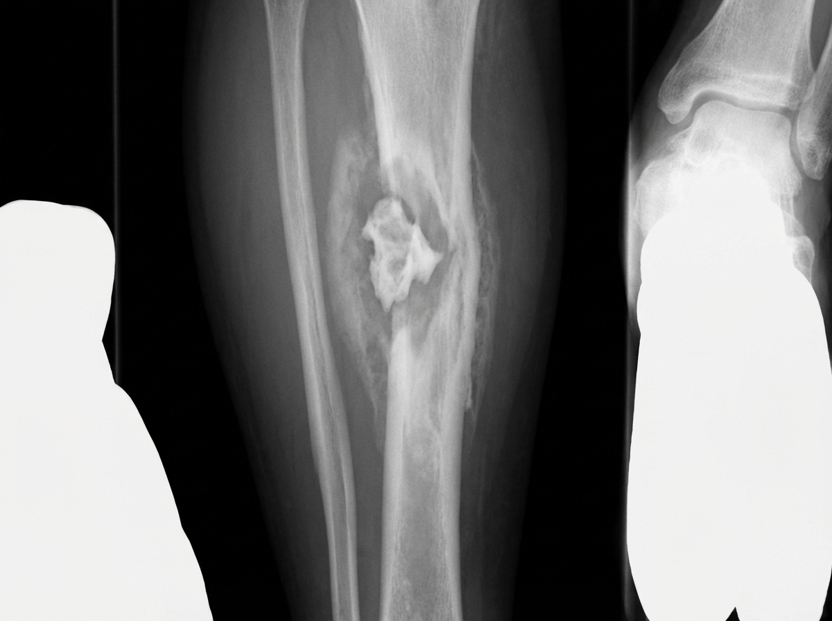
A patient with a history of Road Traffic Accident (RTA) two years ago developed pain and swelling at the same site in the leg. X-ray shows certain features. What is the most likely diagnosis?
What is the most common site of Garre's osteomyelitis?
An 8-year-old boy presents with a gradually progressing swelling and pain over the upper tibia for the past 6 months. Radiography reveals a lytic lesion with sclerotic margins in the upper tibial metaphysis. What is the most likely diagnosis?
What is an involucrum?
Practice by Chapter
Septic Arthritis
Practice Questions
Osteomyelitis
Practice Questions
Tuberculosis of Bones and Joints
Practice Questions
Fungal and Parasitic Infections
Practice Questions
Diabetic Foot Infections
Practice Questions
Prosthetic Joint Infections
Practice Questions
Reactive Arthritis
Practice Questions
Management of Joint Infections
Practice Questions
Prevention of Orthopaedic Infections
Practice Questions
Biofilms in Orthopaedic Infections
Practice Questions
Antibiotic Prophylaxis
Practice Questions
Implant-Related Infections
Practice Questions
Want unlimited practice?
Get full access to all questions, explanations, and performance tracking.
Scan to download app